NMNH Ostreopsis caribbeanus type specimen
Description:
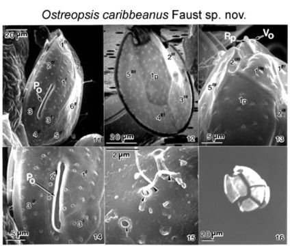
Figs 11-16. Cells of sp. nov. Figs. 11-15. Scanning electron microscopy. Fig. 11. Morphology of epithecal plates and position of apical pore plate (Po). Fig. 12. Hypothecal plates. Note long centrally situated Ip plate. Fig. 13. Cell in antapical view: antapical plate 1" is triangular; plate 2" is narrow and very small. The location of the ventral pore (Vo) and ridged plate (Rp) is illustrated. Fig. 14. Apical pore plate (Po) located off-center; note its morphology. Thecal surface smooth with round pores. Fig. 15. Ejected trichocyst emerges from thecal pores (arrowheads). Fig. 16. Epifluorescence microscopy of partially separated hypothecal plates. EMu:HOLOTYPE SEM NEGATIVE # 174097; SEM STUB # 174; FIELD # Morton-Clones; ACCESSION # ; CATALOG # 1545 ; FIGURE # 11
Included On The Following Pages:
This image is not featured in any collections.
Source Information
- license
- cc-by-nc-sa-3.0
- copyright
- National Museum of Natural History, Smithsonian Institution
- original
- original media file
- visit source
- partner site
- NMNH Marine Dinoflagellates
- ID