Plate 31
Description:
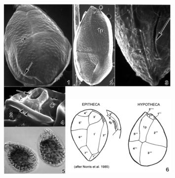
Plate 31. Ostreopsis heptagona. Figs. 1-4. SEM. Fig. 1. Epithecal view: cells broadly oval, oblong and pointed. Long curved apical pore plate, Po, off-center (arrow). Plate 1' heptagonal and distinctive. Fig. 2. Hypothecal view: plate 1p pentagonal and dorso-ventrally elongate. Fig. 3. Po long, narrow and curved. Narrow mucilage strands cover cell surface. Fig. 4. Ventral view: location of ventral opening (arrow), ventral plate (asterisk), and rigid plate (asterisk) within cingulum. Fig. 5. LM. Two cells. Fig. 6. Line drawing: thecal plate arrangement.
Included On The Following Pages:
- Life (creatures)
- Cellular (cellular organisms)
- Eukaryota (eukaryotes)
- SAR (Stramenopiles, Alveolates, Rhizaria)
- Alveolata (alveolates)
- Dinophyceae
- Ostreopsidaceae
- Ostreopsis
- Ostreopsis heptagona
- Gonyaulacales
- Dinoflagellata (dinoflagellates)
This image is not featured in any collections.
Source Information
- license
- cc-publicdomain
- bibliographic citation
- Faust, Maria A. and Rose A. Gulledge. Identifying Harmful Marine Dinoflagellates. Smithsonian Contributions from the United States National Herbarium, volume 42: 1-144 (including 48 plates, 1 figure and 1 table).
- original
- original media file
- visit source
- partner site
- NMNH Marine Dinoflagellates
- ID