NMNH Ostreopsis labens type specimen
Description:
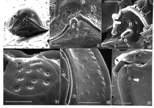
FIGS. 8-13. Ostreopsis labens sp. nov. FIG. 8. Ventral view is showing bi-convexity of the cell. Scale bar = 25 µm. FIG. 9. The 2" is very small (arrowhead). The ventral opening with a protuberant ridge and a curved plate (Rp) are situated in the cingulum adjacent to plate 1" and plate 1". Cingulum is smooth. Scale bar = 5 µm. FIG. 10. The ventral opening (Vo) is situated on the ventral plate (Vp). Scale bar = 5 µm. FIG. 11. Thecal surface is smooth; evenly spaced around trichocyst pores with smooth edges. Scale bar = 5 µm. FIG. 12. The apical pore (Po) is long, curved, and narrow associated with plate 2'. Row of marginal pores similar in size to thecal pores. Scale bar = 5 µm. FIG. 13. Right ventral view is unusual, recessed in sulcus. Flagellar pore opening is narrow (arrow). EMu: : HOLOTYPE SEM NEGATIVE # 170058; SEM STUB # 170; FIELD # 745-94; ACCESSION # 410840; CATALOG # 984; FIGURE # 4
Included On The Following Pages:
- Life (creatures)
- Cellular (cellular organisms)
- Eukaryota (eukaryotes)
- SAR (Stramenopiles, Alveolates, Rhizaria)
- Alveolata (alveolates)
- Dinophyceae
- Ostreopsidaceae
- Ostreopsis
- Ostreopsis labens
- Gonyaulacales
- Dinoflagellata (dinoflagellates)
This image is not featured in any collections.
Source Information
- license
- cc-by-nc-sa-3.0
- copyright
- National Museum of Natural History, Smithsonian Institution
- original
- original media file
- partner site
- NMNH Marine Dinoflagellates
- ID