Plate 33
Description:
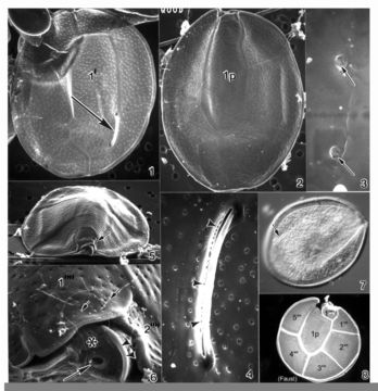
Plate 33. Ostreopsis mascarenensis. Figs. 1-5. SEM. Fig. 1. Epitheca: inner thecal surface. Cell very large, broadly ovate, large plates. Plate 1' elongate and hexagonal. Apical pore plate (Po) nearly straight. Fig. 2. Hypotheca: plate 1p long and wide. Fig. 3. Smooth cell surface with round pores; pores with two small openings (arrows). Fig. 4. Po with long narrow apical pore; small pores line the opening (arrowheads). Figs. 5-6. Ventral view of epitheca. Fig. 5. Cell compressed anterio-posteriorly; cingulum narrow with smooth edge. Small sulcus hidden (arrow). Fig. 6. Location of ventral opening (large arrow), ventral plate (asterisk), and rigid plate (arrowheads) within cingulum. Pores with ejected trichocysts (small arrows). Fig. 7. LM. Epitheca: Po (arrow) and cingulum in focus. Fig. 8. Line drawing: hypotheca plate arrangement.
Included On The Following Pages:
- Life (creatures)
- Cellular (cellular organisms)
- Eukaryota (eukaryotes)
- SAR (Stramenopiles, Alveolates, Rhizaria)
- Alveolata (alveolates)
- Dinophyceae
- Ostreopsidaceae
- Ostreopsis
- Ostreopsis mascarenensis
- Gonyaulacales
- Dinoflagellata (dinoflagellates)
This image is not featured in any collections.
Source Information
- license
- cc-publicdomain
- bibliographic citation
- Faust, Maria A. and Rose A. Gulledge. Identifying Harmful Marine Dinoflagellates. Smithsonian Contributions from the United States National Herbarium, volume 42: 1-144 (including 48 plates, 1 figure and 1 table).
- original
- original media file
- visit source
- partner site
- NMNH Marine Dinoflagellates
- ID