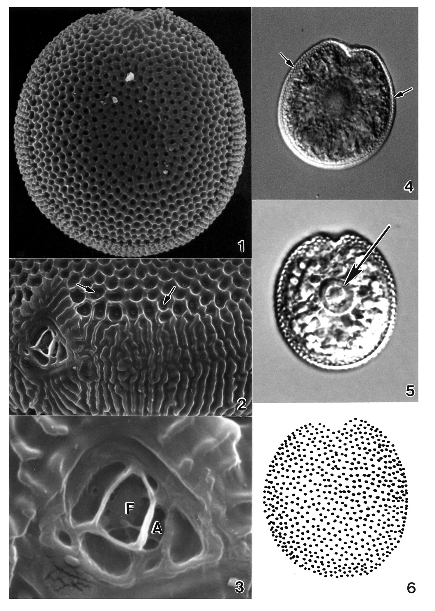
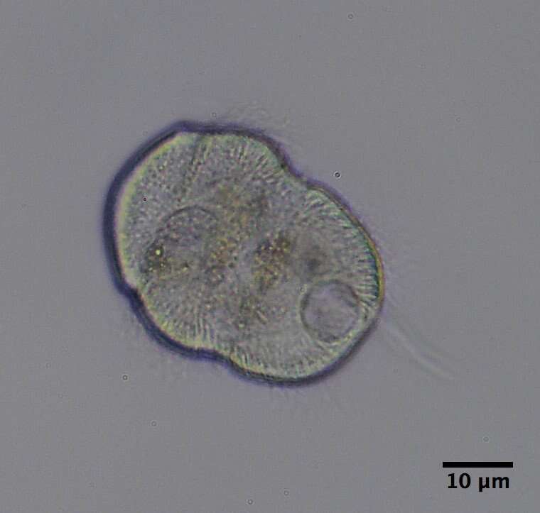
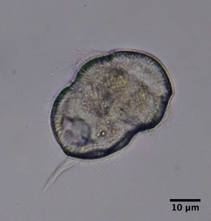
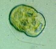
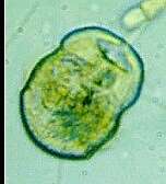
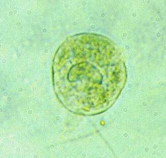
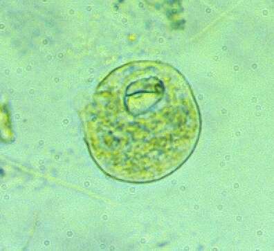
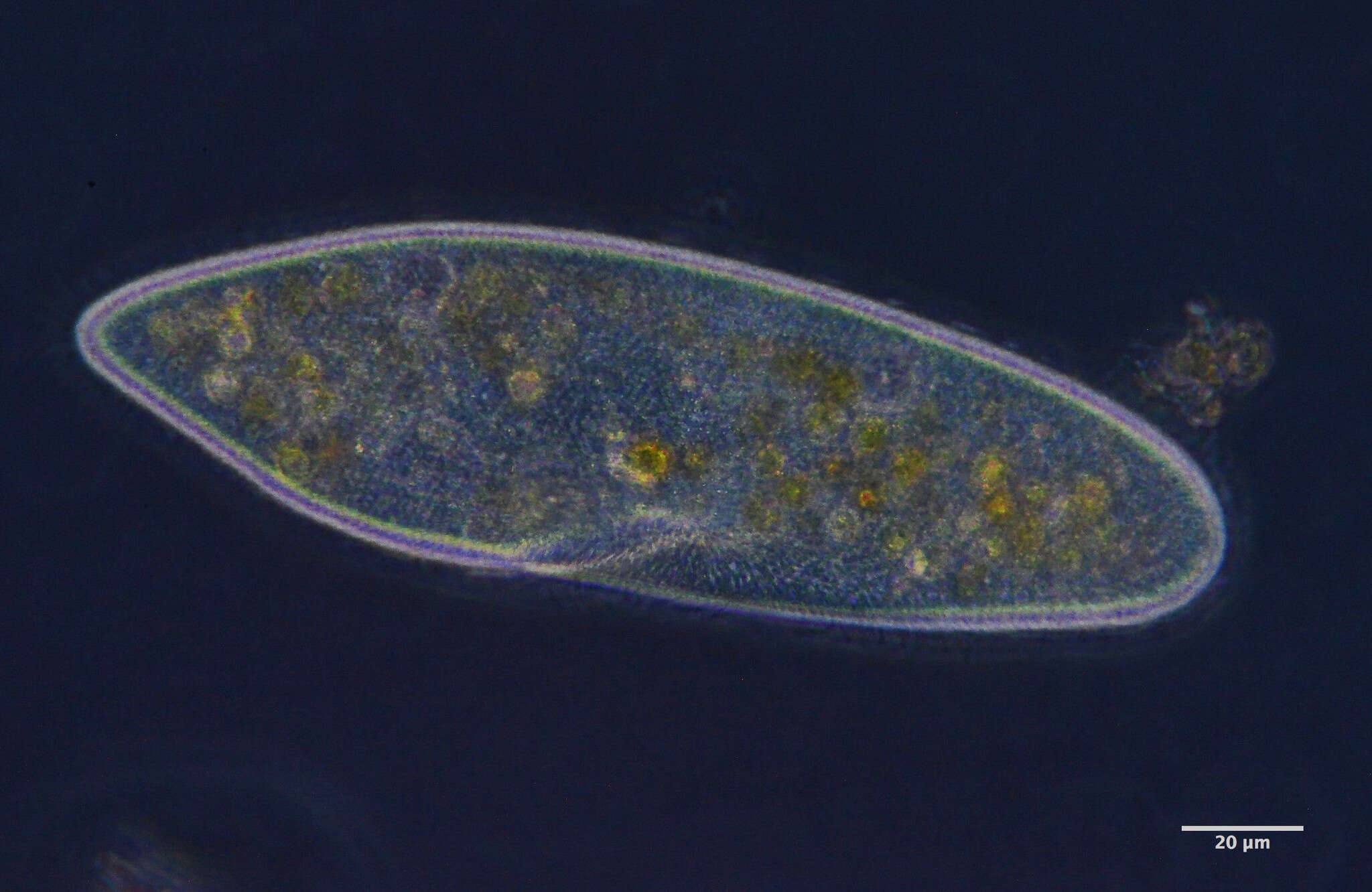
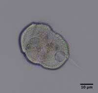
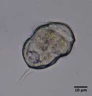
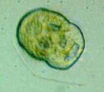
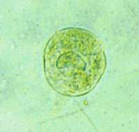
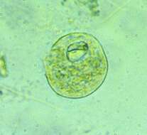
Plate 48. Prorocentrum ruetzlerianum. Figs. 1-3. SEM. Fig. 1. Right valve: cell round to ovoid, covered with pentagonal areolae. Cell surface rugose. Fig. 2. Anterio-lateral view. Each areola with small circular pore at its base (arrows). Intercalary band broad, transversely rugose. Fig. 3. Periflagellar area: small, shallow, unornamented depression on right valve; large flagellar (f) pore and smaller auxiliary (a) pore. Figs. 4-5. LM (M.A. Faust). Right valve: striated valve margins (small arrows); large central pyrenoid (large arrow). Fig. 6. Line drawing: areolae arrangement. (Figs. 1-3,6 after Faust 1990b)