-
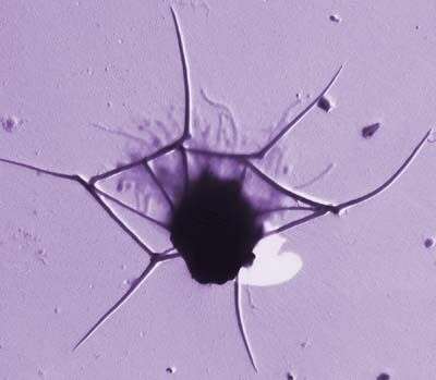
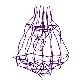
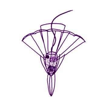
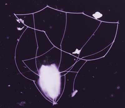
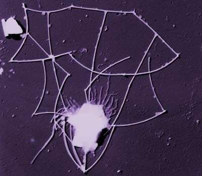
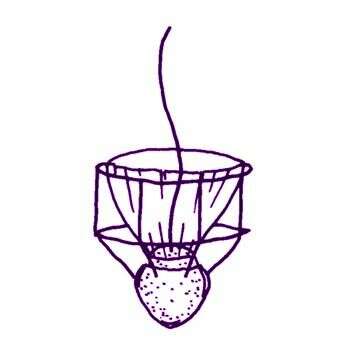
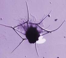
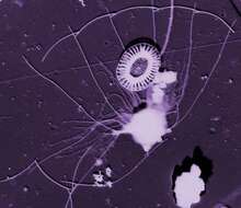
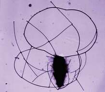
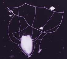
This image was made from samples taken during a scientific cruise in the Pacific. Water was filtered to concentrate the organisms that were present, then dried onto a thin sheet of plastic and then shadowed with a fine layer of metal to provide contrast. The preparation was then observed with an electron-microscope. This technique has been used to document the diversity of marine microbes, especially, protists in the oceans.
-
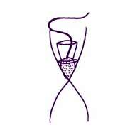
Campyloacantha spinifera (Leadbeater, 1973) Hara and Takahashi, 1987. Solitary choanoflagellate, the lorica is about 12 - 14 microns long with 5 or 6 anterior spines, one transverse costa at the top of the lorica chamber and a short posterior spine.
-
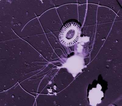
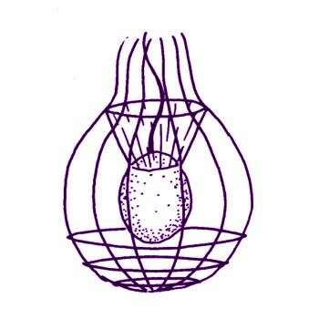
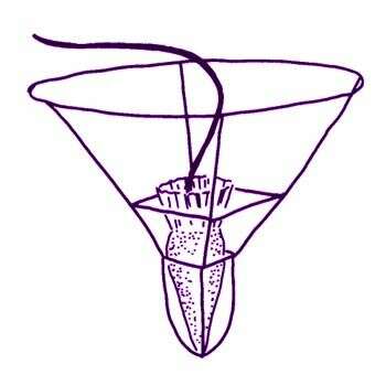
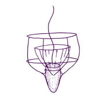
This image was made from samples taken during a scientific cruise in the Pacific. Water was filtered to concentrate the organisms that were present, then dried onto a thin sheet of plastic and then shadowed with a fine layer of metal to provide contrast. The preparation was then observed with an electron-microscope. This technique has been used to document the diversity of marine microbes, especially, protists in the oceans.
-
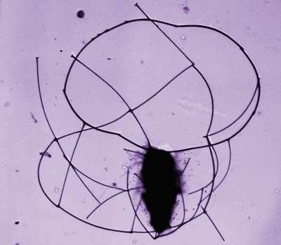
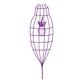
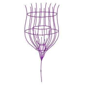
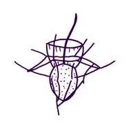
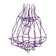
Crinolina isefiordensis Thomsen, 1981. Cell solitary, planktonic, living in skirt-shaped lorica, open anteriorly and posteriorly. Protoplast 8 microns long and 5 microns wide, without chloroplast. Single flagellum 2-3 times protoplast length, surrounded by a collar of tentacles. Height of collar approximately 6 microns, maximum diameter of collar 13 microns Lorica 25-30 microns long, diameter at base 20-30 microns, diameter at neck constriction 10-13 microns Longitudinal costae (12) 15-16, each composed of 6-7 costal strips. Two transverse costae. Longitudinal costae form spines at the anterior end of the lorica. Each spine is composed of two costal strips. Posteriorly the longitudinal costae project slightly beyond the transverse costal ring.
-
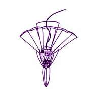
Crucispina cruciformis (Leadbeater, 1974) Espeland in Espeland and Throndsen, 1986. Cell body obovoid, 3-5 microns long x 2-5 microns wide, single flagellum 14-20 microns long, funnel-shaped collar of about 30 tentacles 1-3 microns long. Lorica 12-17 microns high consisting of one chamber formed by one transverse costa and several longitudinal costae converging posteriorly to join with two divergent tapered spines. Two tapered spines projecting anteriorly from the transverse costa.
-
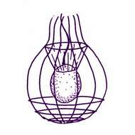
Diaphanoeca grandis Ellis, 1930. Choanoflagellates with a lorica composed of 12-14 longitudinal costae each formed by 7-8 strips of equal length. Three transverse rings encircle the base of the lorica and one ring, consisting of paired strips, encircles the longitudinal costae towards the anterior end. The protoplast, which is situated centrally within the lorica, bears an anterior collar of tentacles and a single flagellum. Accumulations of detached strips are frequently found at either end of the cell within the lorica. These strips are equal in length to those forming the costae.
-
Diaphanoeca grandis minor Throndsen, 1974. Similar to Diaphanoeca grandis but lorica length 18-22 microns, lorica diameter 15 microns Number of longitudinal costae 10.
-
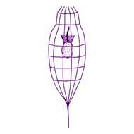
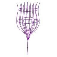
Diaphanoeca multiannulata Buck, 1980. Choanoflagellates with barrel-shaped lorica, 55-60 microns high excluding posterior spine, 24-25 microns wide (maximum), composed of 12 (or 11) longitudinal costae converging at posterior end and five transverse costal rings. Longitudinal costa consists of 12 (or 13) costal strips, each 6 microns long, one and a third costal strips project as free end spines beyond the anterior transverse costal ring. Transverse costal rings consist of 10-11 costal strips each, about 6 microns long, and spaced at intervals of one and a third, four and a half, six, eight and nine costal strips from anterior end of longitudinal costae. Overall length 61-66 microns
-
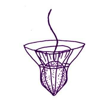
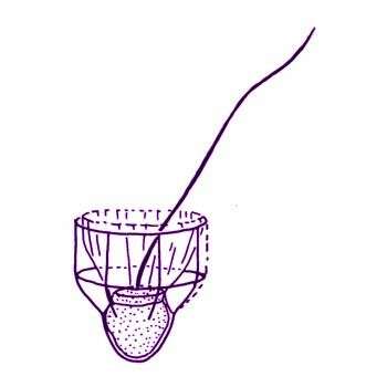
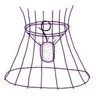
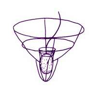
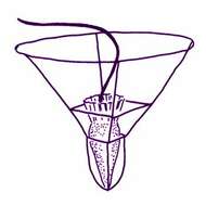
Diaphanoeca pedicellata Leadbeater 1972 (also referred to as Campanoeca pedicellata (Leadbeater, 1972) Throndsen, 1974). The subspherical protoplast (4-5 microns long, 4-5 microns wide) bears an anterior ring of 20-30 tentacles. The funnel-shaped lorica chamber is formed by 20-30 longitudinal costae (about 10 microns long) which at the upper end project as spines beyond the anterior transverse costa. The lorica is 45 microns high consisting of one chamber formed by about 14 longitudinal costae and three transverse costae(one towards the anterior end of the lorica, one just below the mid-lorica region, and one towards the posterior end of the lorica chamber). Posteriorly about seven longitudinal costae converge and join with a posterior stalk (about 11 microns long) formed by at least two or three costal strips attached end-to-end. All the costal strips forming the lorica are approximately equal in length and width.
-
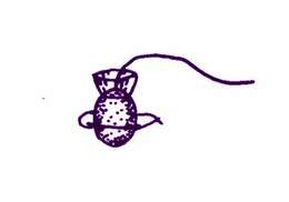
Monocosta fennica Thomsen, 1979. Cell solitary, planktonic. Protoplast (2.5-3.3 microns long) with an anteriorly inserted flagellum (9.2-11.7 microns long) surrounded by a ring of tentacles. Five costal strips forming a ring encircle the protoplast. Doubling of single costal strips may occur, giving a total of six or seven strips. Costal strip length varies between 2.9 and 3.3 microns
-
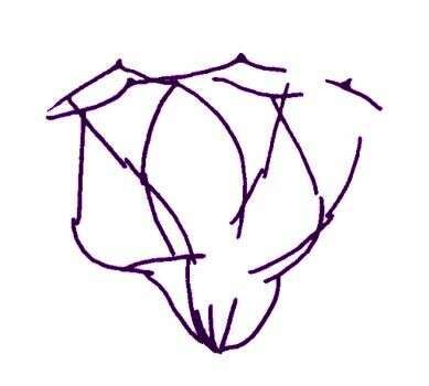
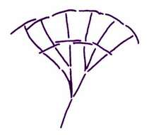
Nannoeca minuta (Leadbeater, 1972) Thomsen, 1988. The lorica about 4.5-7 microns long and tightly surrounds the protoplast and the rather short collar, whereas the flagellum (5-10 microns long) projects over the lorica in a conspicuous manner. The distinct hairpoint is also visible under the light microscope. The conical lorica is constructed from about 25 costal strips arranged in 6-10 longitudinal costae and two transverse costae. One transverse costa is located at the anterior opening of the lorica and the other is positioned in the mid-lorica region. The distance between the two transverse costae corresponds with the half-length or (less frequently) approximately the entire length of an anterior longitudinal costal strip. Lorica diagram (right) emphasizing longitudinal costae.
-

Parvicorbicula circularis Thomsen, 1976. Cell when dried approximately 3 x 5 microns The single flagellum, up to 15 microns long, protrudes above the lorica. The lorica is constructed of 4 longitudinal costae composed of 3 costal strips each and two transverse costae. One transverse costa (8 strips) delimits the lorica anteriorly. The bifurcated anterior tips of the longitudinal costae attach to the middle of every second transverse costal strip ( T -joints). The second transverse costa (6-8 strips) is located at the level of the joints between the first and the second longitudinal costal strip. In specimens with 8 costal strips in this costa, the longitudinal costae cross at every other joint between transverse costal strips. In specimens with 6-7 transverse costal strips, the crossings between longitudinal and transverse elements are less symmetric. The two posterior-most costal strips from each longitudinal costa overlap. The free ends of the penultimate longitudinal costal strips suspend a sheet which encompasses the cell.
-
Parvicorbicula manubriata Tong, 1997. Cells solitary or colonial. Cell 3.5-5.5 x 4.5-7 microns, flagellum 13-19 microns long surrounded by a collar of tentacles ~8 microns long. Lorica conical, ~8 microns long, consisting of a single transverse costa at the anterior of the lorica, and 8-10 longitudinal costae. The transverse costa consists of 10 or 11 slightly curved costal strips, one of which is duplicated. The longitudinal costae have no regular means of attachment to the transverse costa, joints may be in the middle or at the ends of the transverse costal strips. Longitudinal costae undergo numerical reduction, so that three to eight strips form the handle at the posterior of the lorica. All costal strips are narrow rods. Division is tectiform.
-
Parvicorbicula quadricostata Throndsen, 1970. The length of lorica is about 18 microns and its width is 24 microns
-
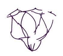
Parvicorbicula socialis (Meunier, 1910) Deflandre, 1960. The funnel-shaped lorica of this species is about 10 microns in diameter, with 10 longitudinal costae converging posteriorly, and two transverse costae. Each longitudinal costa consists of two costal strips. The anterior ring is made up of ten strips, each with a longitudinal costa attached to its middle part forming T-junctions. Drawing from a flattened lorica.
-
Parvicorbicula socialis socialis Deflandre, 1960. Lorica length 10 microns, lorica width 12 microns, longitudinal costae 10, transverse costae 2. Usually forms colonies.
-
This image was made from samples taken during a scientific cruise in the Pacific. Water was filtered to concentrate the organisms that were present, then dried onto a thin sheet of plastic and then shadowed with a fine layer of metal to provide contrast. The preparation was then observed with an electron-microscope. This technique has been used to document the diversity of marine microbes, especially, protists in the oceans.
-
Pleurasiga minima Throndsen, 1970. Lorica 9.5-16 microns long with 2 transverse costae and 7 longitudinal costae, all of which extend to the posterior of the lorica. The transverse costae are of about the same width, and the lorica narrows below the second costa to form a pointed base. Cell located at the posterior of the lorica, with flagellum protruding above it.
-
Pleurasiga minima minima Throndsen, 1970. The different varieties differ mainly in size. The lorica of P. minima minima is about 10 microns long and wide.
-
Pleurasiga reynoldsii Throndsen, 1970. The lorica is 23 microns long and 22 microns wide, respectively.
-
Pleurasiga echinocostata Espeland in Espeland and Throndsen, 1986. Cell 2.5-4.5 microns long, with single smooth flagellum surrounded basally by a pseudopodial collar. Lorica bell-shaped with 7 longitudinal costae, occasionally reduced to 6 or 5 in the posterior part, consisting of 3 costal strips, the 2 most anterior with a distal anterior dilatation. A single anterior transverse lorica ring consisting of 7 costal strips each with a terminal anteriorly pointing spine. Anterior longitudinal costa joining the middle of each coastal strip of the tranverse costa. Lorica length 7.5-10 microns, anterior diameter 5-8.3 microns Thje drawing is of a damaged lorica.