-
-
-
Phase contrast micrograph of a cell associated with detritus that attached to a submerged slide in the Lake.
-
-
-
-
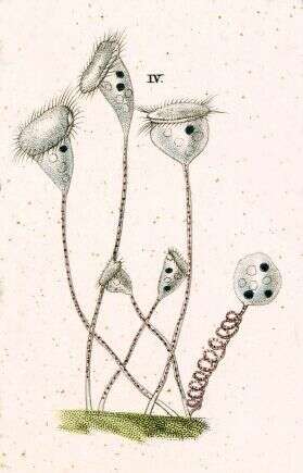
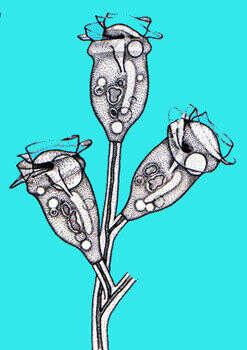
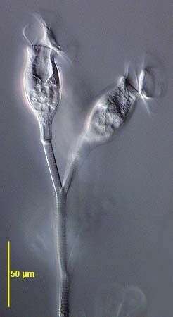
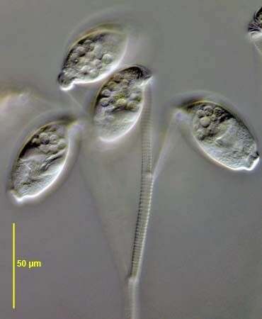
Zoothamnium (zoo-tham-knee-um) is a colonial peritrich ciliate. The feeding cells in sessile peritrich ciliates have lost all of the somatic cilia and only have the feeding cilia. The feeding cilia form a wreath which extends around the front of the cell and descends into a narrowing buccal cavity. This cavity ends at the cytostome where food is packaged into food vacuoles. If the cells become unhappy, they produce a temporary wreath of basal cilia (trochal cilia), break away from their stalk and use these to swim. The contractile elements of all associated cells of Zoothamnium colonies are interconnected so that if one cell contracts, all will tend to contract together. Differential interference contrast.
-
-
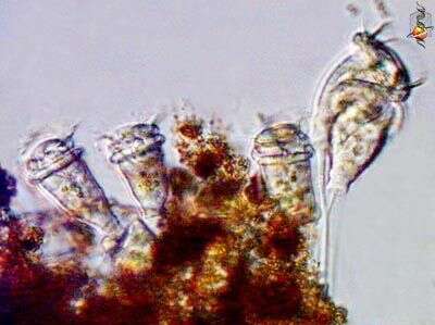
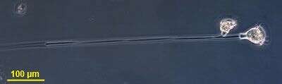
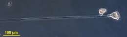
Small colony, two cells, attached to long stalk. The contractile spasmoneme is common to both cells.
-
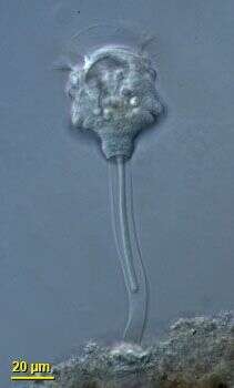
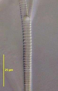
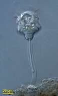
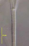
This is a single cell of the usually colonial Zoothamnium, showing the contractile spasmoneme within the stalk.
-
-
-
-
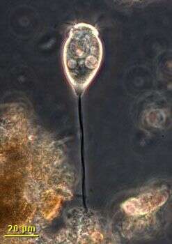
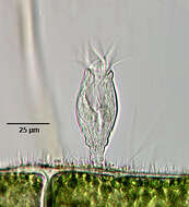
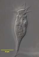
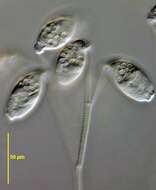
Portrait of the sessile solitary peritrich ciliate, Propyxidium (Corliss, 1979). The zooid is an elongate inverted bell shape. The short stalk is noncontractile. There is no distinct peristomal lip (unlike the similar Rhabdostyla). The prominent tilted peristomal disc is elevated on a broad stalk. The C-shaped macronucleus is transversely oriented (the two ends of the macronucleus are seen in cross-section here).The single contractile vacuole is in the posterior half of the cell. Propyxidium replaced the preoccupied Pyxidiella. Collected from a freshwater pond near Boise, Idaho. Brightfield illumination.
-
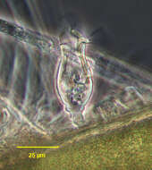
Portrait of the solitary peritrich ciliate, Propyxidium (Corliss, 1979). The zooid is an elongate inverted bell shape. The short stalk is noncontractile. There is no distinct peristomal lip (unlike the similar Rhabdostyla). The prominent tilted peristomal disc is elevated on a broad stalk (seen well here). The C-shaped macronucleus is transversely oriented (not well seen in this image). The single contractile vacuole is in the posterior half of the cell. Propyxidium replaced the preoccupied Pyxidiella. Collected from a freshwater pond near Boise, Idaho. Phase contrast illumination.
-
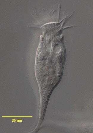
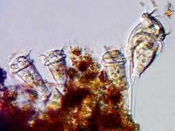
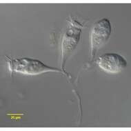
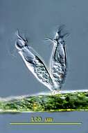
Zooid of Opercularia coarctata (CLAPARÃDE & LACHMANN, 1858) ROUX, 1901. Collected from an ephemeral puddle on a flood-irrigated lawn in Boise, Idaho. July 2007. DIC.
-
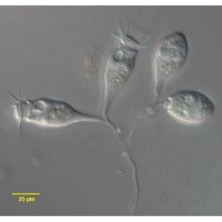
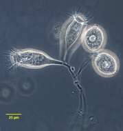
Opercularia coarctata (CLAPARÃDE & LACHMANN, 1858) ROUX, 1901. Collected from an ephemeral puddle on a flood-irrigated lawn in Boise, Idaho. July 2007. DIC. Collected from an ephemeral puddle on a flood-irrigated lawn in Boise, Idaho. July 2007. DIC.
-
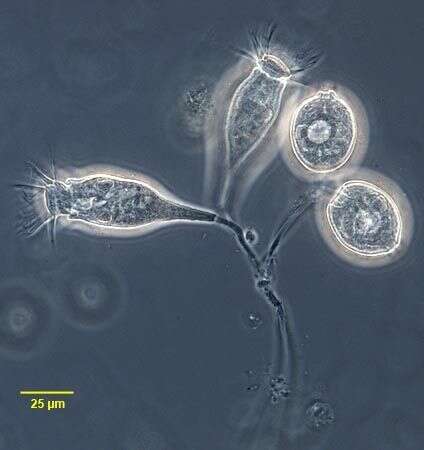
Opercularia coarctata (CLAPARÃDE & LACHMANN, 1858) ROUX, 1901. Collected from an ephemeral puddle on a flood-irrigated lawn in Boise, Idaho. July 2007. DIC.
-
in vivo portrait of Opercularia nutans (EHRENBERG, 1831) STEIN, 1854.Phase contrast.
-
Opercularia nutans (EHRENBERG, 1831) STEIN, 1854.DIC.
-
Detail view of the stalk of Opercularia nutans (EHRENBERG, 1831) STEIN, 1854. DIC.
-
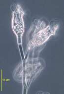
Contracted zooids of Opercularia nutans (EHRENBERG, 1831) STEIN, 1854.DIC.
-
Originally described by Ehrenberg under the name Epistylis nutans.
-
Opercularia (owe-perk-you-lair-ee-a) penardi is attached by a short non-contractile stalk to the substrate. The cells are contractile and retracted individuals have a folded pellicula. The macronucleus is band-shaped. This specimen was collected in freshwater ponds near Konstanz, Germany. nDifferential interference contrast.