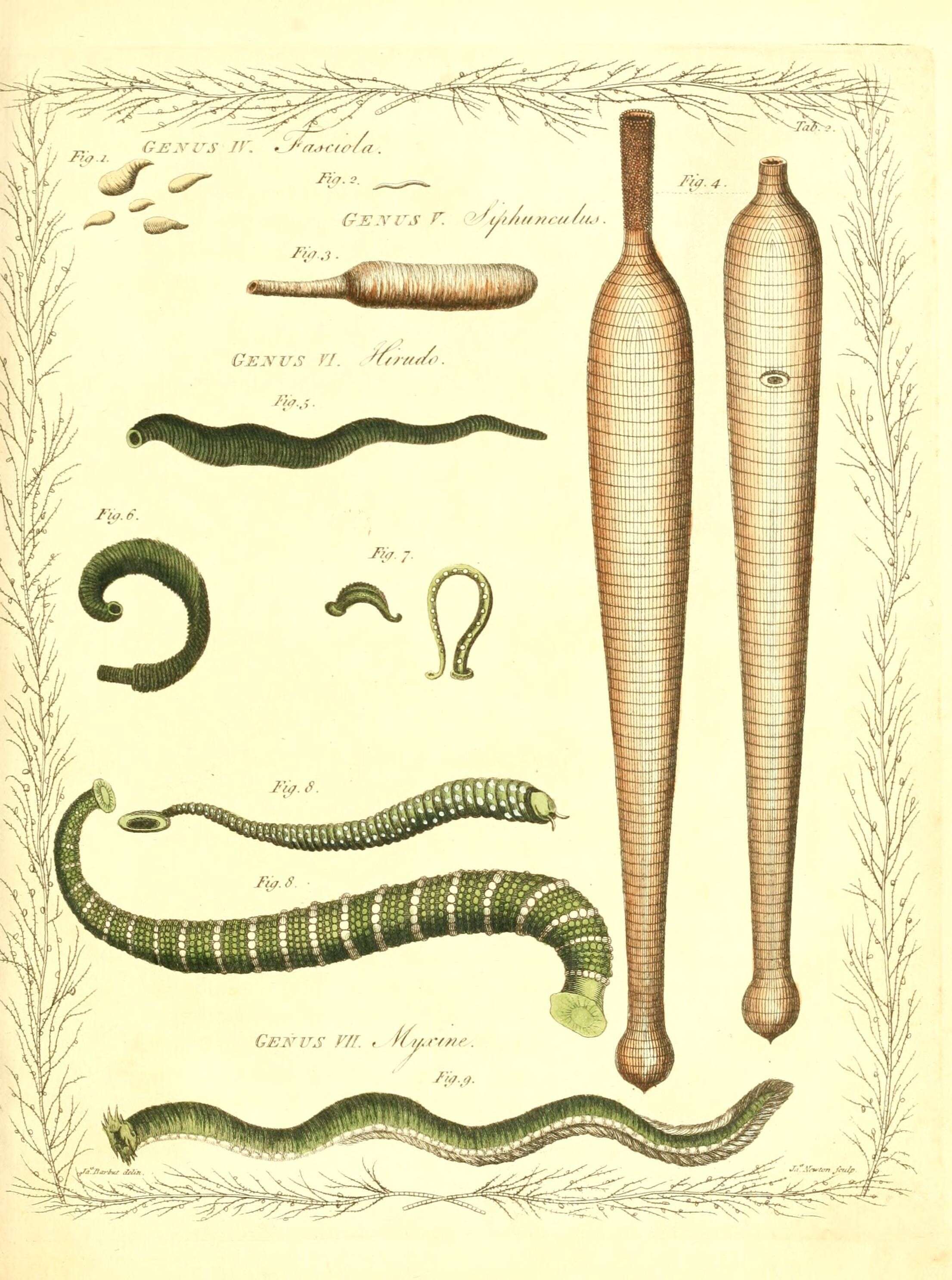
The genera vermium exemplified by various specimens of the animals contained in the orders of the Intestina et Mollusca Linnaei :.
London :Printed for the author by James Dixwell ..., and sold by John Sewell ..., B. White and Son ..., and P. Elmsley ...,1783-1788..
biodiversitylibrary.org/page/29389634
Description: English: Clonorchis showing stoma, pharynx, suckers, ceca, and a bit of overary. Date: 30 September 2020. Source: Own work. Author: The Other 95%.
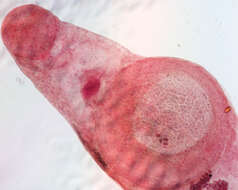
Description: English: The anterior end of Clonorchis, a.k.a, the Chinese liver fluke. The imae shows the stoma, oral sucker, pharyngeal bulb, and the two branches of the digestive tract. Low power on a stained specimen. Date: 11 February 2013, 22:31:24. Source: Own work. Author: Circa24.
Description: English: Clonorchis sinensis egg. Parasite. Date: 1973. Source: : This media comes from the Centers for Disease Control and Prevention's Public Health Image Library (PHIL), with identification number #659. Note: Not all PHIL images are public domain; be sure to check copyright status and credit authors and content providers. العربية | Deutsch | English | македонски | slovenščina | +/−. Author: Photo Credit: Content Providers(s): CDC/Dr. Mae Melvin. Permission(Reusing this file): PD-USGov-HHS-CDC English: None - This image is in the public domain and thus free of any copyright restrictions. As a matter of courtesy we request that the content provider be credited and notified in any public or private usage of this image.
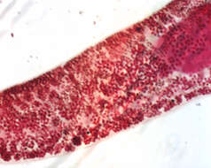
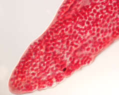
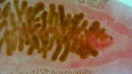
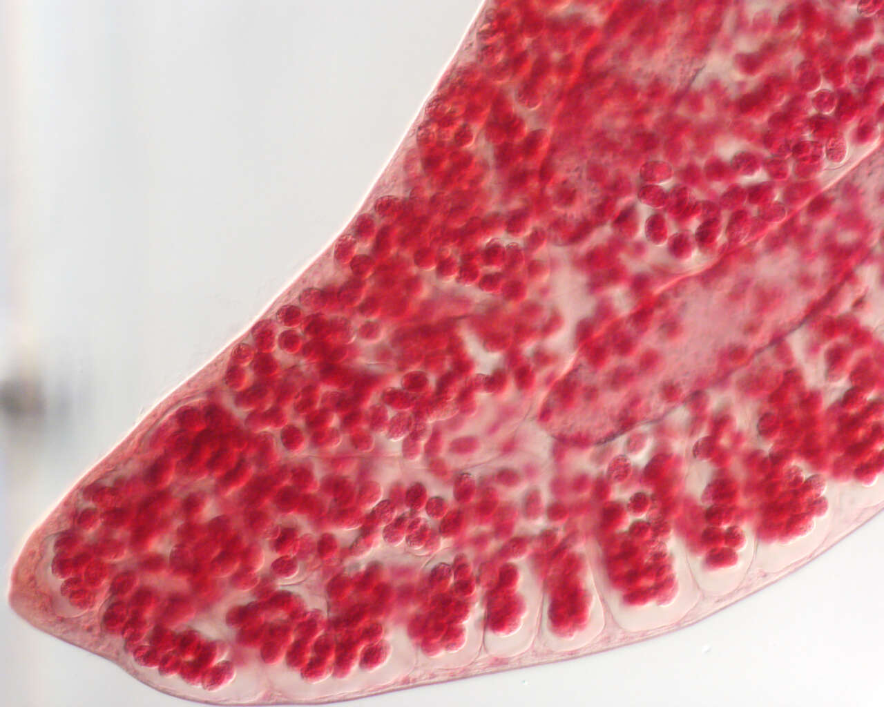
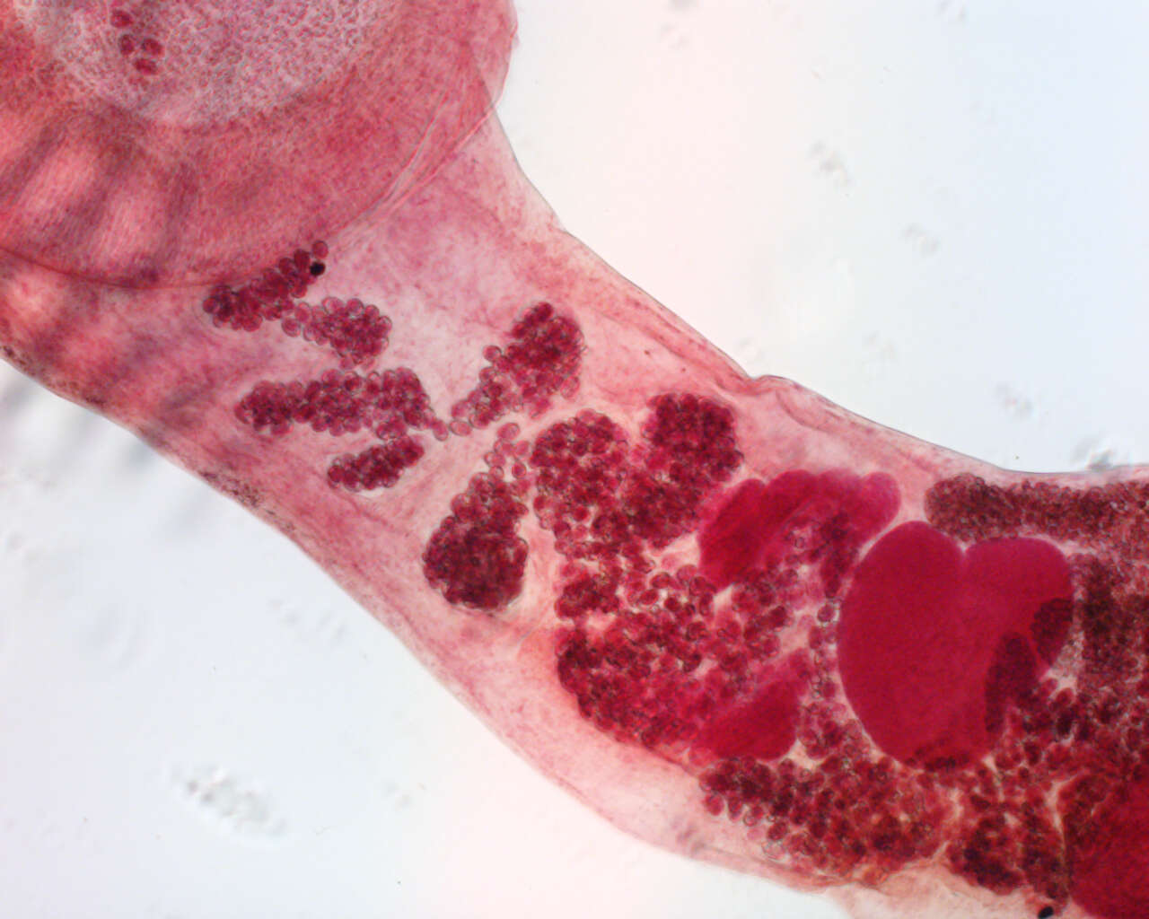
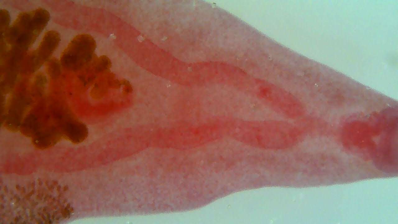
Description: English: Clonorchis wm. Vitelline Glands, vitelline ducts, uterus, and ceca. Date: 30 September 2020. Source: Own work. Author: The Other 95%.
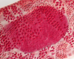
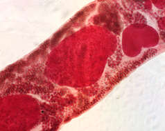
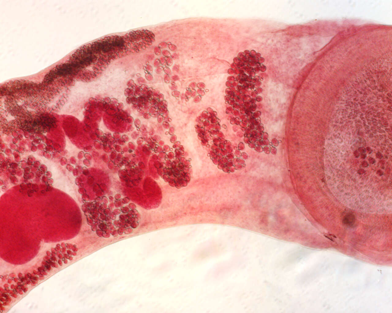
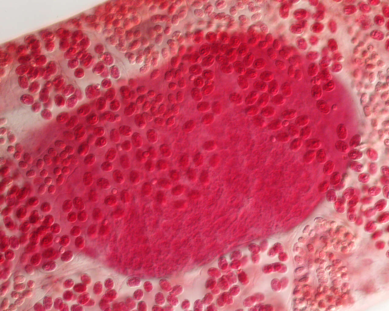
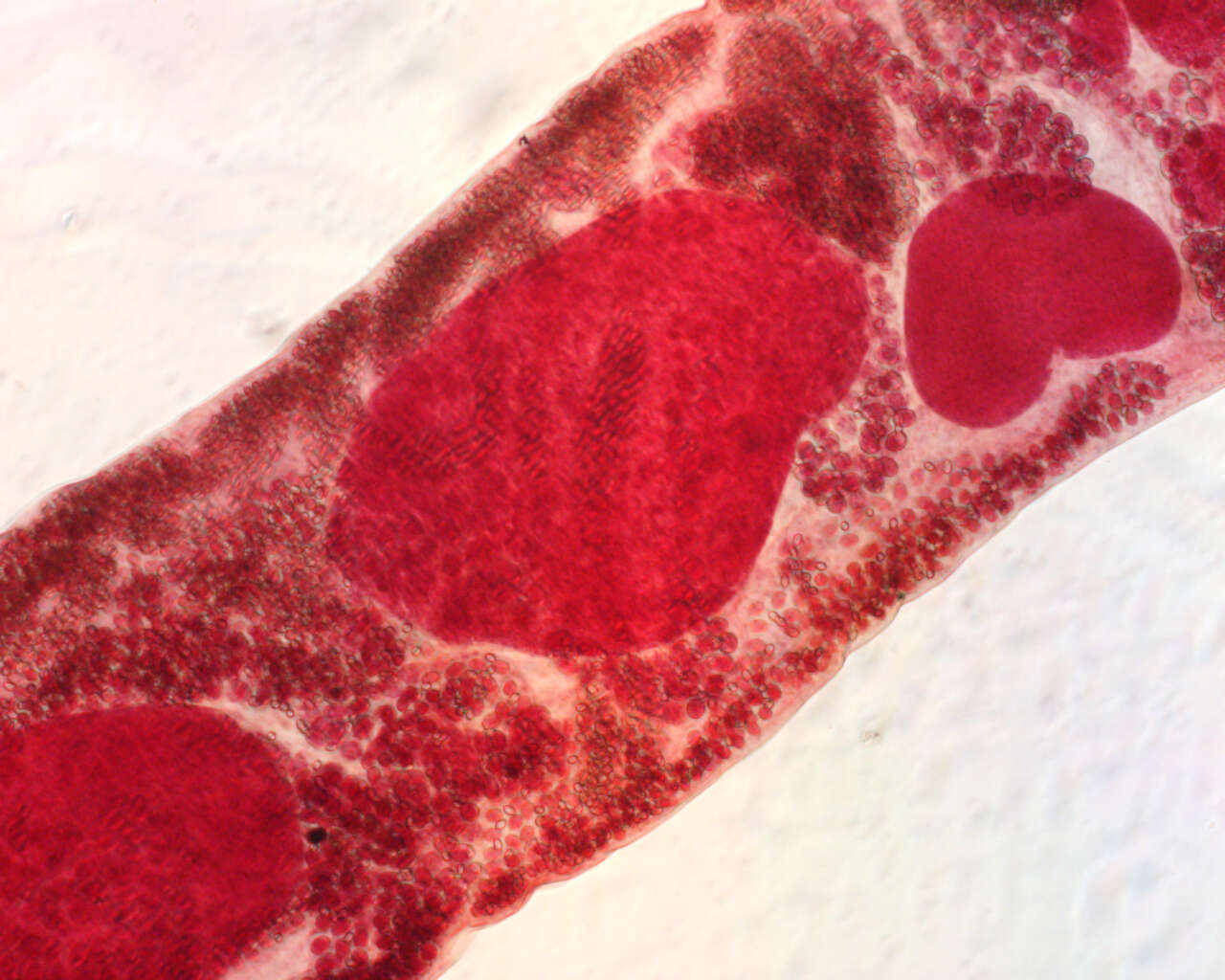
Description: English: Clonorchis. Uterus to right; germarium (ovary) center. Vitelline duct; seminal recepticle with sperm mass; and (faintly) seminal vesicle (middle of ovaries to right), and testies to left (posterior). Laurer's canal. Date: 30 September 2020. Source: Own work. Author: The Other 95%.
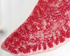
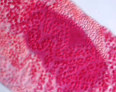
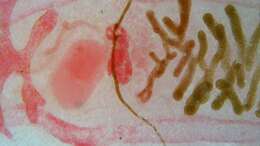
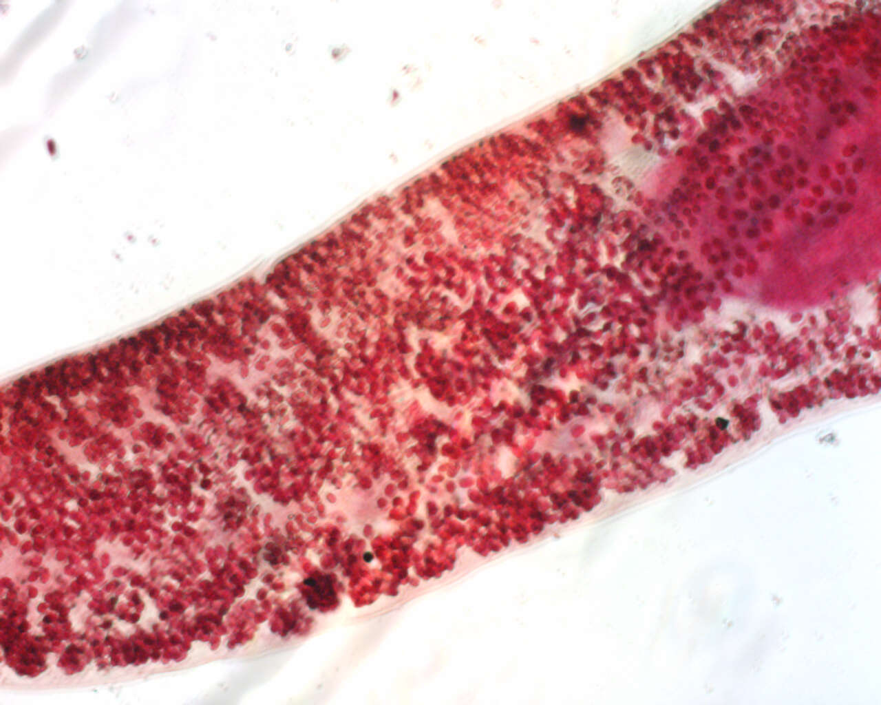
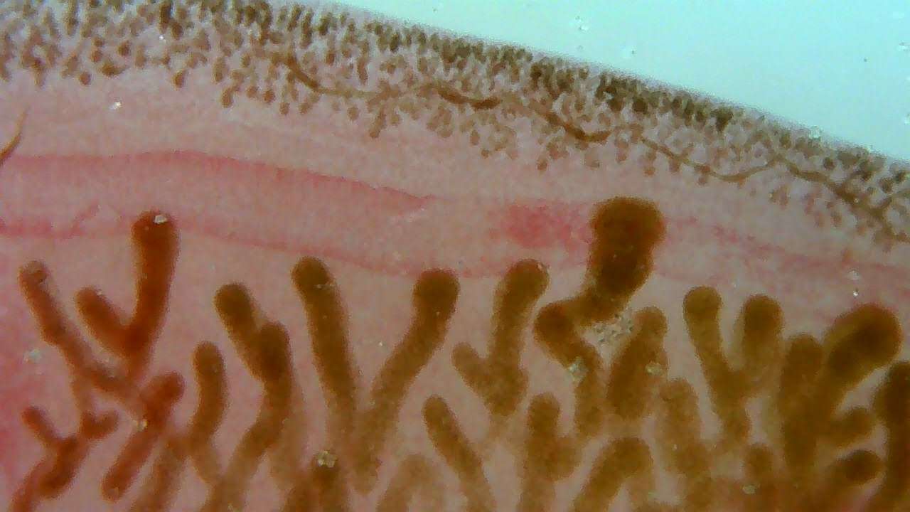
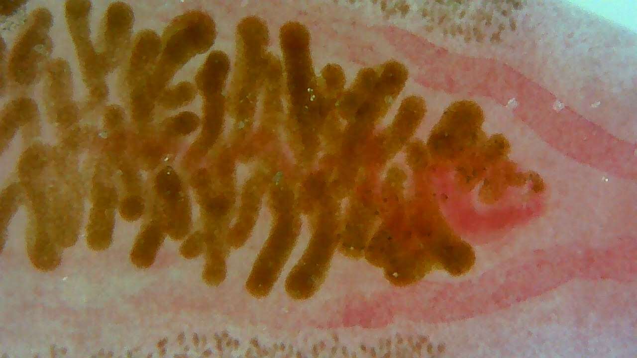
Description: English: Anterior wm of a Clonorchis. Pharynx, esophabus, branched ceca. By the uterus, one can t race the path to the gonopore, just anterior to the ventral sucker. Vitellene gland is visible on lower left. Date: 30 September 2020. Source: Own work. Author: The Other 95%.
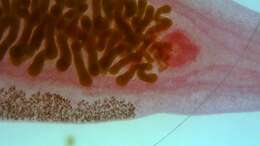
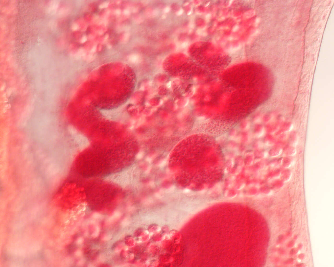
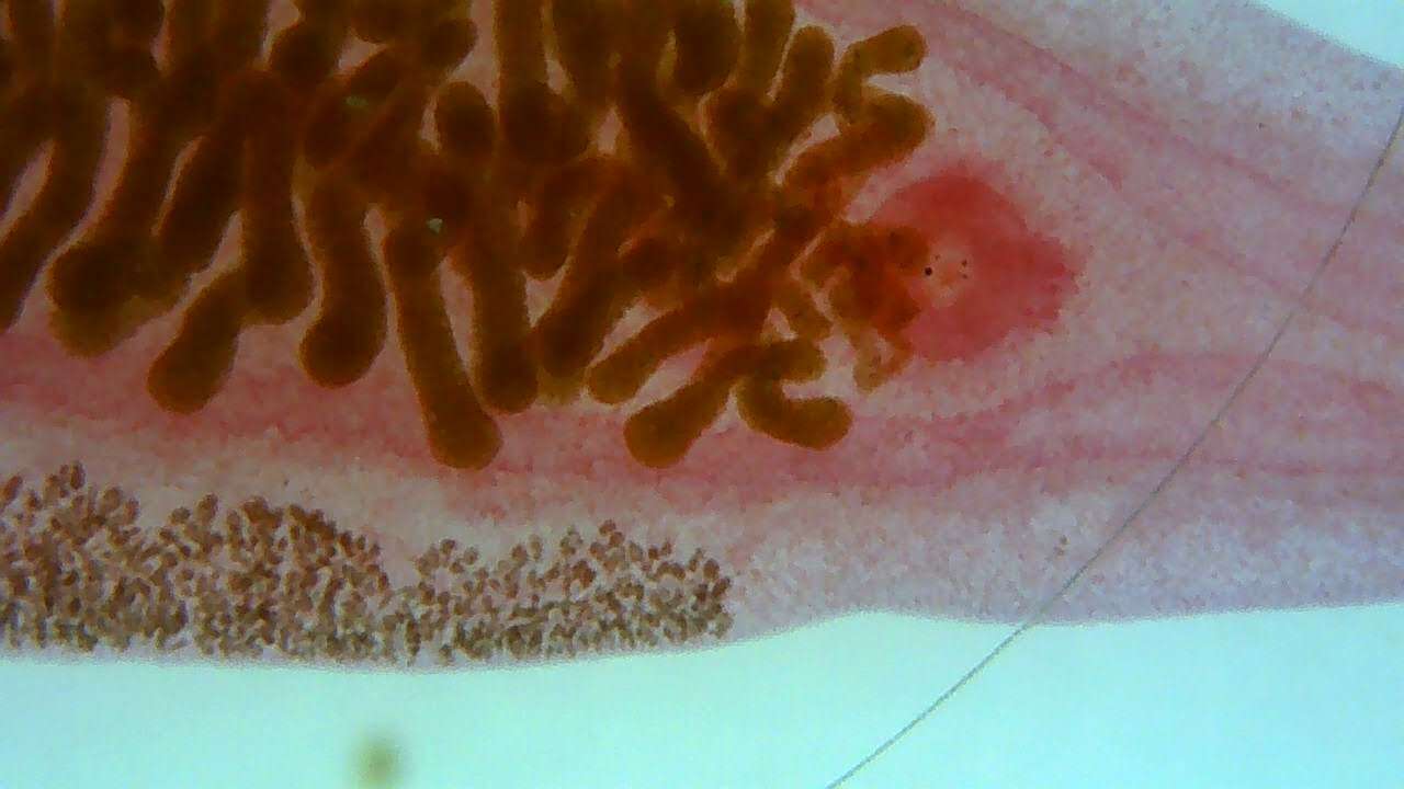
Description: English: Anterior wm of a Clonorchis. , branched ceca. By the uterus, one can trace the path to the gonopore, just anterior to the ventral sucker. Vitellene gland is visible at the top and the bottom. The seminal vessicle shows as a fait pink zig-zag line down the centre of the uterus. Date: 30 September 2020. Source: Own work. Author: The Other 95%.
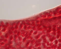
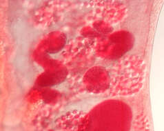
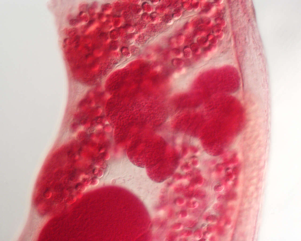
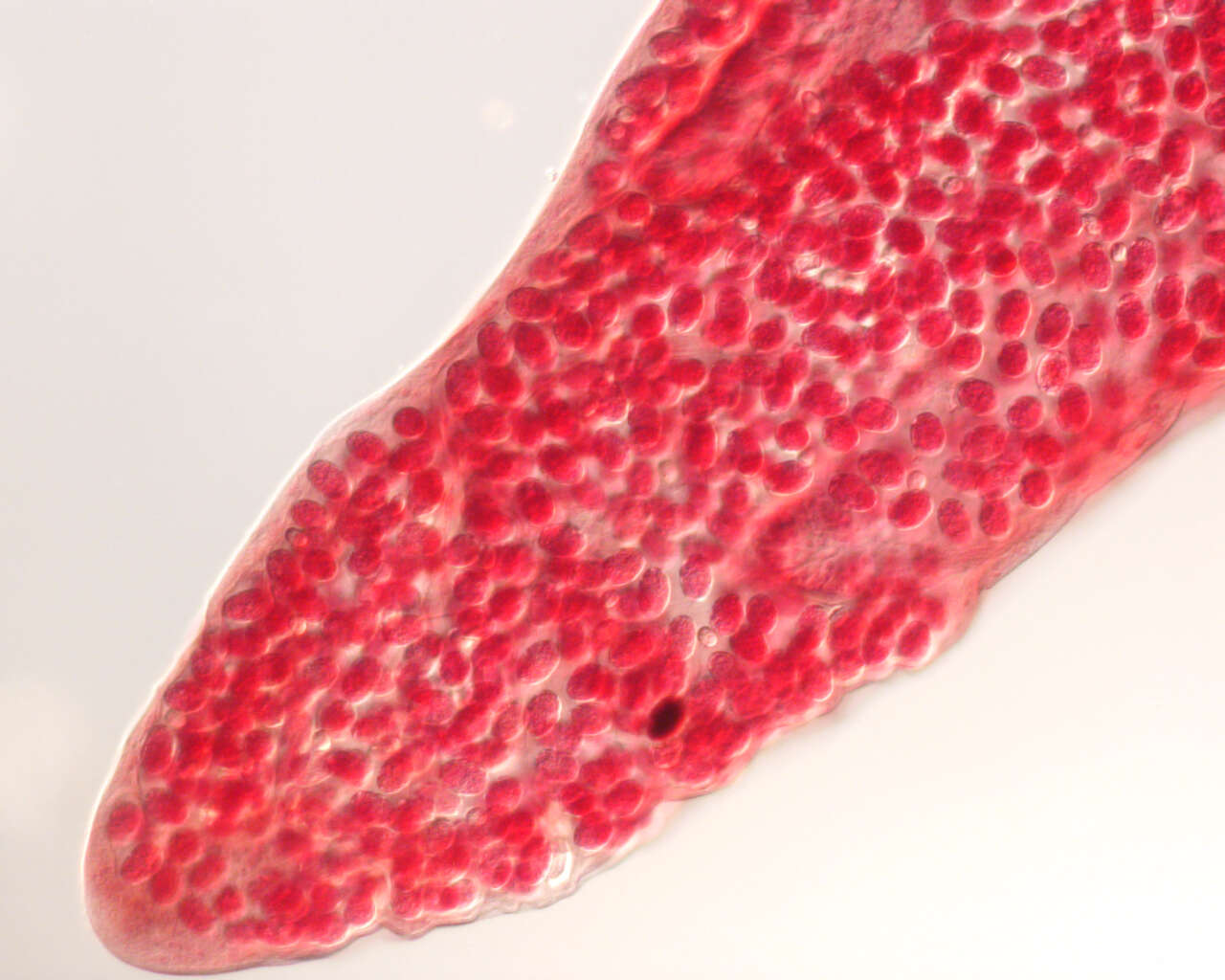
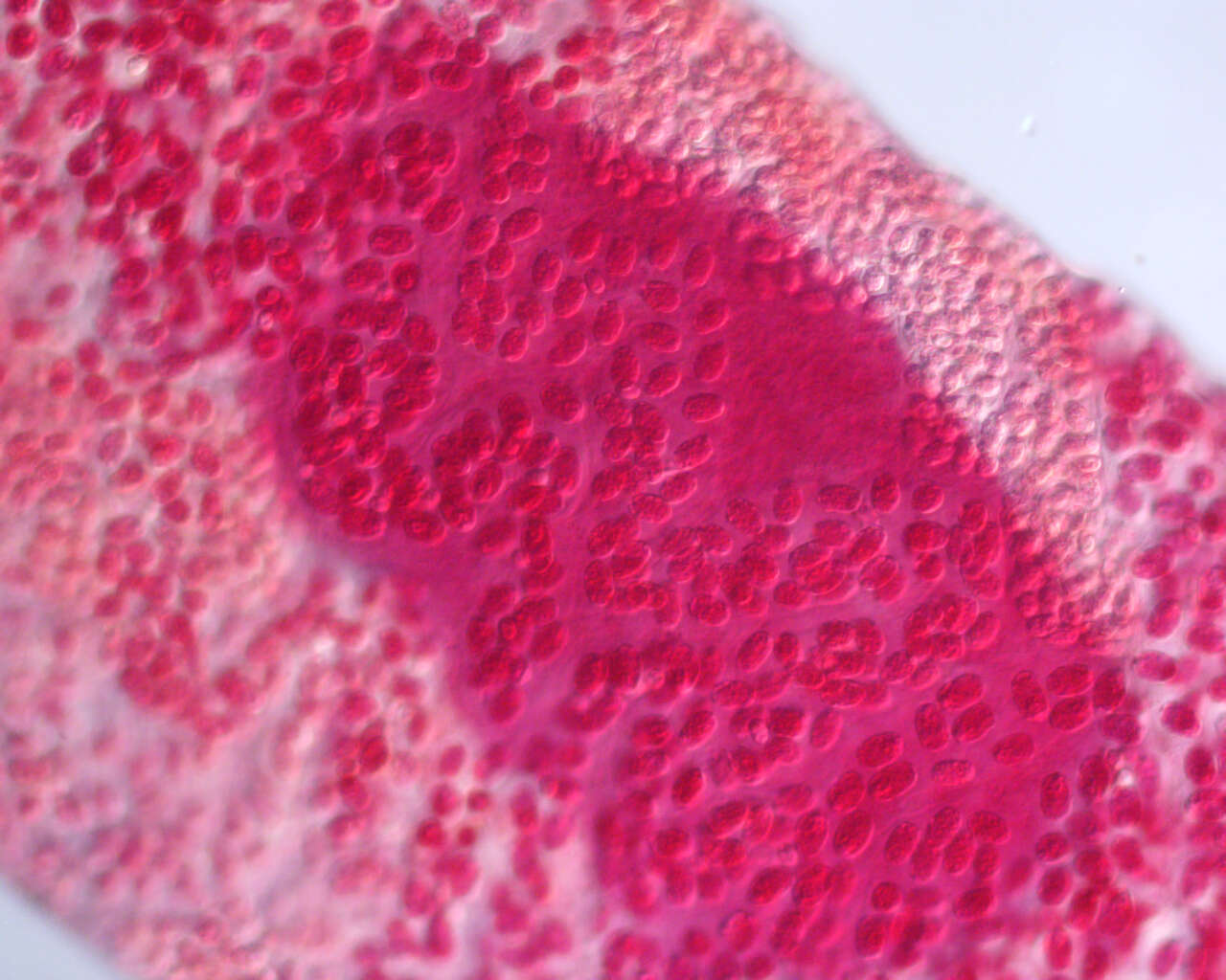
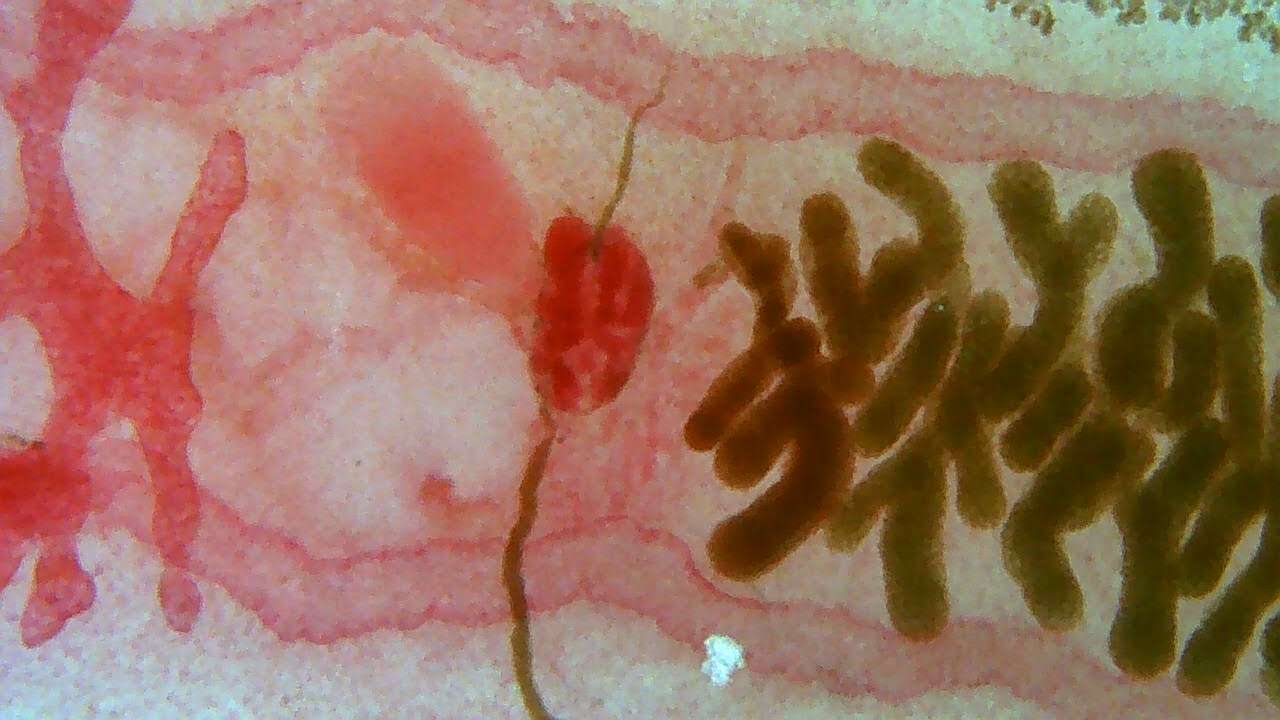
Description: English: Shows ceca, ventral sucker, anterior ovaries with embryonated eggs, and yolk glands. Date: 30 September 2020. Source: Own work. Author: The Other 95%.