Nematodes within the Secernentea have phasmids, which are unicellular glands. Phasmids likely function as chemoreceptors. Females may produce pheromones to attract males.
Nematodes in general have papillae, setae and amphids as the main sense organs. Setae detect motion (mechanoreceptors), while amphids detect chemicals (chemoreceptors).
Communication Channels: tactile ; chemical
Other Communication Modes: pheromones
Perception Channels: tactile ; chemical
Adult T. cati migrate to the intestine, and begin producing eggs after just a few weeks. The eggs are excreted with the host's feces, and require a variable amount of time in the environment to mature. Depending on temperature and weather conditions, maturation takes on average about 2-3 weeks during the summer (Prociv, 1989). During this time, the juvenile passes through its first stage (J1), and into its second infective stage (J2) (Roberts and Janovy, 2000). After this time, any organism (from worms and rodents to humans) that ingests these eggs is at risk of infection. Stimulated by the stomach acid, the eggs hatch and the J2 larvae begin their migration through the host's body. Many move from the stomach into the stomach wall, to the liver and lungs, and then back into the stomach wall again. After having passed through four juvenile stages, the mature T. cati migrate to the small intestine where they reproduce and begin shedding eggs (Prociv, 1989).
Anywhere from 8-42% of cats may be infected with T. cati (O'Lorcain, 1994). Routes of transmission are either transmammary (directly to the newborn kittens), or by accidental ingestion of eggs found within paratenic hosts, or within the environment.
Cats are not the only source of infection. Every year 3,000-4,000 cases of human infection are also reported to the Centers for Disease Control and Prevention and state public health departments (CDC, 2000). Children are most often the recipient of these worms by accidently swallowing the eggs when they lick their fingers, or orally clean out the dirt underneath their fingernails. As with other paratenic hosts, the J2 larvae hatch and migrate throughout the human's body. The tissues they migrate to determine the symptoms of the child's infection.
Infection with the worm, T. cati, is called Toxocariasis. The two forms of this disease are ocular larva migrans (OLM) and visceral larva migrans (VLM). OLM is an eye disease caused when the worm enters the eye causing inflammation and scaring of the retina. This may lead to blindness. VLM, on the other hand, is due to repeated infections of T. cati, and effects of its continual movement throughout the body. The organs and tissues swell from the immune response to this foreign invader, and the central immune system may be affected. Symptoms of VLM include fever, coughing, ashma, or pneumonia. VLM is easily treated with antiparasitic drugs and anti-inflammatory medications. OLM, on the other hand is more difficult to treat, and the focus is usually on prevention versus cure (CDC, 2000).
Negative Impacts: injures humans (causes disease in humans ); causes or carries domestic animal disease
Toxocara cati are found in domestic as well as wild cats around the world.
Ecosystem Impact: parasite
Species Used as Host:
Toxocara cati reside in the lumen of the small intestines. However, because they do not attach to the wall of its host's gut, it becomes apparent that these worms do not suck blood. Instead they feed primarily off of the liquid contents in the intestine (Roberts and Janovy, 2000), often resulting in malnutrition for the host.
Pharyngeal glands and intestinal epithelium produce digestive enzymes to feed on the hosts’ body fluids. Extracellular digestion begins within the lumen and is finished intracellularly (Barnes, 1987).
Animal Foods: body fluids
Primary Diet: carnivore (Eats body fluids)
Toxocara cati are found in domestic as well as wild cats around the world. Because cats have become more domesticated, this species is also more concentrated in those areas where humans reside. However, the spread of T. cati is dependent on the ingestion of its eggs, which are excreted with the hosts feces. Climate is a major factor in not only maintaining the eggs, but the feces that surrounds it. Therefore it has been observed that areas with mild temperate climates are the most favorable environment for T. cati.
Biogeographic Regions: nearctic ; palearctic ; oriental ; ethiopian ; neotropical ; australian
Other Geographic Terms: cosmopolitan
Toxocara cati hosts are predominantly within the genus Felis, the cats. Most research, however, has been focused on the house cat, Felis domesticus because of its close contact with humans. Studies excluding strays cited 8% of the cats with Toxocara cati while another study including strays found 42% of cats had the parasite (O'Lorcain, 1994).
There are also many other paratenic hosts. These include earthworms, cockroaches, birds, rodents, dogs and humans (Schierenberg, 1997). Within the paratenic hosts, T. cati remain juveniles and, depending on where they migrate, cause immune reactions throughout their host's body. Therefore during the other stages of its life, T. cati is found in limited areas of the cat. As J1s, they remain in the egg, in the environment outside of potential hosts. If ingested, as J2s they may be found in the stomach wall, or if they undergo further migration - in the liver, lungs, bronchi, trachea, pharynx, stomach, and small intestine. Finally, as J3s, J4s, and adults, T. cati is found in the lumen of the intestine of cats only. Eggs are produced within this area, and excreted with the cat's feces (Prociv, 1989).
Toxocara cati are mainly found in kittens because of the transmammary route of transmission. In female kittens, they are most prevalent from 24-48 weeks, while in male kittens they are mainly found in those aging 12-24 weeks. The reason for this sexual variation is possibly due to the different timing of their immune system development (O'Lorcain, 1994).
Habitat Regions: temperate ; tropical ; terrestrial
Terrestrial Biomes: desert or dune ; savanna or grassland ; chaparral ; forest ; rainforest ; scrub forest ; mountains
Other Habitat Features: urban ; suburban ; agricultural
Toxocaara cati, as well as most nematodes, do not have appendages, eyes, segments, or other features that would cause these small creatures to stand out. The hydroskeleton is a single chamber, similar to a balloon filled with water. Movement depends on the pressure exerted in the hydroskeleton over the parasite's body. By contracting its longitudinal muscles, which are separated into four quadrants, the worm squeezes its "balloon" and moves through the hosts body in "contractile waves" (Schierenberg, 1997). Adult T. cati also have a complete digestive system with a mouth at one end, made up of three fleshy lips, and an anus near the posterior end (Roberts and Janovy, 2000).
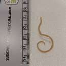
Toxocara cati are dioecious, and like most nematodes, the females in this species, at about 10 cm, are larger than the males which can be up to 6 cm (Uga et al., 2000). The posterior end of the males is curved with paired spicules, small pointy structures that help him to "feel" a female with which he can mate (Roberts and Janovy, 2000).
The rough, pitted eggs produced, on average, measure 75 X 65 µm and may survive for years in the environment (Uga et al., 2000). These appear very dark under the microscope because the one-celled zygote fills the entire interior of the egg, but due to their ovoid shape, the albuminous shell of the egg becomes visible as well.
Four larval instars precede the well-known "arrowhead" adult nematode. Both males and females have clear thickened cuticles, alae, along the sides of the anterior end of the worm, which end abruptly and give them the appearance of an arrow. These alae may provide stability and help in its movement through the host's body.
Range length: 6 to 10 cm.
Other Physical Features: ectothermic ; heterothermic ; bilateral symmetry
Sexual Dimorphism: female larger; sexes shaped differently
These parasites are usually not preyed on directly, but are ingested from host to host.
Adult T. cati migrate to the intestine, and begin producing eggs after just a few weeks. The eggs are excreted with the host's feces, and require a variable amount of time in the environment to mature. Depending on temperature and weather conditions, maturation takes on average about 2-3 weeks during the summer (Prociv, 1989). During this time, the juvenile passes through its first stage (J1), and into its second infective stage (J2) (Roberts and Janovy, 2000). After this time, any organism (from worms and rodents to humans) that ingests these eggs is at risk of infection. Stimulated by the stomach acid, the eggs hatch and the J2 larvae begin their migration through the host's body. Many move from the stomach into the stomach wall, to the liver and lungs, and then back into the stomach wall again. After having passed through four juvenile stages, the mature T. cati migrate to the small intestine where they reproduce and begin shedding eggs (Prociv, 1989).
Paratenic hosts (those hosts that carry the worm, but the worm will not further develop) are especially important in T. cati infection of cats because of their predatory nature. If a mouse, for example, ingests a T. cati egg - the egg hatches, but the J2 larvae does not undergo further development. When this mouse is eaten by the cat, the J2 enters the wall of the stomach, and molts (by shedding its external cuticle and replacing it with the new one formed underneath) into J3 (Schrierenberg, 1997). When the juvenile procedes to migrate into the intestine lumen, it undergoes its last molt, into J4. These juveniles then molt into immature adults, and finally mature into adults capable of reproducing (Prociv, 1989).
Another important method of transmission is through the mammary glands of a pregnant female cat. Because T. cati do not migrate through the bloodstream and into the placenta, kittens are born free of worms, even if their mother was infected. However, after about 3-4 weeks, the newly hatched J2s migrate to the mother's mammary glands, where they are ingested by the feeding kittens (O'Lorcain, 1994). These enter the stomach, and then intestine of the kitten where they undergo the rest of their development into mature adults (Prociv, 1989).
Key Reproductive Features: sexual ; fertilization (Internal ); oviparous
Parental Investment: pre-fertilization (Provisioning)