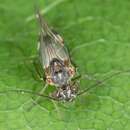
-
Figure 3.Photograph of hypopharynx filaments (arrows) of Libanopsyllipsocus alexanderasnitsyni gen. et sp. n., holotype, male.
-
Michel P. Valim, Ricardo L. Palma
Zookeys
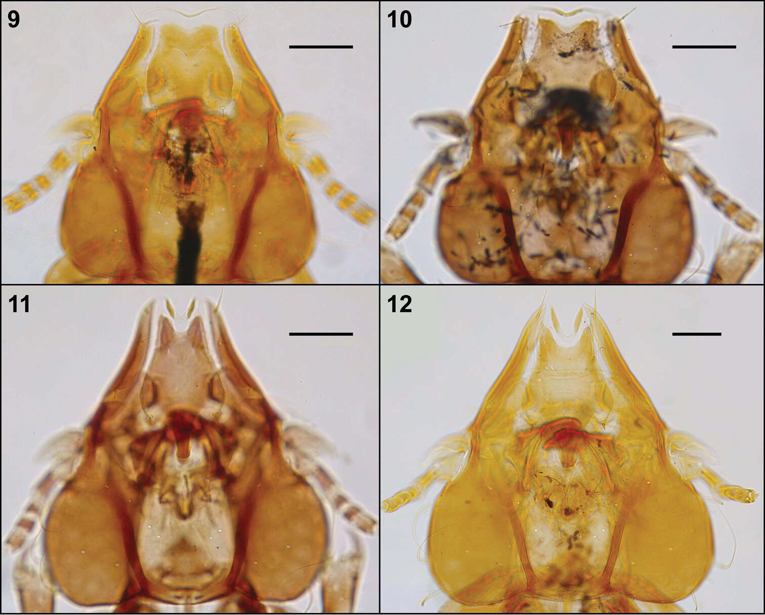
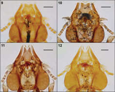
Figures 9–12.Head, dorsal view: Philopteroides beckeri female paratype (9); Philopteroides pilgrimi female holotype (10); Philopteroides fuliginosus female (11); Philopteroides macrocephalus female holotype (12). Scale bars = 0.1 mm
-
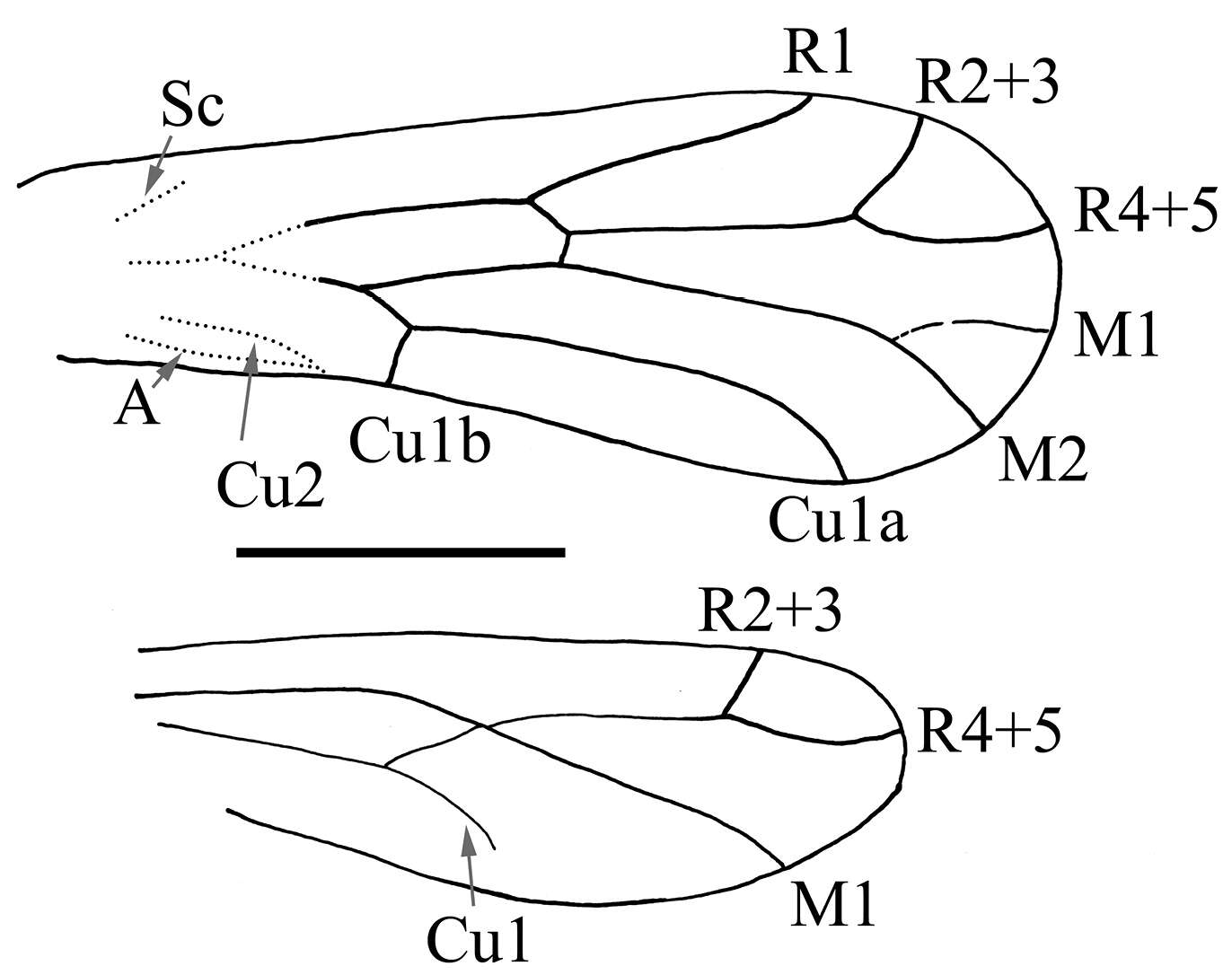
Figure 4.Drawing of wings of Libanopsyllipsocus alexanderasnitsyni gen. et sp. n., holotype, male, scale bar = 0.3 mm.
-
Michel P. Valim, Ricardo L. Palma
Zookeys
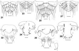
Figures 21–28.Dorsal terminalia: Philopteroides fuliginosus female (21); Philopteroides fuliginosus male (23); Philopteroides macrocephalus female (25); Philopteroides macrocephalus male (27). Female ventral terminalia: Philopteroides fuliginosus (22); Philopteroides macrocephalus (26); Philopteroides macrocephalus intraspecific variation (26a). Male subgenital plate: Philopteroides fuliginosus (24); Philopteroides macrocephalus (28). Scale bars = 0.1 mm.
-
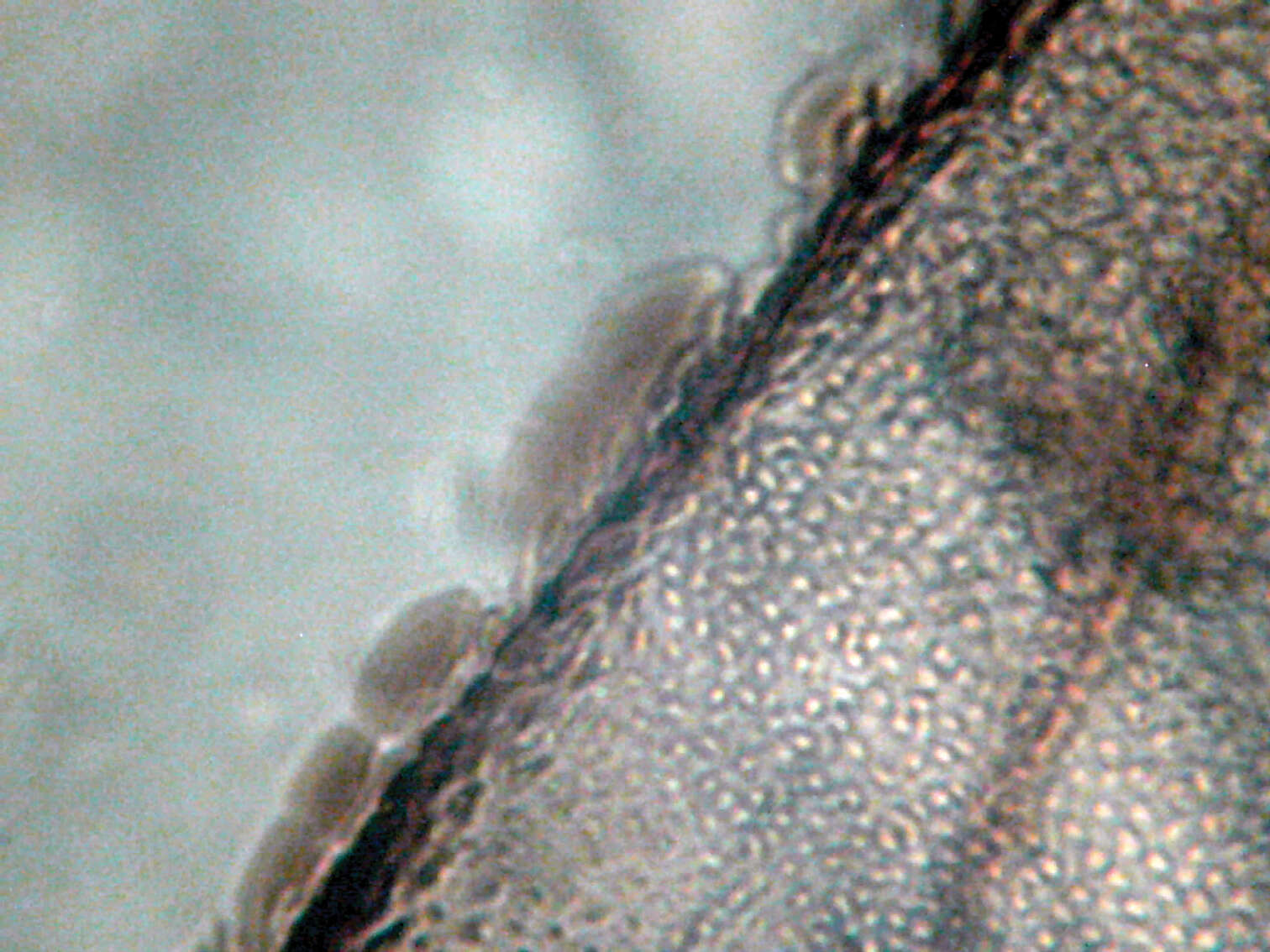
Figure 5.Microphotograph of nodulus, arrow showing the meeting area of Cu2 and A.
-
Michel P. Valim, Ricardo L. Palma
Zookeys
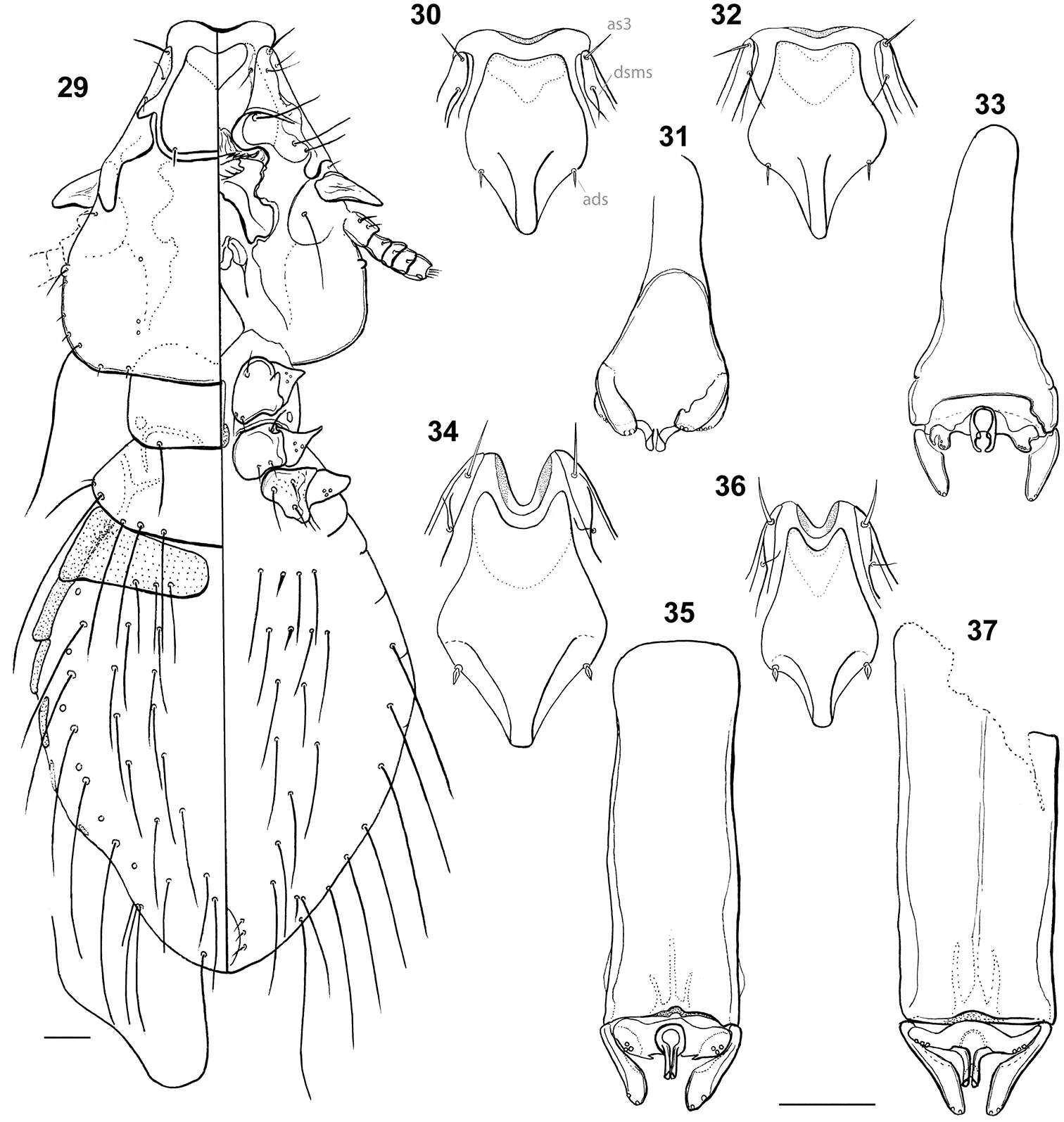
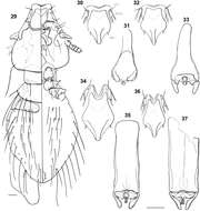
Figures 29–37.Habitus: Philopteroides beckeri nymph II, dorso-ventral view (29). Hyaline margins and anterior dorsal plates: Philopteroides beckeri (30); Philopteroides pilgrimi (32); Philopteroides fuliginosus (34); Philopteroides macrocephalus (36). Male genitalia: Philopteroides beckeri (31); Philopteroides pilgrimi (33); Philopteroides fuliginosus (35); Philopteroides macrocephalus (37). Scale bars = 0.05 mm
-
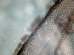
Figure 6.Microphotograph of structure of forewing margin.
-
Michel P. Valim, Ricardo L. Palma
Zookeys
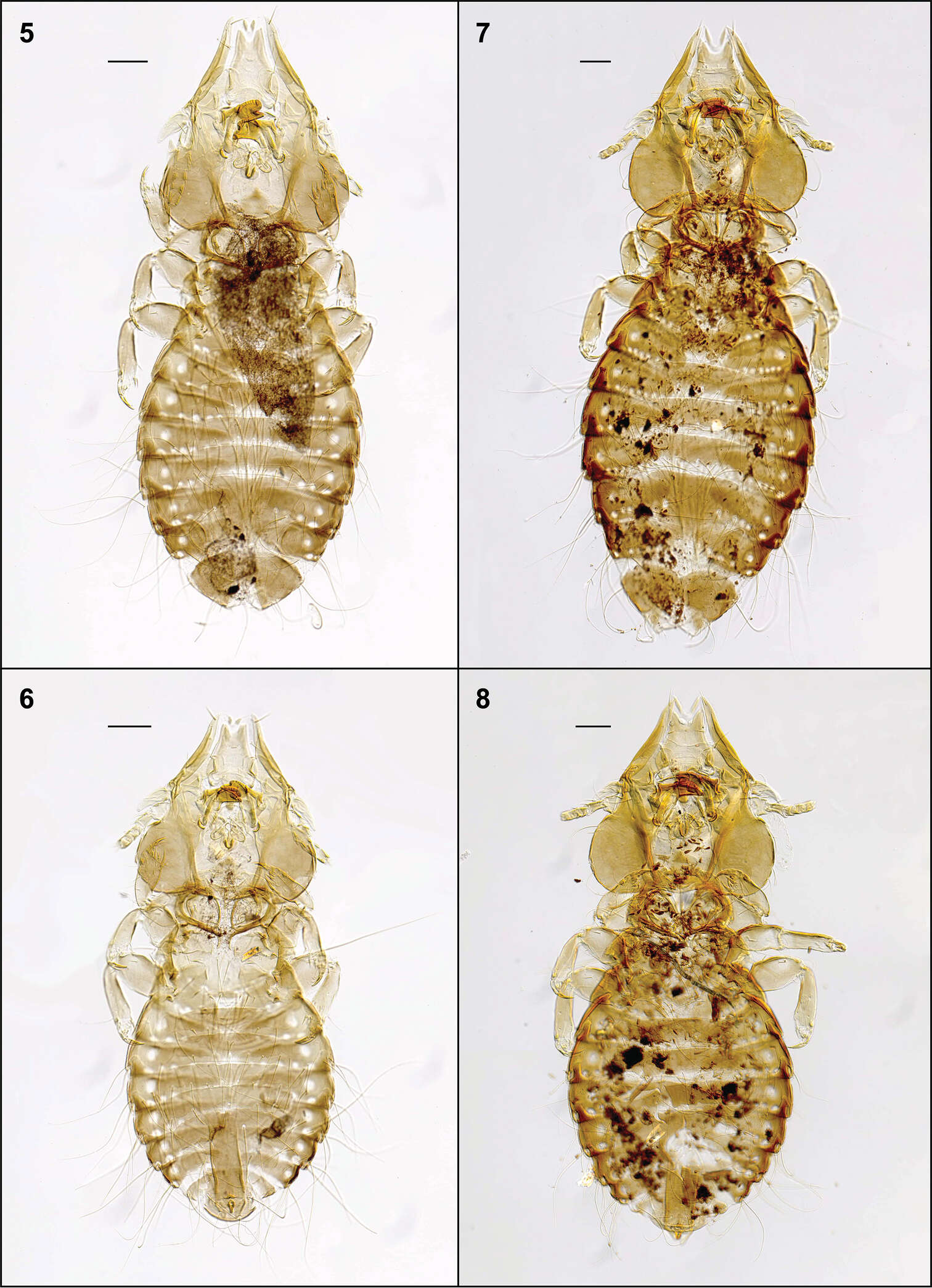
Figures 5–8.Habitus, ventral views: Philopteroides fuliginosus female holotype (5); Philopteroides fuliginosus male paratype (6); Philopteroides macrocephalus female holotype (7); Philopteroides macrocephalus male paratype (8). Scale bars = 0.1 mm
-
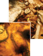
Figure 7.Microphotograph of hind leg coxal rasp (Pearman’s organ).
-
Michel P. Valim, Ricardo L. Palma
Zookeys
Figures 9–12.Head, dorsal view: Philopteroides beckeri female paratype (9); Philopteroides pilgrimi female holotype (10); Philopteroides fuliginosus female (11); Philopteroides macrocephalus female holotype (12). Scale bars = 0.1 mm
-
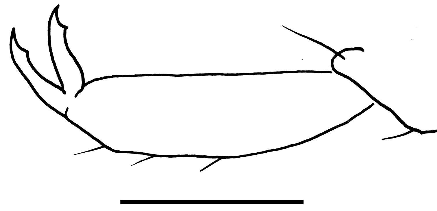
Figure 8.Drawing of pretarsal claw of Libanopsyllipsocus alexanderasnitsyni gen. et sp. n., holotype, male, scale bar = 0.03 mm.
-
Michel P. Valim, Ricardo L. Palma
Zookeys
Figures 21–28.Dorsal terminalia: Philopteroides fuliginosus female (21); Philopteroides fuliginosus male (23); Philopteroides macrocephalus female (25); Philopteroides macrocephalus male (27). Female ventral terminalia: Philopteroides fuliginosus (22); Philopteroides macrocephalus (26); Philopteroides macrocephalus intraspecific variation (26a). Male subgenital plate: Philopteroides fuliginosus (24); Philopteroides macrocephalus (28). Scale bars = 0.1 mm.
-
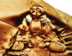
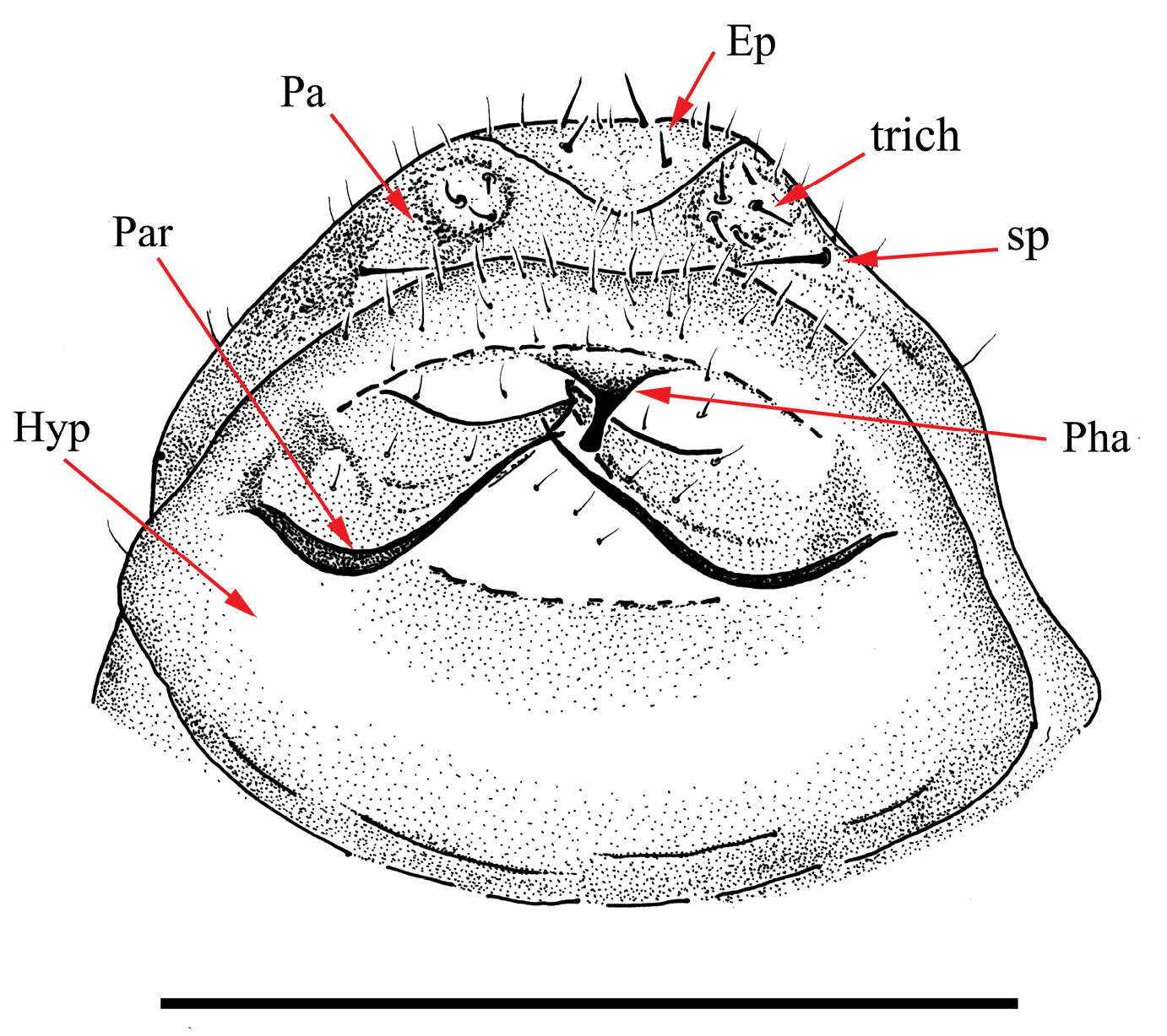
Figure 9.Drawing of aedeagus of Libanopsyllipsocus alexanderasnitsyni gen. et sp. n., holotype, male; Ep = epiproct, Hyp = hypandrium, Par = paraproct, par = paramers, Pha = phallosome, sp = anal spine, trich = trichobothria; scale bar = 0.3 mm.
-
Michel P. Valim, Ricardo L. Palma
Zookeys
Figures 29–37.Habitus: Philopteroides beckeri nymph II, dorso-ventral view (29). Hyaline margins and anterior dorsal plates: Philopteroides beckeri (30); Philopteroides pilgrimi (32); Philopteroides fuliginosus (34); Philopteroides macrocephalus (36). Male genitalia: Philopteroides beckeri (31); Philopteroides pilgrimi (33); Philopteroides fuliginosus (35); Philopteroides macrocephalus (37). Scale bars = 0.05 mm
-
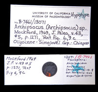
2016 University of California Museum of Paleontology
CalPhotos
-
-
-
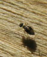
Contact photographer for info.
-
Please contact photographer for info.
-
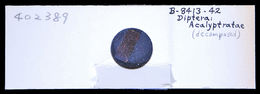
2016 University of California Museum of Paleontology
CalPhotos
-
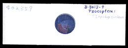
2017 University of California Museum of Paleontology
CalPhotos
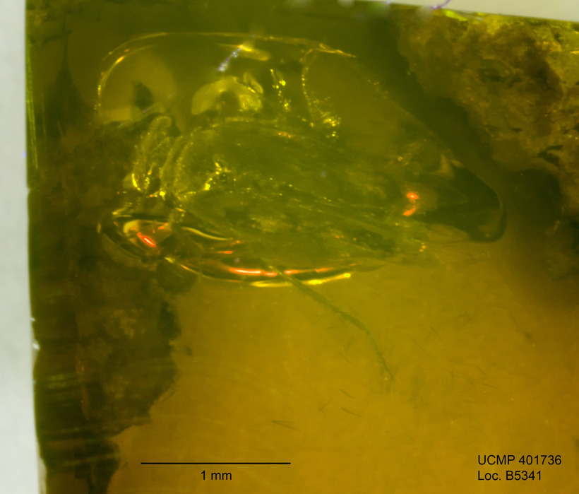
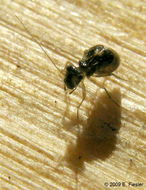
Image taken using a StackShot rail with Helicon Remote software and rendered using Helicon Focus software.
-
-
-