-
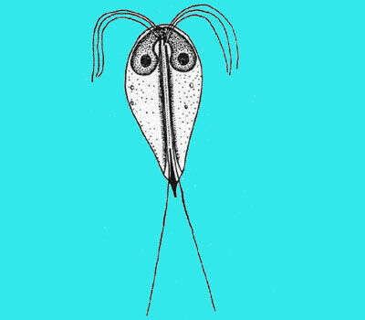
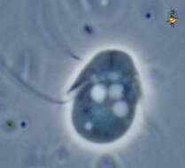
Hexamita (hex-a-mite-a), a free living diplomonad flagellate. Diplomonads are so-called because most members of the group have two nuclei, and have clusters of up to 4 flagella emerging from opposing sides of the cell. They arise in grooves, which are believed to correspond to the ventral grooves of the excavate flagellates. Normally associated with anoxic habitats. Diplomonads are probably best known because one their members, Giardia, is significant as a parasite of the intestinal system, and because it is prominent in studies on the evolution of eukaryotic cells - "clinging resolutely" to the base of the eukaryotic tree as our best candidate for the most primitive eukaryote. The two nuclei are at the anterior (top) and the junction between them is seen as a dark line. The flagellar grooves and some of the flagella are evident. Phase contrast.
-
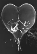
This scanning electron micrograph (SEM) depicted a Giardia lamblia protozoan that was about to become two separate organisms, as it was caught in a late stage of cell division, producing a heart-shaped form. Note the intimate intertwining of two of the organisms eight flagella that will facilitate their motility.Created: 1986
-
Hexamita (hex-a-mite-a), a free living diplomonad flagellate. Diplomonads are so-called because most members of the group have two nuclei, and have clusters of up to 4 flagella emerging from opposing sides of the cell. They arise in grooves, which are believed to correspond to the ventral grooves of the excavate flagellates. Normally associated with anoxic habitats. Diplomonads are probably best known because one their members, Giardia, is significant as a parasite of the intestinal system, and because it is prominent in studies on the evolution of eukaryotic cells - "clinging resolutely" to the base of the eukaryotic tree as our best candidate for the most primitive eukaryote. The two nuclei are at the anterior (top) and the junction between them is seen as a dark line. The flagellar grooves and some of the flagella are evident. Phase contrast.
-
Hexamita (hex-a-mite-a), a free living diplomonad flagellate. Diplomonads are so-called because most members of the group have two nuclei, and have clusters of up to 4 flagella emerging from opposing sides of the cell. They arise in grooves, which are believed to correspond to the ventral grooves of the excavate flagellates. Normally associated with anoxic habitats. Diplomonads are probably best known because one their members, Giardia, is significant as a parasite of the intestinal system, and because it is prominent in studies on the evolution of eukaryotic cells - "clinging resolutely" to the base of the eukaryotic tree as our best candidate for the most primitive eukaryote. All flagella can be seen with an eye of faith. Phase contrast.
-
-
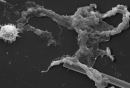
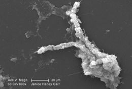
This scanning electron micrograph (SEM) of an untreated water specimen extracted from a wild stream mainly used to control flooding during inclement weather, revealed the presence of unidentified organisms, which included bacteria, protozoa, and algae. Occupying most of the field of view, an unidentified amorphous mucoidal biofilm was featured, which appeared to have enmeshed numbers of amoeboid organisms, while on the left was a strangely-beautiful microorganism displaying an outer surface studded with numerous projections, making it appear like a microscopic sea urchin. See PHIL 11715 for a colorized version of this image.Created: 2009
-
-
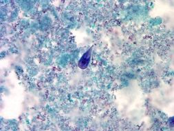
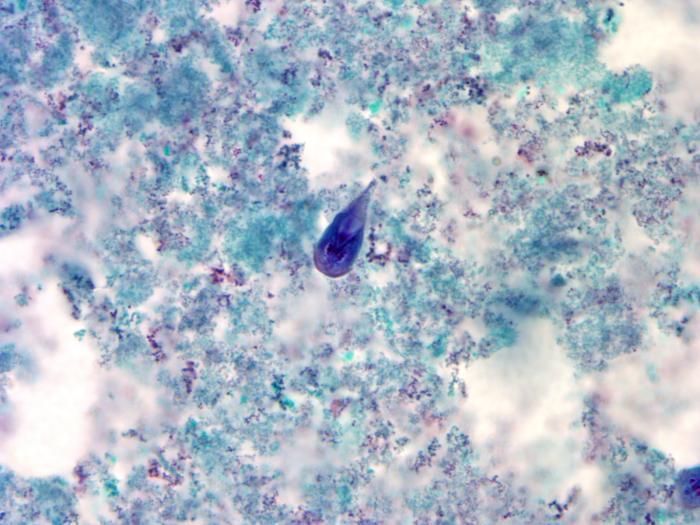
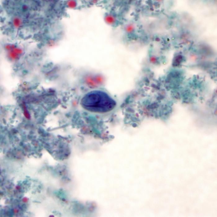
At a magnification of 1000X, this trichrome-stained photomicrograph revealed the morphologic characteristics of a blue-stained Giardia intestinalis protozoan trophozoite (center). In the small intestine, the protozoan cysts release trophozoites, with each cyst producing two trophozoites. Trophozoites multiply by longitudinal binary fission, remaining in the lumen of the proximal small bowel where they can be free, or attached to the mucosa by a ventral sucking disk. As the trophozoites mature, they are simultaneously migrating towards the colon, whereupon, they once again become thick-walled cysts, and are in this way, passed in the hosts stool into the environment. As cysts, these protozoan parasites can survive for many months until they are accidentally ingested by another unfortunate host.Created:
-
-
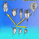
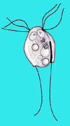
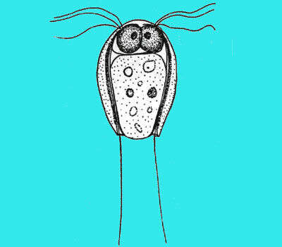
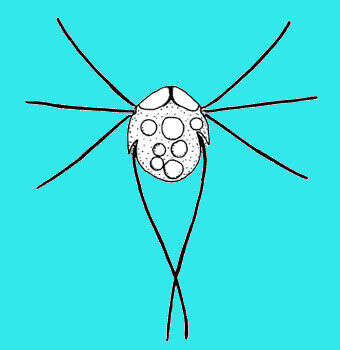
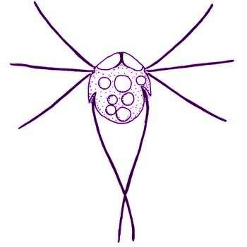
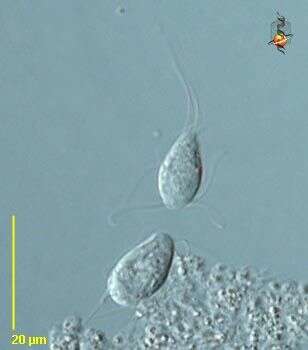
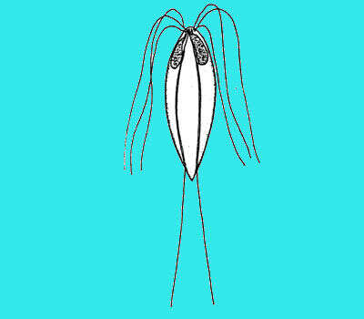
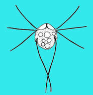
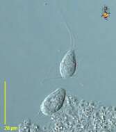
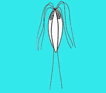
Hexamita (hecks-a-mite-a) inflata. Dujardin, 1838. Cells are roundish to ovoid and about 6 - 10 microns long. Several contractile vacuoles are seen. The posterior end of the cell may be pointed or rounded. Two nuclei are located anteriorly, and two longitudinal cytostomal tubes are visible below the equator of the cell and on the ventral side of the cell. There are two sets of 4 flagella (total eight flagella), two medium flagella are about 1.3 - 1.5 times the cell length, four short flagella are about the cell length. These flagella insert anterio-laterally into a small depression on the anterior part of the cell. Two long flagella emerge from the cytostomal tube and are about 2 times the length of the cell. The long flagella normally cross each other. The cells move by skidding or swimming. Relatively common.
-
Hexamita inflata Dujardin, 1838. Cells are roundish to ovoid and about 6 - 10 microns long. Several contractile vacuoles are seen. The posterior end of the cell may be pointed or rounded. Two nuclei are located anteriorly, and two longitudinal cytostomal tubes are visible below the equator of the cell and on the ventral side of the cell. There are two sets of 4 flagella (total eight flagella), two medium flagella are about 1.3 - 1.5 times the cell length, four short flagella are about the cell length. These flagella insert anterio-laterally into a small depression on the anterior part of the cell. Two long flagella emerge from the cytostomal tube and are about 2 times the length of the cell. The long flagella normally cross each other. The cells move by skidding or swimming.
-
This scanning electron micrograph (SEM) of an untreated water specimen extracted from a wild stream mainly used to control flooding during inclement weather, revealed the presence of unidentified organisms, which included bacteria, protozoa, and algae. In this particular image, an unidentified amorphous mucoidal biofilm was featured, which appeared to have enmeshed numbers of amoeboid organisms.Created: 2009
-
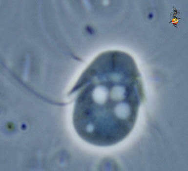
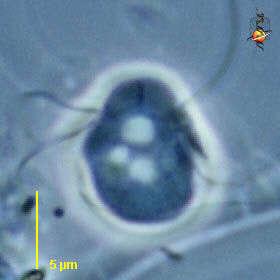
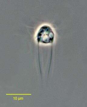
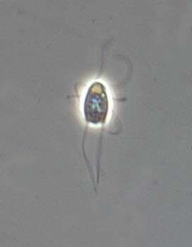
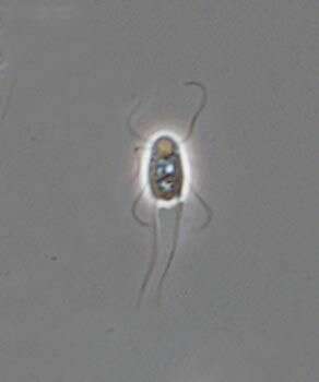
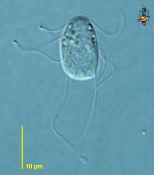
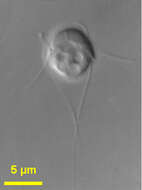
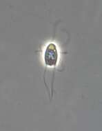
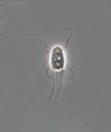
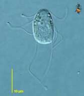
Portrait of the diplomonad Hexamita inflata (Dujardin 1838).Collected from sapropelic bottom sediments of a freshwater pond near Boise, Idaho. Phase contrast.
-
Using a trichrome stain, this photomicrograph revealed the morphologic characteristics of a blue-stained Giardia intestinalis protozoa cyst (center). The Giardia parasite lives in the intestine of infected humans or animals. Millions of cystic protozoa can be released in a bowel movement from an infected human or animal. Giardia is found in soil, food, water, or surfaces that have been contaminated with the feces from infected humans or animals. You can become infected after accidentally swallowing the parasite; you cannot become infected through contact with blood.Created:
-
Portrait of the diplomonad Hexamita inflata (Dujardin 1838).
-
Portrait of the diplomonad Hexamita inflata (Dujardin 1838).Collected from sapropelic bottom sediments of a freshwater pond near Boise, Idaho. Phase contrast.
-
Portrait of the diplomonad Hexamita inflata (Dujardin 1838).Collected from sapropelic bottom sediments of a freshwater pond near Boise, Idaho. Phase contrast.
-
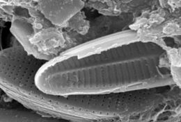
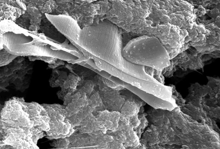
This scanning electron micrograph (SEM) of an untreated water specimen extracted from a wild stream mainly used to control flooding during inclement weather, revealed the presence of unidentified organisms, which included bacteria, protozoa, and algae. In this particular image, unidentified species of diatoms are seen to be caught up in an amorphous gelatinous biofilm, which had entrapped stream particulates as well.Created: 2009
-
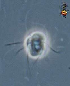
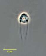
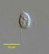
Spironucleus (spire-owe-new-clee-us) is a diplomonad flagellate. Like most genera in the group, there are two anterior nuclei, and arising in association with each nucleus is a group of four flagella. The flagella insert at the head of a lateral groove. Most members of this genus are parasites. Differential interference contrast.
-
Spironucleus (spire-owe-new-clee-us)is a diplomonad flagellate. Like most genera in the group, there are two anterior nuclei, and arising in association with each nucleus is a group of four flagella. The flagella insert at the head of a lateral groove. Most members of this genus are parasites. Differential interference contrast.
-
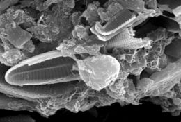
This scanning electron micrograph (SEM) of an untreated water specimen extracted from a wild stream mainly used to control flooding during inclement weather, revealed the presence of unidentified organisms, which included bacteria, protozoa, and algae. In this particular image, unidentified species of diatoms are seen to be caught up in an amorphous gelatinous biofilm, which had entrapped stream particulates as well. In the center, youll note what may have been an amoeboid organism.Created: 2009
-
-
-
This scanning electron micrograph (SEM) of an untreated water specimen extracted from a wild stream mainly used to control flooding during inclement weather, revealed the presence of unidentified organisms, which included bacteria, protozoa, and algae. In this particular image, unidentified sheets of algae were wrapped in a mass of what appeared to be a mucoid amorphous biofilm. See PHIL 11713 for a colorized version of this image.Created: 2009