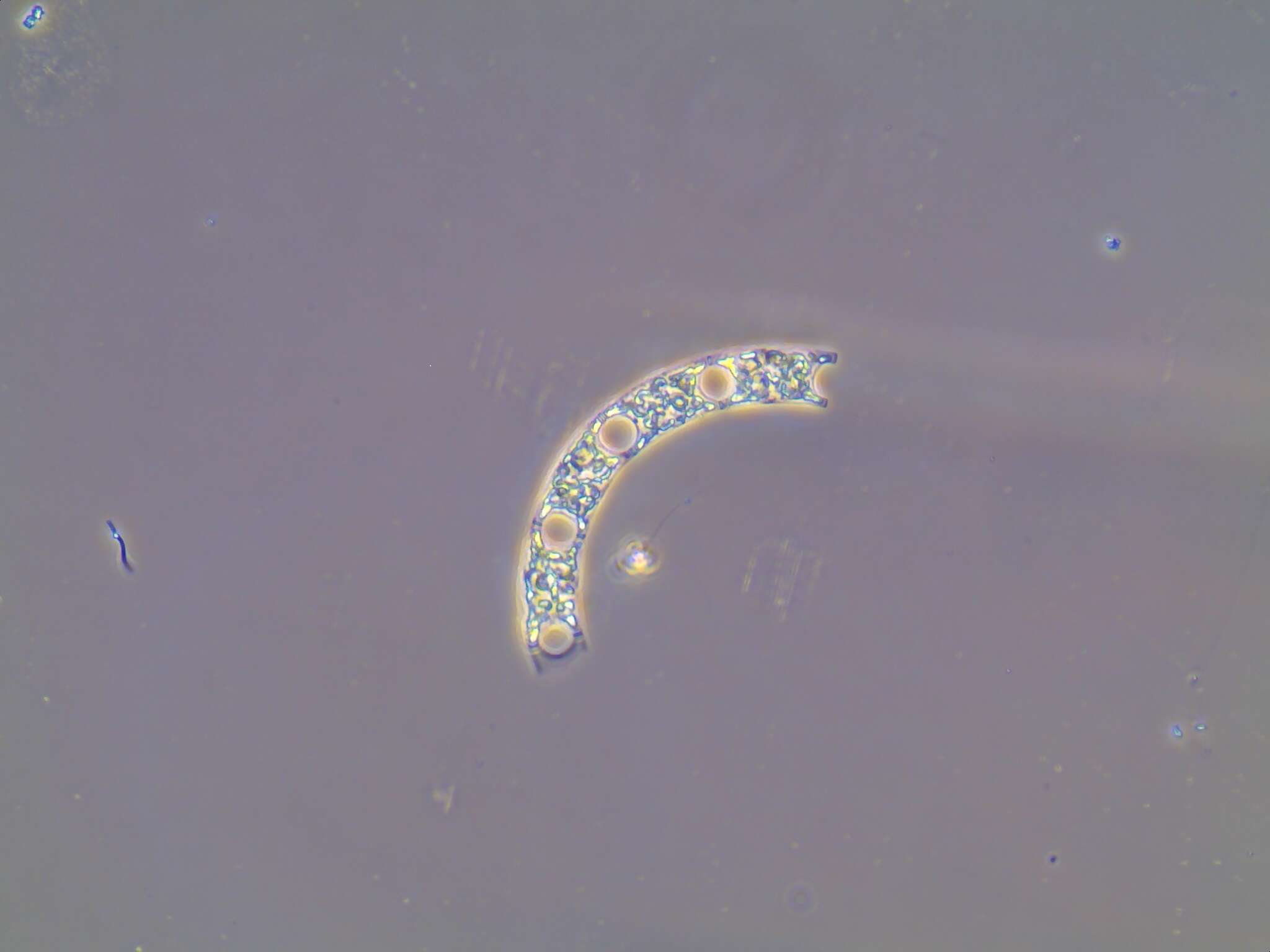
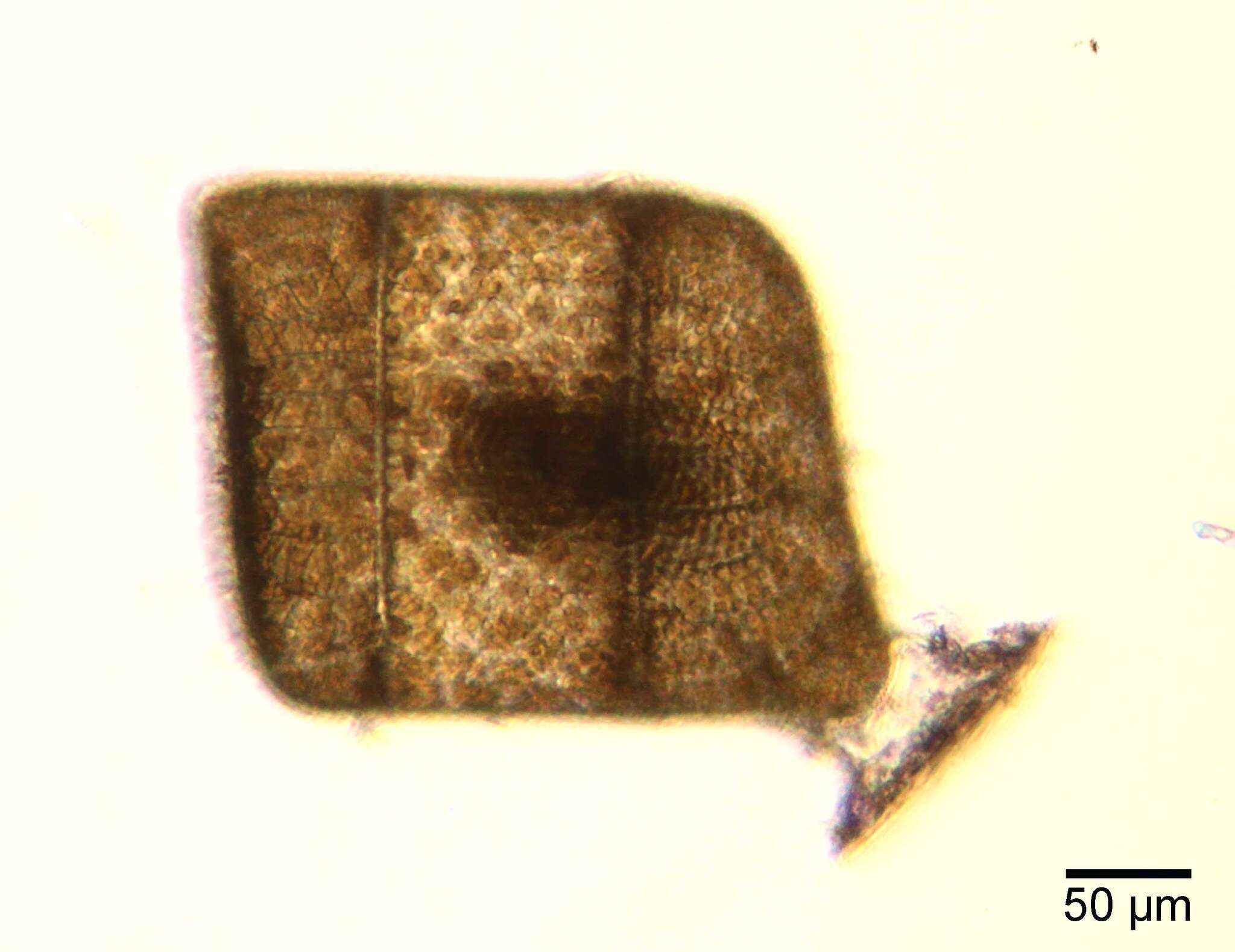
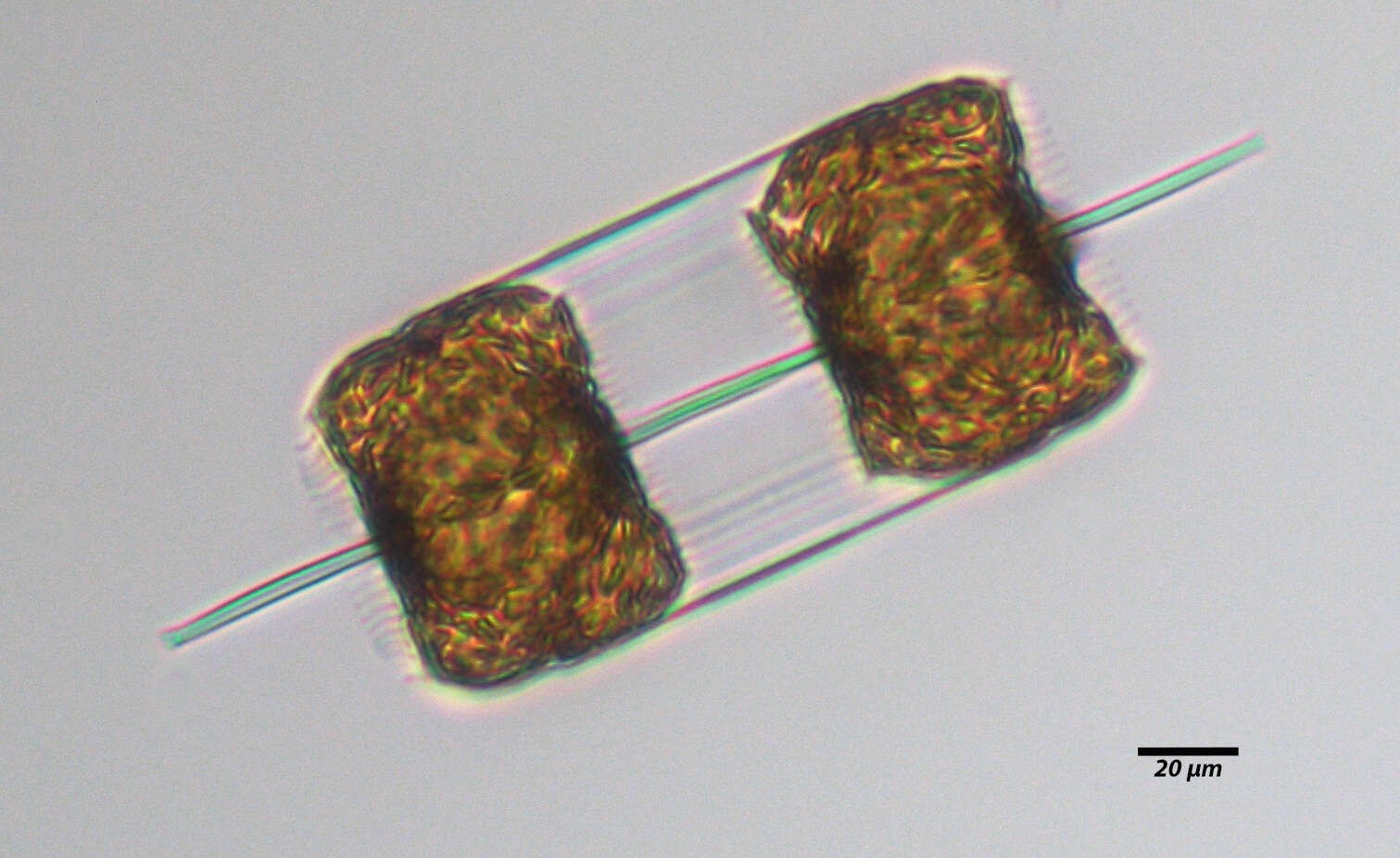
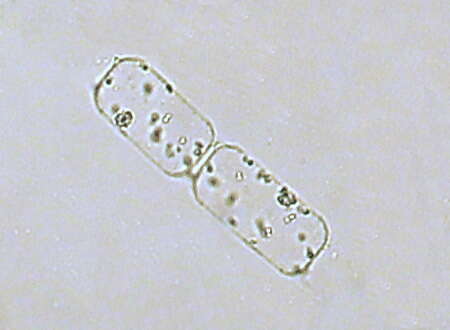
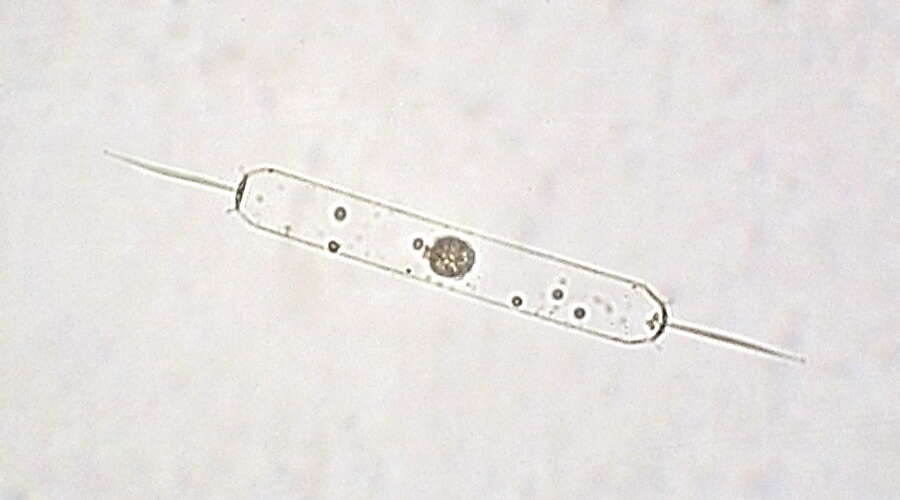
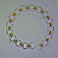
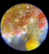
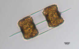
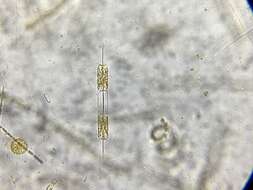
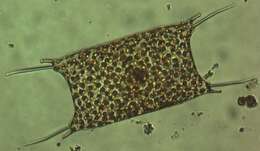
Description: English: Odontella mobiliensis preserved by lugol solution. A field sample from Long Island Sound. Date: 17 September 2013. Source: flickr. Author: NOAA Photo Library.
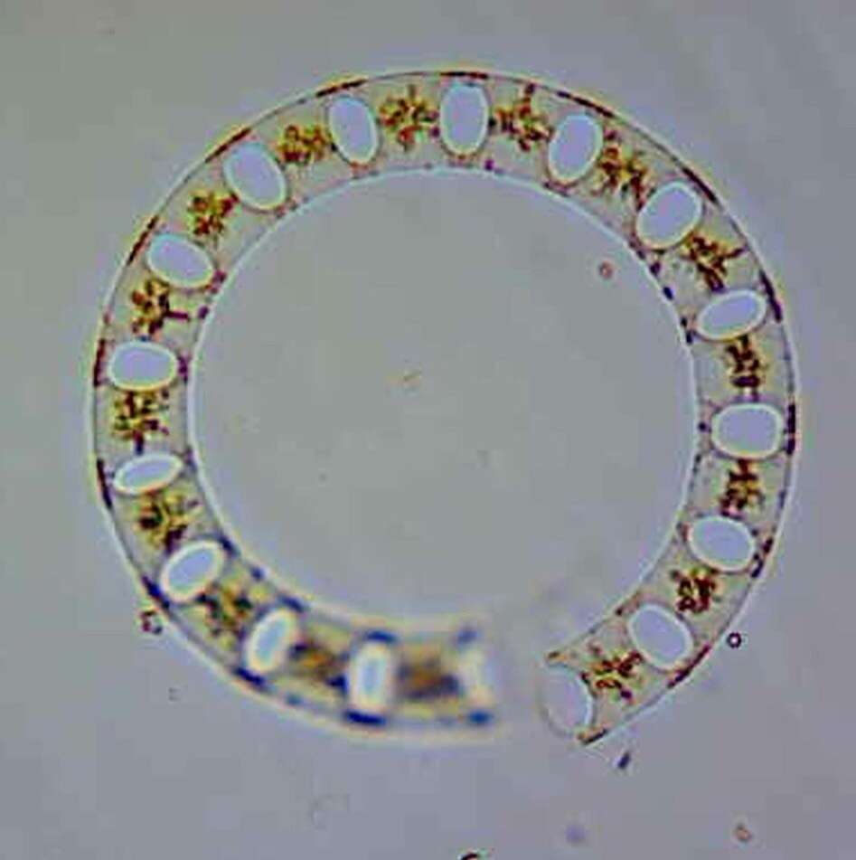
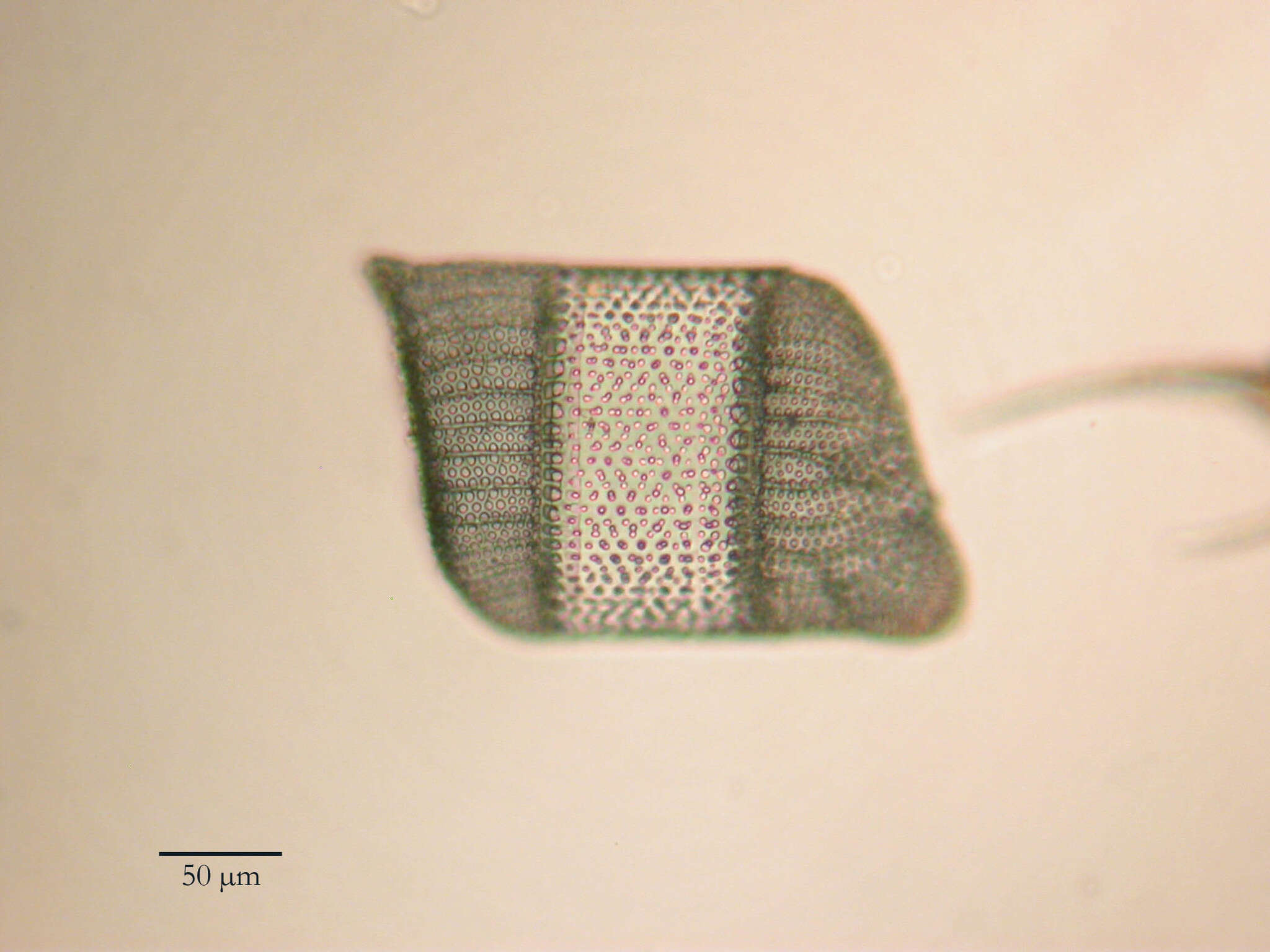
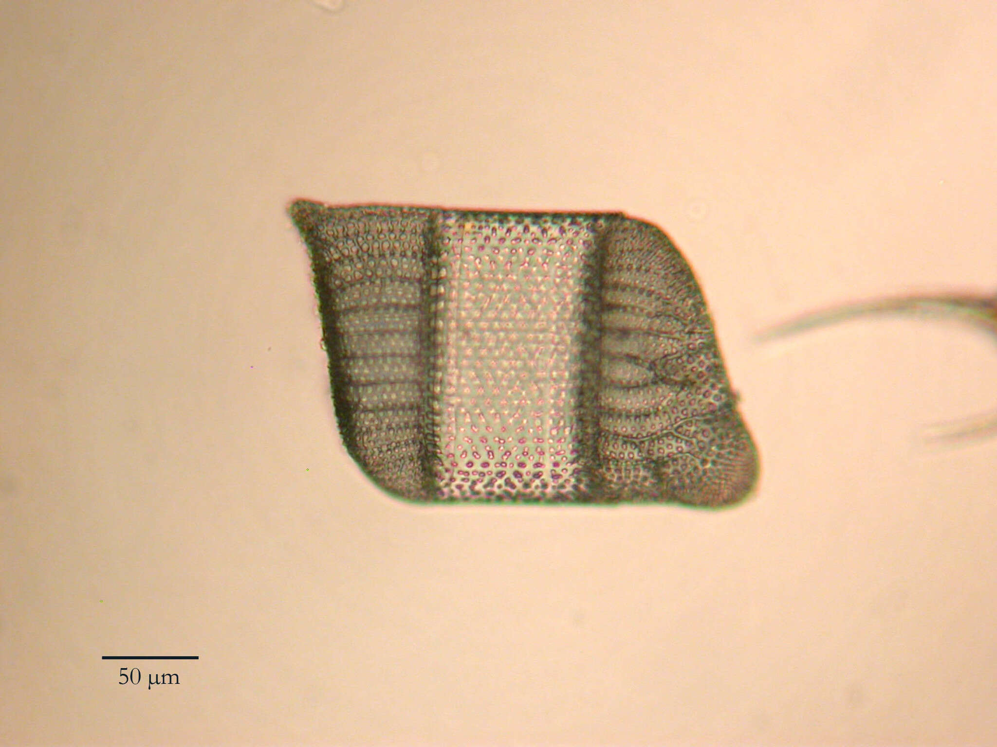
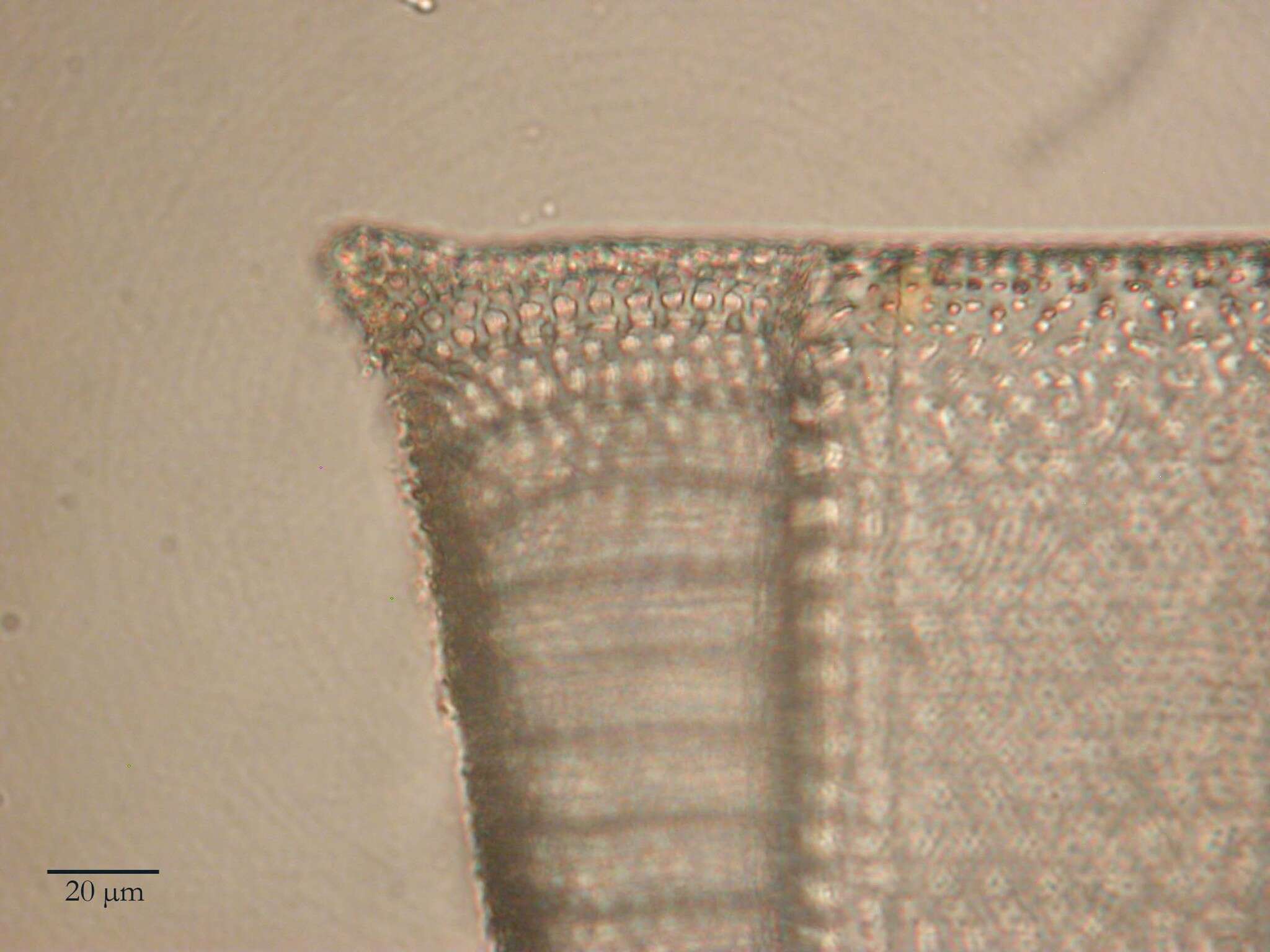
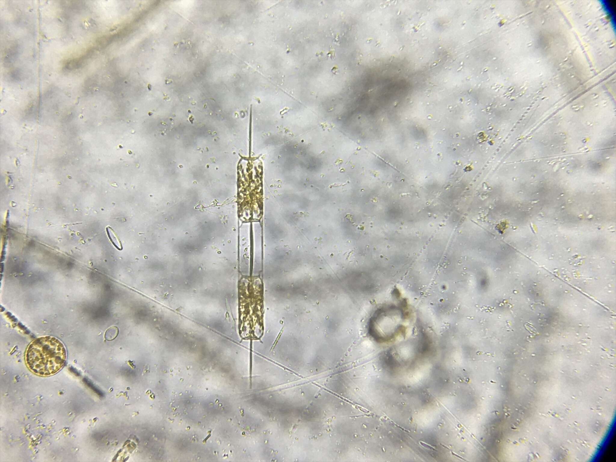
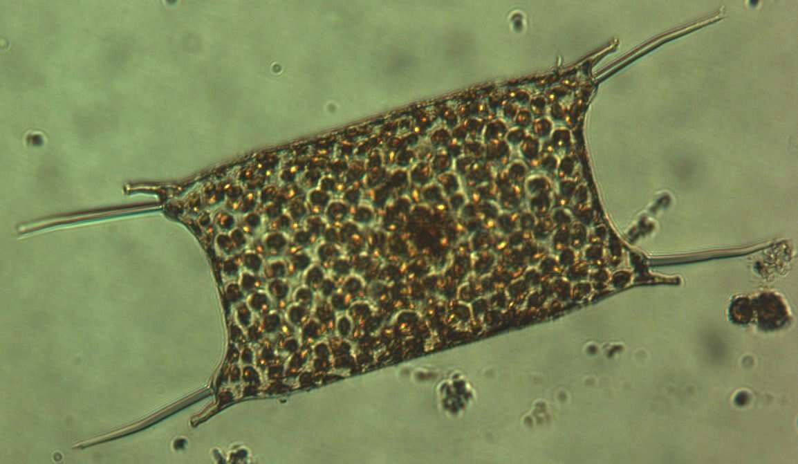
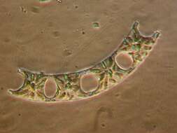
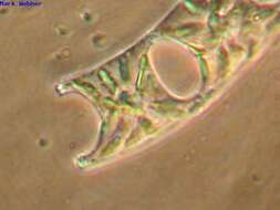
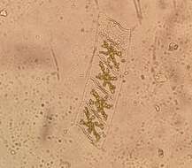
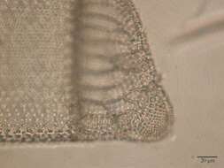
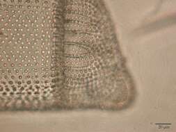
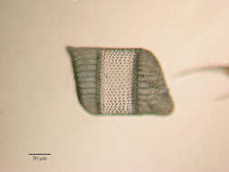
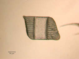
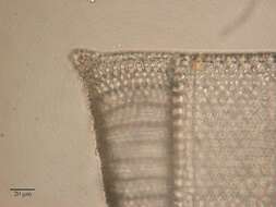
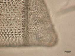
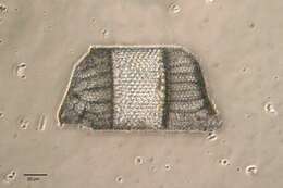
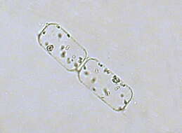
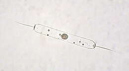
Description: Ditylum Brightwellii (West) Grun. 1860 (Triceratium Brightwellii T. West 1860); Biddulphiaceae, Biddulphiales, Centrophyceae, Bacillariophyta English: North-West Black Sea, coastal waters, at a depth of 0.5 metre Русский: Дитилум Брайтвелла; Северо-Запад Чёрного моря, прибрежные воды, на глубине 0,5 м. Date: 18 March 2008. Source: Own work. Author: Minami Himemiya.