-
-
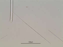
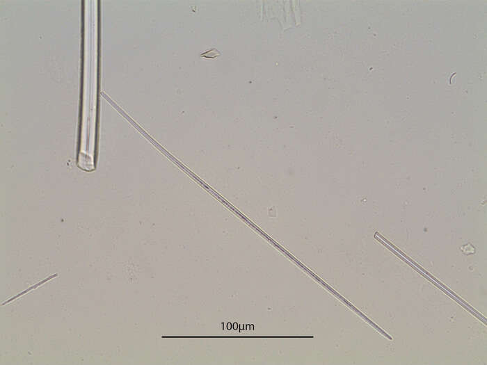
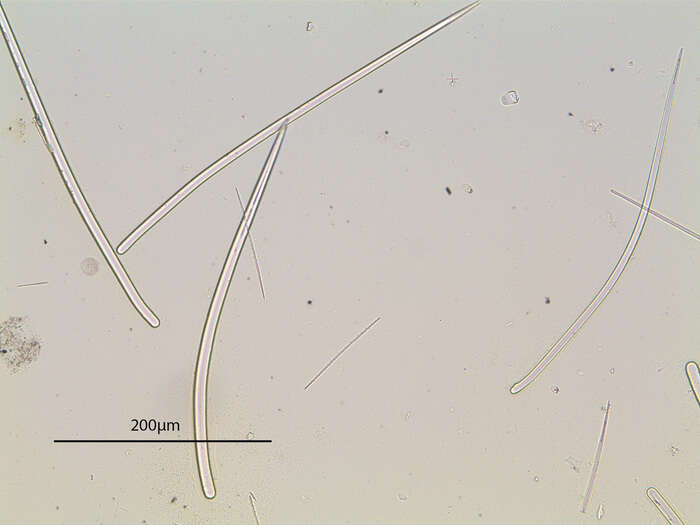
Photograph of spicules from the original slide
-
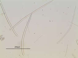
Photograph of spicules from the original slide
-
Photograph of spicules from the original slide
-
-
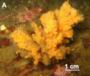
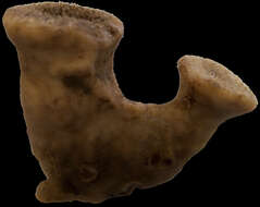
holotype CNPGG–2302. tubular habitus (picture: Patricia Gómez)
-
holotype CNPGG?1534, ficiform habitus. (picture: Patricia Gómez)
-
-
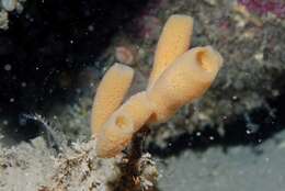
Chenal d'Ile Verte, Roscoff, W coast of France, June 1982, 2 m depth
-
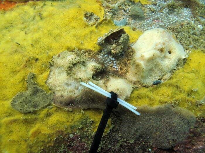
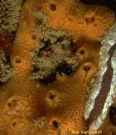
Rathlin Island, Co Antrim, Ireland 55°17'25.861" N 6°15'5.101" W 2007-06-12
-
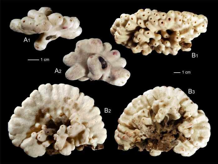
(a,b): Paratype specimen MNHN-IP-2019-3; (a): lateral view; (b): top view. (c–e): Holotype specimen MNHN-IP-2019-2; (c): top view, (d,e): lateral views
-
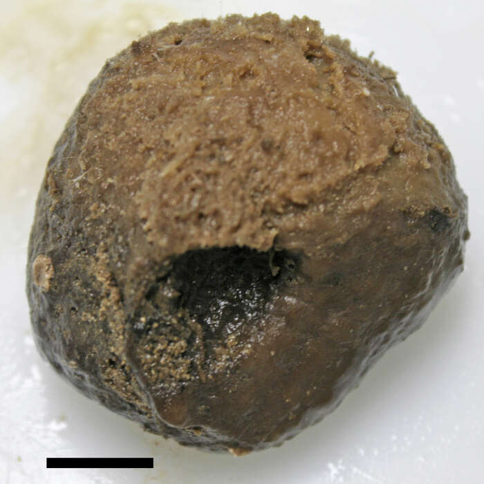
Holotype, MNHN-IP-2019-7 (picture: Andrzej Pisera).
-
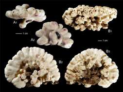
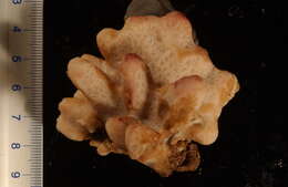
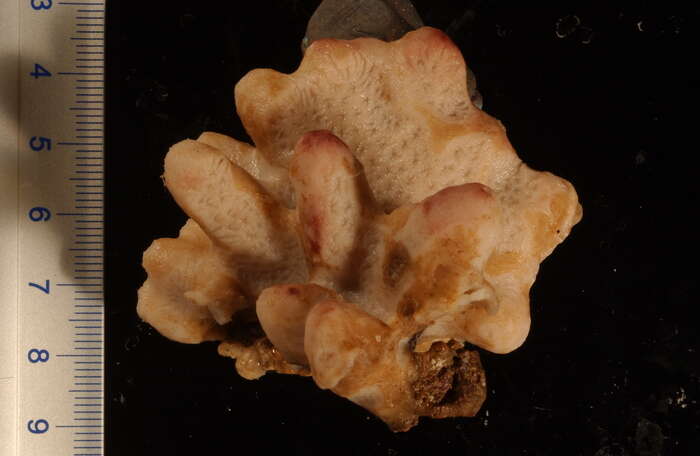
Holotype UFSPOR 1130 (picture: Danilo Almeida) scale = 1 cm
-
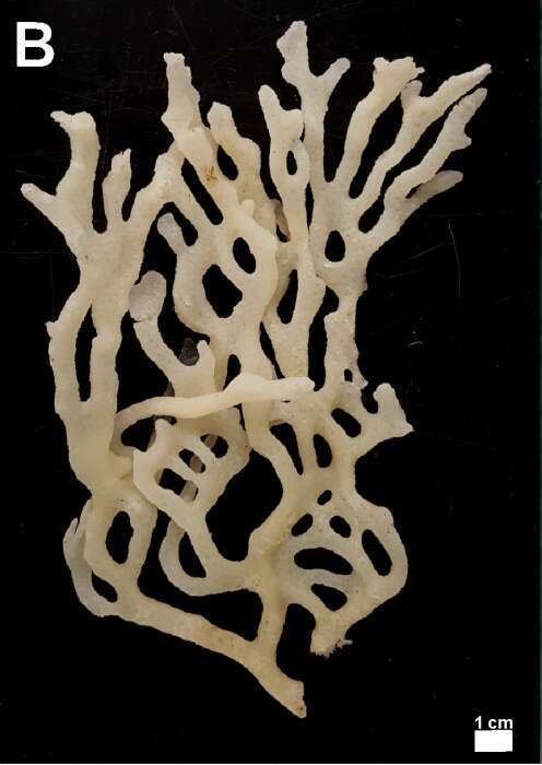
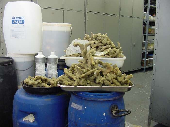
Petrosia (Petrosia) revizee Rocha, Moraes, Salani & Hajdu, 2021. MNRJ 44461. The holotype, MNRJ 22190, was sorted out from among these many broken branches.
-
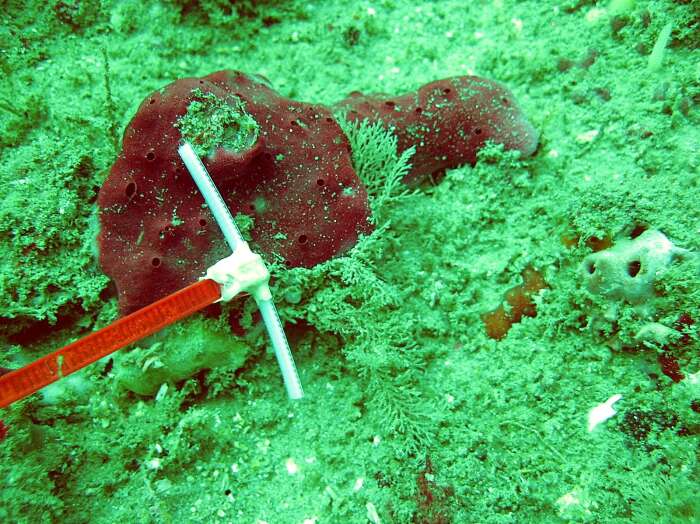
MNRJ 13119, paratype of Petrosia (Petrosia) revizee Rocha, Moraes, Salani & Hajdu, 2021 in situ. Scale = 5 cm.
-
Holotype of Xestospongia dorigo Rocha, Moraes, Salani & Hajdu, 2021 (MNRJ 15405). Scale = 5 cm.
-
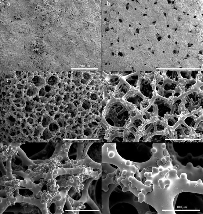
Holotype a–b. overview of the outer ectosomal surface showing oxeas and phyllotriaenes; c. overview of choanosomal skeleton; d. choanosomal desmas with short, rounded, and blunt tubercles; e. details of desmas; f. desma zygosis. (SEM pictures: Lin Gong).
-
Holotype ARC 81577. (A) In vivo appearance. (B) Skeleton. (C) Oxea. (D) Microrhabd. (E) Orthotriaenes/Dichotriaenes. (F) Aspidaster. (G) Aspidaster surface. (H) Strongylasters. (plate: C. Goodwin)
-
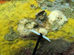
MNRJ 11272 at Isla Tortuga (Ancash Region, Peru), 8 m depth
-
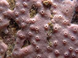
Topotypical paratype (MNRJ 12892), scale 5 cm wide
-
Topotypical paratype (MNRJ 12892)
-
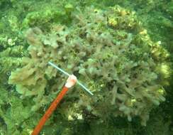
MNRJ 11262 - Isla Tortuga (Ancash Region, Peru), 6.3 m depth, coll. E. Hajdu, 2007-IX-23
-
MNRJ 12140 (Playa Catarindo, Mollendo, Arequipa Region, Peru), 4-5 m depth, coll. Y. Hooker, U. Zanabria & Ph. Willenz, 2008.XI.26
-

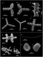
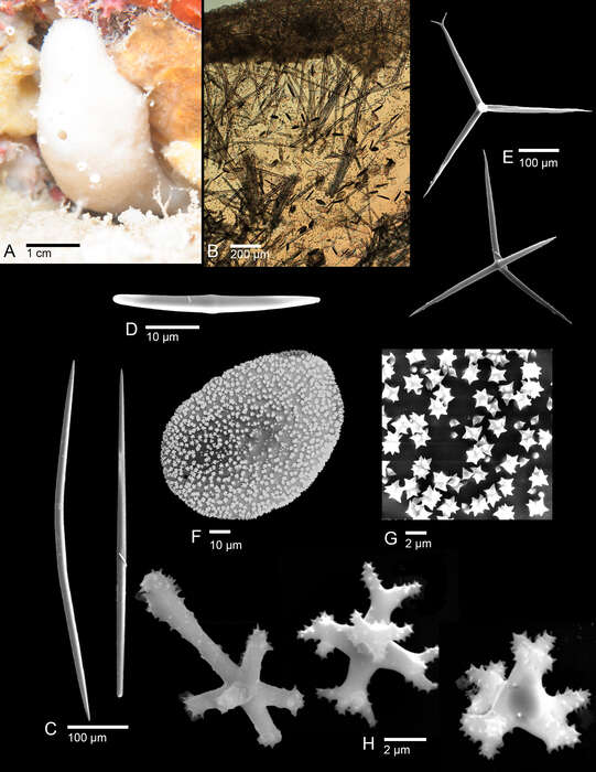
Figure 5 from original publication. Spicule composition of Alectona ricardi sp. nov. present in the red coral sample. SEM images: ages: (a,b) triactines covered by triangular spines; (c) magnification of a ray showing the compound (a,b) triactines covered by triangular spines; (c) magnification of a ray showing the compound spines; spines; (d) slightly spined triactine with spines clearly organised in verticils; (e) smooth triactine; (f) (d) slightly spined triactine with spines clearly organised in verticils; (e) smooth triactine; (f) triactine triactine with four rays; (g) nodular amphiaster; (h) nodular amphiaster (in the background) and with four rays; (g) nodular amphiaster; (h) nodular amphiaster (in the background) and thinner thinner amphiaster; (i) very small amphiaster; (j) oxea of an armed larva; (k) outer surface of a amphiaster; (i) very small amphiaster; (j) oxea of an armed larva; (k) outer surface of a quadrangular quadrangular discotriaene; (l) inner surface of a quadrangular discotriaene. discotriaene; (l) inner surface of a quadrangular discotriaene.