-
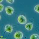
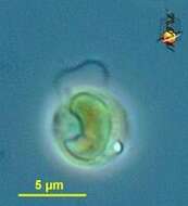
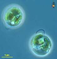
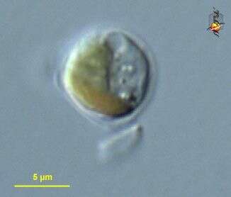
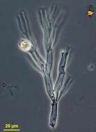
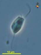
Chrysamoeba (cry-as-a-me-ba) mikrokonta, a chrysophyte (stramenopile) with plastids and often occurring as an amoeboid organisms. It produces fine pseudopodia. Phase Contrast microscopy.
data on this strain.
-
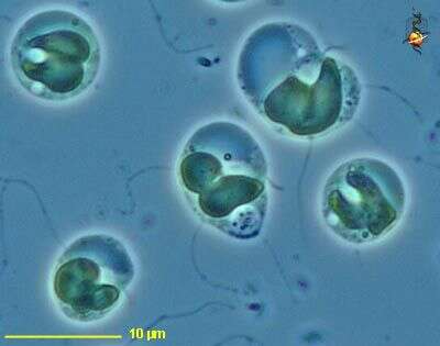
Chrysamoeba (cry-as-a-me-ba) mikrokonta, , a chrysophyte (stramenopile) with plastids and often occurring as an amoeboid organism. This detail of a flattened cell shows the two-lobed plastid, the carbohydrate (leucosin) storage vacuole (large irregular vacuole), a short flagellum and several fine pseudopodia. Phase contrast microscopy.
data on this strain.
-
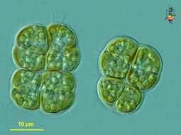
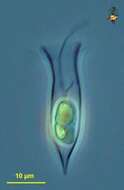
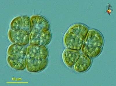
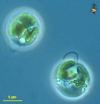
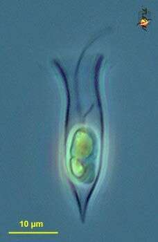
Chrysocapsa (cry-so-cap-sa) vernalis, a colonial chrysophyte (stramenopile) that forms a loose aggregate of cells inside a gelatinous matrix. Cells have a single golden bi-lobed chloroplast. Differential interference microscopy.
data on this strain.
-
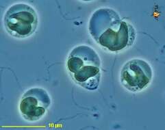
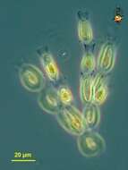
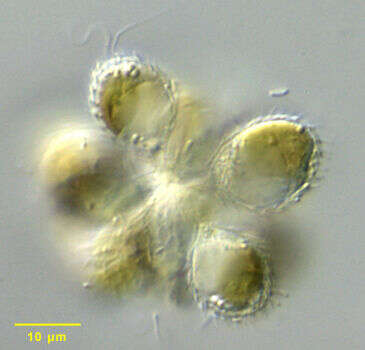
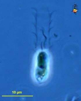
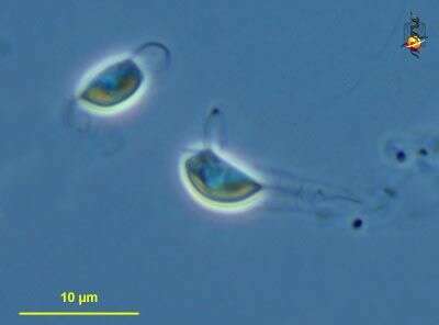
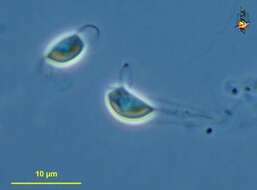
Ochromonas (ock-roe-moan-ass) sphaerocystis, iconic genus of the chrysophytes, body elongated or rounded, with two emergent flagella, golden plastids, sometimes with extrusible bodies under the cell surface. Phase contrast microscopy.
data on this strain.
-
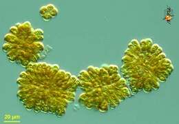
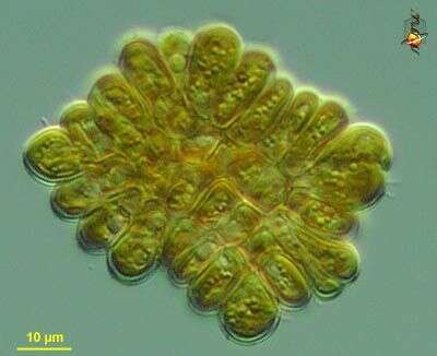
Phaeoplaca (fay-owe-plack-a) thallosa, a chrysophyte in which cells adhere to each other to form irregular rectangular sheets. Differential interference microscopy.
data on this strain.
-
Phaeoplaca (fay-owe-plack-a) thallosa, a chrysophyte in which cells adhere to each other to form irregular rectangular sheets. Differential interference microscopy.
data on this strain.
-
Phaeoplaca (fay-owe-plack-a) thallosa, a chrysophyte in which cells adhere to each other to form irregular rectangular sheets. Differential interference microscopy.
data on this strain.
-
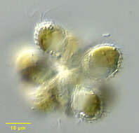
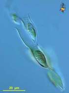
Synura sphagnicola, a colonial chrysophyte. Synonymous with Skadovskiella. Cells are joined at their posterior ends in the center of the colony. Cells are ovoid with two golden chloroplasts. There are two equal flagella less than 1 cell length. Siliceous scales with a unique ring form from which there is a rod-like projection cover the cell. A detached scale is seen in the center of this image. The scale structure is diagnostic for the species. A stigma is absent. Large vacuoles containing the glucopyranoside storage polymer, chrysolaminarin (leucosin) accumulate in the cytoplasm . The colonies swim with a slow rolling motion.From a polysaprobic temporary freshwater farm pond near Boise, Idaho. Differential interference contrast.Differential interference contrast optics.
-
Synura sphagnicola, a colonial chrysophyte. Synonymous with Skadovskiella. Cells are joined at their posterior ends in the center of the colony. Cells are ovoid with two golden chloroplasts. There are two equal flagella less than 1 cell length. The cell is covered by siliceous scales the morphology of which is diagnostic for the species (see accompanying image). A stigma is absent. Large vacuoles containing the glucopyranoside storage polymer, chrysolaminarin (leucosin) accumulate in the cytoplasm. The colonies swim with a slow rolling motion.From a polysaprobic temporary freshwater farm pond near Boise, Idaho. Differential interference contrast.
-
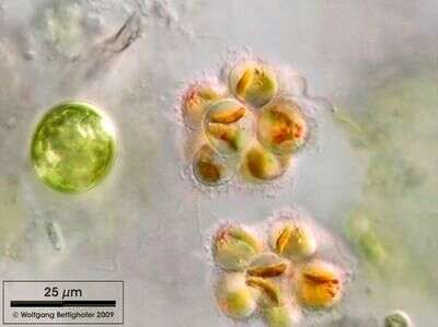
Synura sphagnicola can be preferentially found in sphagnum ponds. Typical are the reddish oil droplets they bear in plasma. Sample from sphagnum pond Dosenmoor near Neumuenster (Schleswig-Holstein, Germany). This image was taken using Zeiss Universal with Olympus C7070 CCD camera.
-
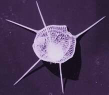
This image was made from samples taken during a scientific cruise in the Pacific. Water was filtered to concentrate the organisms that were present, then dried onto a thin sheet of plastic and then shadowed with a fine layer of metal to provide contrast. The preparation was then observed with an electron-microscope. This technique has been used to document the diversity of marine microbes, especially, protists in the oceans.
-
Chrysophyte. This cell was not identified to genus. It contains a plastid with chlorophylls a and c, a contractile vacuole is evident at the top of the cell, and the cell is surrounded in an organic wall. although these cells were common in some locations, no motile forms corresponding to these cells were seen. Differential interference contrast. Material from Nymph Creek and Nymph Lake, thermal sites within Yellowstone National Park, photograph by Kathy Sheehan and David Patterson.
-
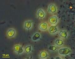
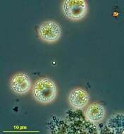
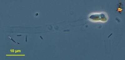
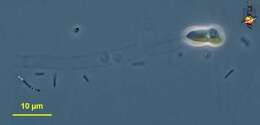
Chromulina (crumb-you-line-a) nebulosa, a small chrysophyte (stramenopile) with a single emergent flagellum, and with a golden plastid. Phase contrast microscopy.
data on this strain.
-
Chromulina (crumb-you-line-a) nebulosa, a small chrysophyte (stramenopiles) with a single emergent flagellum. A second short flagellum is visible because cell division is beginning to occur (note the two chloroplasts) and a second flagellum has formed. -Phase contrast microscopy.
data on this strain.
-
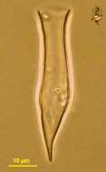
Dinobryon (dine-owe-bry-on) is a loricate chrysophyte (stramenopile) flagellate. This image shows a single lorica. Phase contrast.
-
Dinobryon (dine-owe-bry-on) is a loricate chrysophyte (stramenopile) flagellate. Cells have one long and one short flagellum, and there is an eye-spot at the front end of the plastid. Differential interference contrast.
-
Dinobryon (dine-owe-bry-on) is a loricate chrysophyte (stramenopile) flagellate. Cells have one long and one short flagellum, and there is an eye-spot at the front end of the plastid. Neck with a number of ridges. Phase contrast.
-
Dinobryon (dine-owe-bry-on) is a loricate chrysophyte (stramenopile) flagellate. Cells have one long and one short flagellum, and there is an eye-spot at the front end of the plastid. Cells can encyst within the lorica, and form a pored cyst called a stomatocyst, one is visible here. Phase contrast.
-
-
Dinobryon (dine-oh-bry-on) a mixotrophic stramenopile (chrysophyte) with one long flagellum and one short flagellum. When feeding heterotrophically, the beating of the long flagellum draws food towards the cell where it may be ingested. The cell also has brownish chloroplasts. It forms a flimsy tubular lorica. Phase contrast microscopy.
-
Dinobryon (dine-oh-bry-on) a mixotrophic stramenopile (chrysophyte) with one long flagellum and one short flagellum. When feeding heterotrophically, the beating of the long flagellum draws food towards the cell where it may be ingested. The cell also has brownish chloroplasts. It forms a flimsy tubular lorica which is emphasised in this image. Phase contrast microscopy.
-
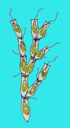
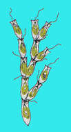
Dinobryon (die-know-bry-on) sertularia, a loricate chrysophyte (stramenochrome) alga, the vase-shaped lorica is organic, most species are usually found with the loricae attached to each other to form arborescent colonies. With two flagella, one longer one drawing water towards the cell and a shorter one. With golden plastid. Phase contrast microscopy.
data on this strain.
-
Dinobryon (die-know-bry-on) sertularia, a loricate chrysophyte (stramenochrome) alga, the vase-shaped lorica is organic, most species are usually found with the loricae attached to each other to form arborescent colonies. Differential interference microscopy.
data on this strain.