-
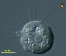
Nuclearia (new-clee-air-ee-a), a nucleariid (cristi-discoid) filose amoeba. Exclusively with thin pseudopodia without skeletal supports. More of these project from the front of the cell (to the right), whereas they appear a little more crumpled at the posterior end. Eats bacteria, algae, detritus. Phase contrast.
-
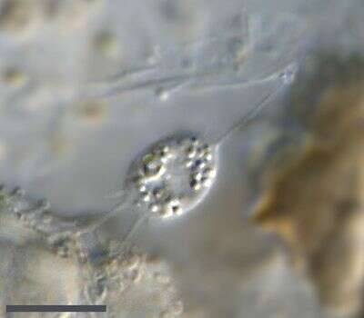
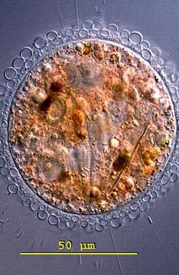
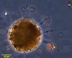
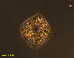
Nuclearia (this species also reported widely under the name Astrodisculus radians), nucleariid filose amoebae with thin pseduopodia. This species with a layer of surrounding mucus, and with bacteria attached to the outer surface of the mucus. Differential interference contrast.
-
-
Nuclearia (new-clee-air-ee-a), a nucleariid (cristi-discoid) filose amoeba. Exclusively with thin pseudopodia without skeletal supports. More of these project from the front of the cell (to the right), whereas they appear a little more crumpled at the posterior end. Eats bacteria, algae, detritus. Phase contrast.
-
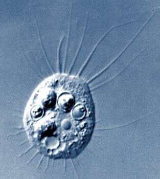
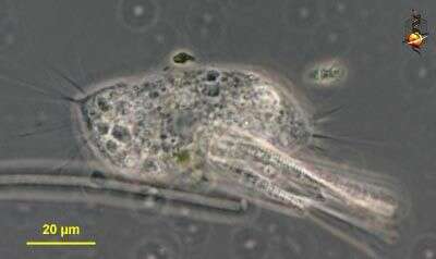
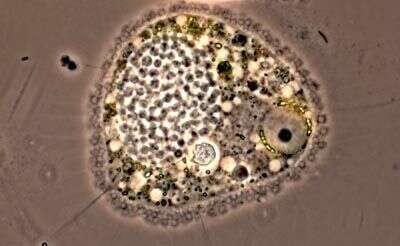
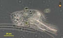
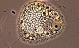
Nuclearia, filose amoeba. Differential interference contrast. This cell has been feeding on yeast cells. Thin filose pseudopodia, without any evident internal skeletal elements, extend from the front of the cell, and are pulled in at the back. The contractile vacuole with surrounding vesicles is located at the rear of the cell. The nucleus with the round nucleolus lies just anterior to the contractile vacuole.
-
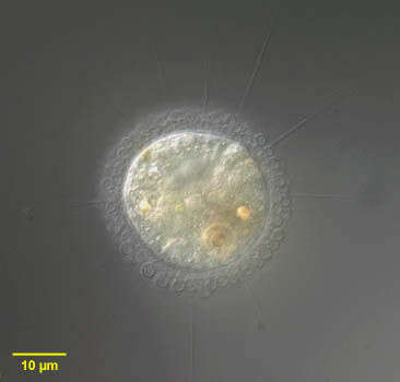
Nuclearia (nuke-lee-air-ee-a) is a naked filose amoeba. It is referred to as naked because there is no lorica and wall, and filose because of the thread-like appearance of the pseudopodia. Normally consume algae and detritus. Phase contrast. Material from Nymph Creek and Nymph Lake, thermal sites within Yellowstone National Park, photograph by Kathy Sheehan and David Patterson.
-
-
-
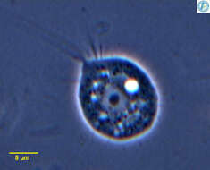
Nuclearia , nucleariid filose amoebae with thin pseduopodia. This is one of the smaller species, either N. moebiusi or N. simplex. There is a large nucleus with a dark spherical nucleolus and to the right is a contractile vacuole. The fine pseudopodia extend from the front of the cell (upper left) and are withdrawn from the back of the cell. From Lake Donghu, China. Phase contrast micrograph.
-
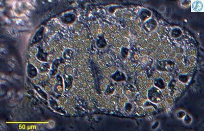
Nuclearia , nucleariid filose amoebae with thin pseduopodia. This is one of the smaller species, either N. moebiusi or N. simplex. It has invaded a colony of Microcystis (cyanobacteria) and is eating the bacterial cells and multiplying within the colony. From Lake Donghu, China Phase contrast micrograph.
-
Nuclearia, one of the naked filose amoebae, producing thin pseudopodia without internal skeletons. This is one of the larger more rounded species, eats detritus and algae. Phase contrast micrograph.
-
Nuclearia isolated from sandy sediments from Little Sippiwissett salt marsh. Micrograph taken by Jeffrey Cole.
-
Nuclearia (new-clee-air-ee-a), a nucleariid (cristi-discoid) filose amoeba. Exclusively with thin pseudopodia without skeletal supports. More of these project from the front of the cell (to the left), whereas they appear a little more crumpled at the posterior end. The structure slightly right and below the centre is the nucleus, with a large nucleolus and the nucleolus having a small empty central region. Eats bacteria, algae, detritus. Differential interference contrast.
-
This cell, like many Nuclearia species, has been consuming algae. The nucleus is the clear area with the dark nucleolus in the center. Nucleariid amoebae have thin non-stiff pseudopodia. Phase contrast image.
-
Cysts, from a culture. Phase contrast.
-
Live cell, phase contrast. The fine pseudopodia are typical of nuclearid filose amoebae. The surrounding material is mostly comprised of yeast cell walls (the species is provided with yeast as food).
-
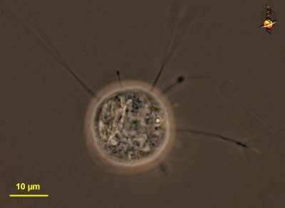
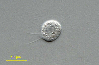
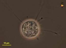
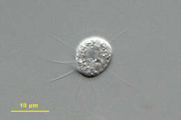
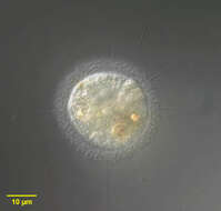
This specimen was isolated from a sample of material coming out of a sphagnum pond in the so called "Dosenmoor" near Neumuenster (Schleswig-Holstein, Germany). The scale bar indicates 10 µm. Images were taken using Zeiss Universal with Olympus C7070 CCD camera.
-
-
-
-
-
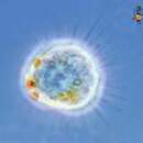
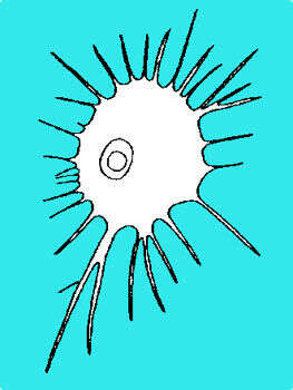
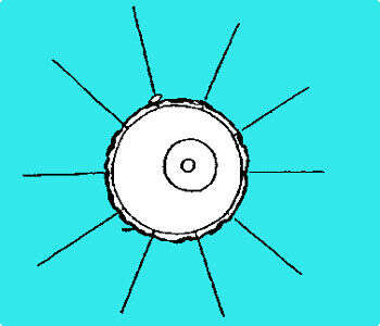
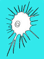
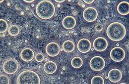
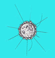
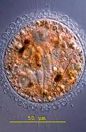
Pompholyxophrys is a filose amoeba. Its cell surface is covered by a layer of delicate siliceous (glass-like) spheres. It has thin, delicate pseudopodia that it uses for movement. The structure to the right in the cell is its nucleus.
-
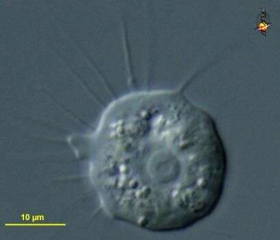
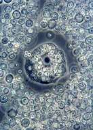
Portrait of Pompholyxophrys, one of the heliozoon-like amoebae previously assigned to the order Rotosphaerida. Pompholyxophrys has a periplast composed of endogenously formed siliceous elements of a single type within a species. These are spherical in the type species P. punicea but may be ovoid (see images of P. ovuligera), discoid or bone-shaped. Radiating spicules are absent. What appear at first glance to be axopodia are, in fact, filopods that lack axonemes and extrusomes. Contracted filopodia may appear granular leading to confusion but close examination of the extended filopodia shows extrusomes are absent. From standing freshwater Typha latifolia marsh near Boise, Idaho. Differential interference contrast
-
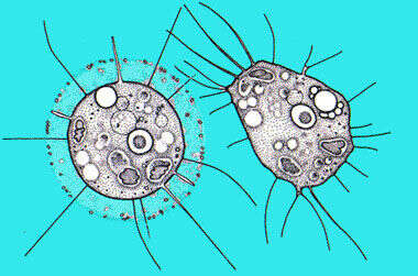
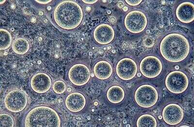
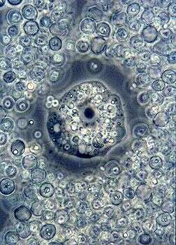
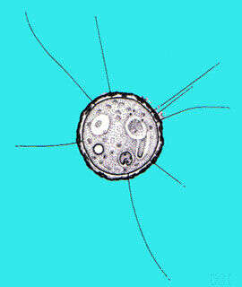
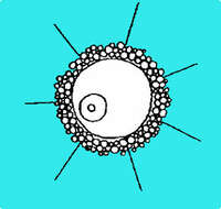
Pompholyxophrys (pom-folly-zoff-riss) punicea. The cytoplasm of the spherical body is colorless or reddish, often interspersed with colored granules and green or brown food particles. The outer periplast is built up from conspicuous minute colourless spherical granules (perles) arranged in concentric layers. The granules are glass like hollow spheres and arranged in concentric layers to form a compact envelope. The large nucleus is located eccentricly. The straight and pointed pseudopodia are tenuous and indistinct. Individuals are found occasionally in ponds and swamps. This specimen was collected in a bog pond near Konstanz, Germany. The outer sphere of colourless spherical perles can be seen to be lying in concentric layers. Differential interference contrast.