-
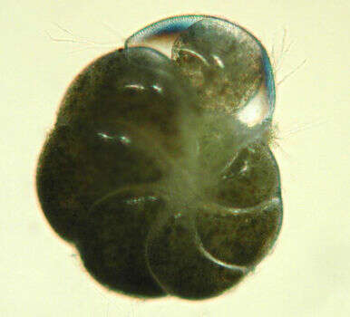
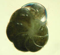
This specimen was collected on Sapelo Island, Georgia. The nearly transparent tendrils extending from the test are the reticulopodia. The green color is caused by chloroplasts that the foram has stolen from diatoms that it eats. Image courtesy of Susan T. Goldstein, University of Georgia.
-
Specimen collected at Archachon, France. Image courtesy of Stefan Revets. This image first appeared in Hansen and Revets, J. Foram. Res. 22:166-180 (1992) and is used with permission.
-
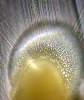
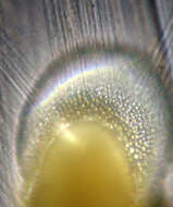
This image shows a closeup of the aperture in the test from which the reticulopodia extend. The actual aperture follows the curved line between the glassy-looking part of the test, which is the youngest chamber, and the yellowish part, which is another part of the test (and out of the plane of focus, which is why it's slightly blurry.) The pods themselves are the transparent ray-like objects. Forams use reticulopods to move, eat, gather materials, build their tests, and do pretty much everything else they do. Image courtesy of Susan T. Goldstein, University of Georgia.
-
Horizontal section through the test. Image courtesy of Stefan Revets. This image first appeared in Hansen and Revets, J. Foram. Res. 22:166-180 (1992) and is used with permission.
-
Sample collected at Hamble Estuary, Hampshire, England. Image courtesy of Elisabeth Alve, University of Oslo. Originally published in the Journal of Foraminiferal Research 31:1; used with permission.
-
Image courtesy of Stefan Revets. This image first appeared in Hansen and Revets, J. Foram. Res. 22:166-180 (1992) and is used with permission.
-
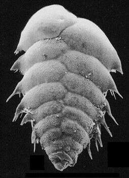
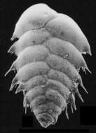
Electron micrograph of an individual recovered from the Santa Barbara Basin, California. This species is found at the most anoxic part of the basin; it can apparently survive for months with no oxygen. Length: about 300 um. Image courtesy of Joan Bernhard, Woods Hole Oceanographic Institute. Originally published in the Journal of Foraminiferal Research 27:4; used with permission.
-
Image courtesy of Stefan Revets. This image first appeared in Hansen and Revets, J. Foram. Res. 22:166-180 (1992) and is used with permission.
-
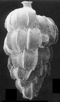
Electron micrograph of an individual recovered from the Santa Barbara Basin, California. Length: about 400 um. Image courtesy of Joan Bernhard, Woods Hole Oceanographic Institute. Originally published in the Journal of Foraminiferal Research 27:4; used with permission.
-
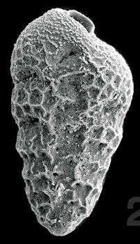
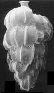
This Eocene fossil specimen was collected near Parnes, France. Image courtesy of Stefan Revets. This image first appeared in Hansen and Revets, J. Foram. Res. 22:166-180 (1992) and is used with permission.
-
Electron micrograph of an individual recovered from the Santa Barbara Basin, California. Length: about 500 um. Image courtesy of Joan Bernhard, Woods Hole Oceanographic Institute. Originally published in the Journal of Foraminiferal Research 27:4; used with permission.
-
Image courtesy of Stefan Revets. This image first appeared in Hansen and Revets, J. Foram. Res. 22:166-180 (1992) and is used with permission.
-
Sample collected at Hamble Estuary, Hampshire, England. Bolivinids are generally found in the estuary only in fall and early winter, when river water flow is low and salinity is relatively high. Image courtesy of Elisabeth Alve, University of Oslo. Originally published in the Journal of Foraminiferal Research 31:1; used with permission.
-
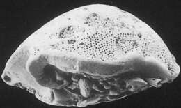
The aperture is at the center right edge. Image courtesy of Stefan Revets. This image first appeared in Hansen and Revets, J. Foram. Res. 22:166-180 (1992) and is used with permission.
-
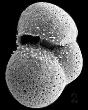
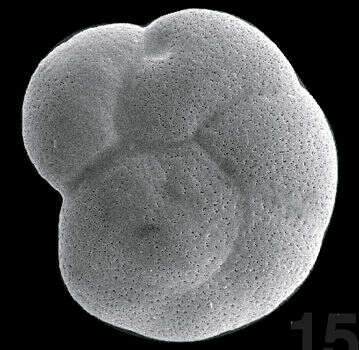
This foram, from a recently described (2000) genus, is named after the estuary in which it was discovered. Sample collected at Hamble Estuary, Hampshire, England. Image courtesy of Elisabeth Alve, University of Oslo. Originally published in the Journal of Foraminiferal Research 31:1; used with permission.
-
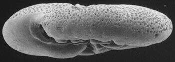
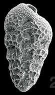
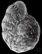
This Recent specimen was collected at the Dry Tortugas, Florida, USA. Image courtesy of Stefan Revets. This image first appeared in Hansen and Revets, J. Foram. Res. 22:166-180 (1992) and is used with permission.
-
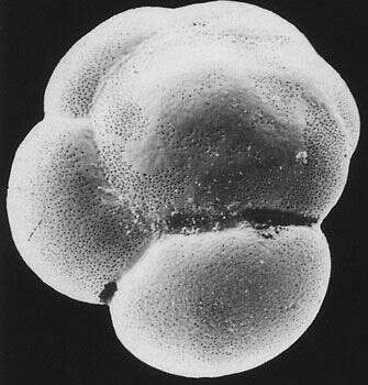
Sample collected at Hamble Estuary, Hampshire, England. Image courtesy of Elisabeth Alve, University of Oslo. Originally published in the Journal of Foraminiferal Research 31:1; used with permission.
-
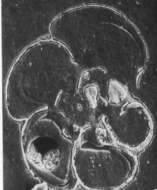
Horizontal section through the test. Image courtesy of Stefan Revets. This image first appeared in Hansen and Revets, J. Foram. Res. 22:166-180 (1992) and is used with permission.
-
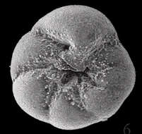
From Laguna Madre, Texas. Image courtesy of Pamela Stephens, Midwestern State University.
-
Image courtesy of Stefan Revets. This image first appeared in Hansen and Revets, J. Foram. Res. 22:166-180 (1992) and is used with permission.
-
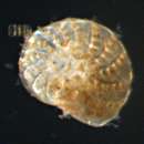
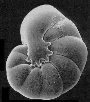
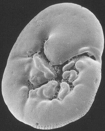
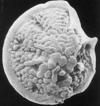
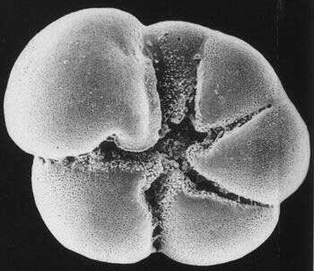
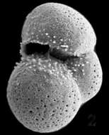
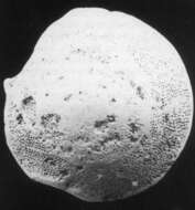
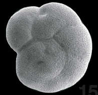
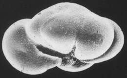
This is the top (spiral) surface of a gamont of Ammonia parkinsoniana. Forams have complex and varied reproductive cycles. Some forms, such as this species, alternate between agamonts/schizonts (which are produced when two gametes fuse) and gamonts (which are produced when an agamont splits up into hundreds of smaller individuals). The two types often look so distinct from each other that they were originally mistaken for different species. Image courtesy of Pamela Stephens, Midwestern State University.
-
Image courtesy of Stefan Revets. This image first appeared in Hansen and Revets, J. Foram. Res. 22:166-180 (1992) and is used with permission.
-
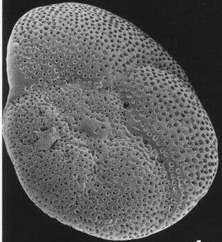
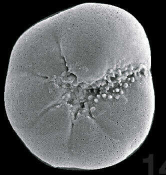
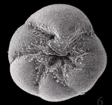
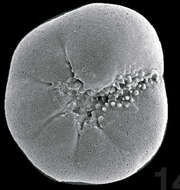
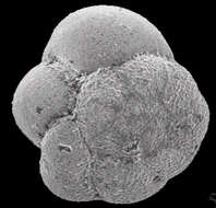
This is the bottom (umbilical) face of an A. parkinsoniana gamont. Image courtesy of Pamela Stephens, Midwestern State University.
-
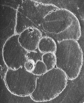
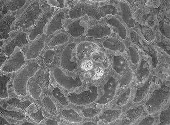
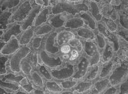
This view shows the transition between the early, spiral growth pattern (the chambers in the center) and the later cyclic pattern. Image courtesy of Carles Ferrandez-Canadell, University of Barcelona. This image first appeared in J. Foram. Res. 28: 135-140 and is used with permission.