-
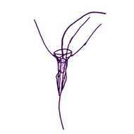
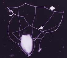
Calliacantha natans (Groentved, 1956) Leadbeater, 1978. Choanoflagellates with a conical lorica, 25 - 27 microns long and with six longitudinal and two transverse costae. One transverse costa is located at the top of the lorica chamber, the other is shortly below it, but is formed of thinner costal strips and may be difficult to see. Three spines project from the top of the lorica and a single posterior spine. The anterior spines are not continuous with the longitudinal costae.
-
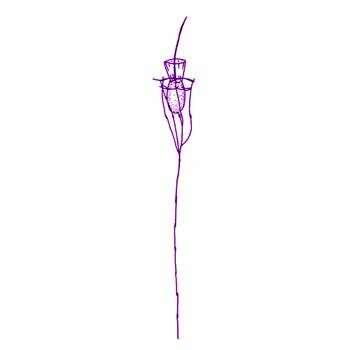
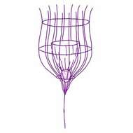
Calliacantha longicaudata (Leadbeater, 1975) Leadbeater, 1978. Choanoflagellates, lorica of which consists of a single chamber, 15-17 microns long x 6-8 microns wide, composed: of 5 (sometimes locally 4 or 6) longitudinal costae, each of 2 linearly attached slender cylindrical costal strips overlapping where they join, delimited anteriorly by 2 transverse costae, one terminal and the other parallel to it at about one sixth I of the length of the chamber behind it, transverse costal strips of both rings joined by their ends to the subtending longitudinal costae, at the posterior end, the longitudinal costae converging to subtend a caudal appendage of exceptional length (up to 100 microns) composed of up to 13 linearly attached costal strips overlapping at their ends and becoming thinner distally. The cell is enclosed by a conical membrane within the lorica chamber, attached to its base and equalling it in length, width when full 3-5 microns, the single flagellum exceeding 20 microns in length, surrounded by a ring of about 40 tentacles up to 6 microns in length, sometimes with terminal swellings.
-
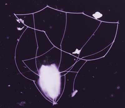
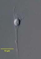
This image was made from samples taken during a scientific cruise in the Pacific. Water was filtered to concentrate the organisms that were present, then dried onto a thin sheet of plastic and then shadowed with a fine layer of metal to provide contrast. The preparation was then observed with an electron-microscope. This technique has been used to document the diversity of marine microbes, especially, protists in the oceans.
-
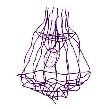
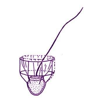
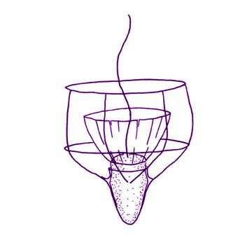
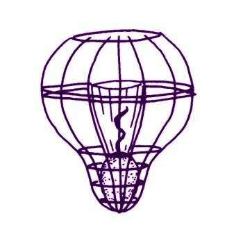
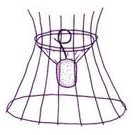
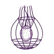
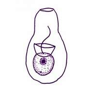
Crinolina isefiordensis Thomsen, 1981. Cell solitary, planktonic, living in skirt-shaped lorica, open anteriorly and posteriorly. Protoplast 8 microns long and 5 microns wide, without chloroplast. Single flagellum 2-3 times protoplast length, surrounded by a collar of tentacles. Height of collar approximately 6 microns, maximum diameter of collar 13 microns Lorica 25-30 microns long, diameter at base 20-30 microns, diameter at neck constriction 10-13 microns Longitudinal costae (12) 15-16, each composed of 6-7 costal strips. Two transverse costae. Longitudinal costae form spines at the anterior end of the lorica. Each spine is composed of two costal strips. Posteriorly the longitudinal costae project slightly beyond the transverse costal ring.
-
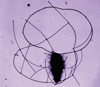
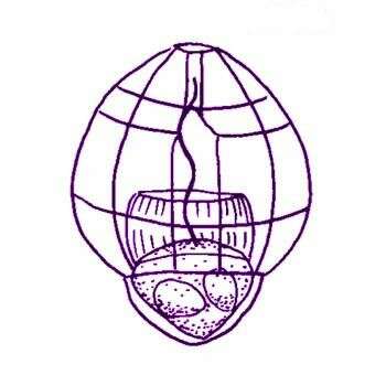
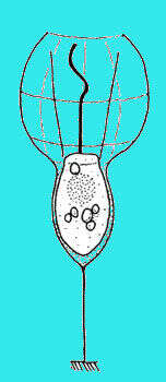
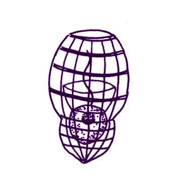
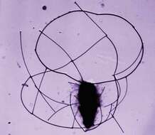
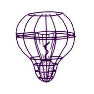
Diaphanoeca grandis Ellis, 1930. Choanoflagellates with a lorica composed of 12-14 longitudinal costae each formed by 7-8 strips of equal length. Three transverse rings encircle the base of the lorica and one ring, consisting of paired strips, encircles the longitudinal costae towards the anterior end. The protoplast, which is situated centrally within the lorica, bears an anterior collar of tentacles and a single flagellum. Accumulations of detached strips are frequently found at either end of the cell within the lorica. These strips are equal in length to those forming the costae.
-
Diaphanoeca grandis minor Throndsen, 1974. Similar to Diaphanoeca grandis but lorica length 18-22 microns, lorica diameter 15 microns Number of longitudinal costae 10.
-
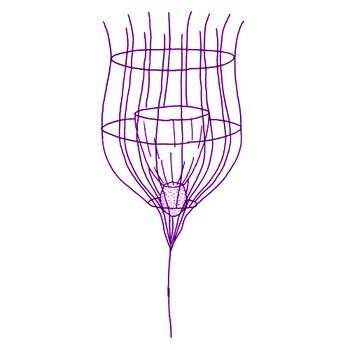
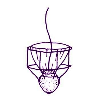
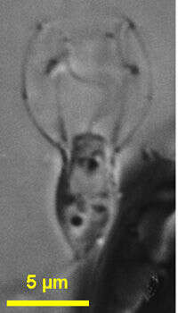
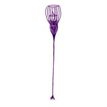
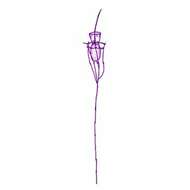
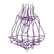
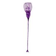
Diaphanoeca pedicellata Leadbeater 1972 (also referred to as Campanoeca pedicellata (Leadbeater, 1972) Throndsen, 1974). The subspherical protoplast (4-5 microns long, 4-5 microns wide) bears an anterior ring of 20-30 tentacles. The funnel-shaped lorica chamber is formed by 20-30 longitudinal costae (about 10 microns long) which at the upper end project as spines beyond the anterior transverse costa. The lorica is 45 microns high consisting of one chamber formed by about 14 longitudinal costae and three transverse costae(one towards the anterior end of the lorica, one just below the mid-lorica region, and one towards the posterior end of the lorica chamber). Posteriorly about seven longitudinal costae converge and join with a posterior stalk (about 11 microns long) formed by at least two or three costal strips attached end-to-end. All the costal strips forming the lorica are approximately equal in length and width.
-
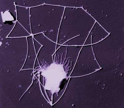
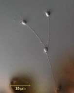
This image was made from samples taken during a scientific cruise in the Pacific. Water was filtered to concentrate the organisms that were present, then dried onto a thin sheet of plastic and then shadowed with a fine layer of metal to provide contrast. The preparation was then observed with an electron-microscope. This technique has been used to document the diversity of marine microbes, especially, protists in the oceans.
-
Pleurasiga minima Throndsen, 1970. Lorica 9.5-16 microns long with 2 transverse costae and 7 longitudinal costae, all of which extend to the posterior of the lorica. The transverse costae are of about the same width, and the lorica narrows below the second costa to form a pointed base. Cell located at the posterior of the lorica, with flagellum protruding above it.
-
Pleurasiga minima minima Throndsen, 1970. The different varieties differ mainly in size. The lorica of P. minima minima is about 10 microns long and wide.
-
Pleurasiga reynoldsii Throndsen, 1970. The lorica is 23 microns long and 22 microns wide, respectively.
-
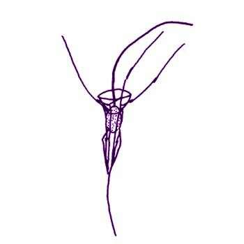
Polyoeca dichotoma Kent, 1880. Lorica of polythecium urceolate, pedicellate, tapering posterioly, slightly constricted at a distance of one-third of the total length from the anterior margin, and then widening out to their greatest diameter, pediceles of each separate lorica straight, slender, varying from the same to two or three times the length of the latter structure, contained animalcules ovate, occupying respectively about one-half of the cavities of the lorica, contour of polythecium subdichotomous, each zooid usually giving rise by trasverse fission to two new ones which attach themselves to opposite sides of the parent lorica. Length of separate lorica 10 microns
-

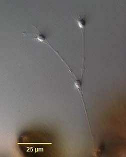
Portrait of the acanthoecid choanoflagellate, Polyoeca dichotoma (Kent, 1881). Cells are solitary or united to form linear or dendroid colonies which attach to the substrate. The lorica is funnel-shaped, constructed of numerous siliceous longitudinal costae, each made up of costal strips. The longitudinal costae terminate as anterior spines (11-17); 2 or more bands of equally spaced transverse costal strips (difficult to see in vivo) encircle the lorica chamber in which the protoplast (cell body) resides. At the apex of the funnel the longitudinal costae unite to form an aggregated pedicel. In dendroid colonies the pedicel of the daughter (anterior) cell attaches to the outside of the lorica of the parent cell. Details of lorica morphology are best seen with scanning electron microscopy. The cell body has one anterior flagellum (seen here) surrounded by a rhizopodial collar (not visible here). Division is nudiform (i.e. naked swarmers are formed which then form a lorica). Collected from a commercial saltwater aquarium in Boise, Idaho February 2004. DIC.
-
Savillea micropora (Norris, 1965) Leadbeater, 1975. Cells spherical to ovoid, with collar and flagellum. Cells located in base of lorica composed of longitudinal and transverse costae. Base of lorica with numerous closely spaced transverse costae, expanded anterior part of lorica composed of approximately 12 longitudinal costae traversed at regular or irregular intervals by 4-6 transverse costae. Terminal transverse costa forming pore with small diameter (approximately 1 microns diameter). Cell 2.5-3 microns diameter, base of lorica 1.6-3.5 microns long, 3 microns diameter, anterior part of lorica 5-6.3 microns long, 5-6.3 microns diameter.
-
Savillea parva (Ellis, 1930) Loeblich III, 1967. Cells are 2.5-4 microns (spherical), lorica 10-10.5 microns in length. The cell body is situated at the bottom of the lorica, the pseudopodial collar extends about two-thirds of the way up the lorica, and the flagellum extends to the top of the lorica. Unlike Savillea micropora, a flagellum is always present and active.
-
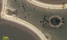
Stephanoeca (steff-ann-owe-eek-a) is a collar flagellate in which the body is located within a lorica, which is made up of siliceous spicules. In this case the spicules cannot be seen individually. One cell (to the left) is active and has a flagellum projecting from the anterior of the cell and the base of the flagellum is surrounded by a collar of fine pseudopodia. The cell to the right seems to have encysted. Phase contrast micrograph.
-
-
Stephanoeca (steff-ann-o-eek-a) diplocostata Ellis, 1929. Collar flagellate with lorica divided into two chambers by a waist at about two fifths of the distance from the base of lorica. Cells have a lorica with transverse and horizontal costae. The cells have a single apical flagellum. The flagellum is as long as the cell body. The cell body is about 10 microns long and fills the posterior chamber of the lorica. The lorica attaches to the substrate with a stalk or with the posterior end of the lorica. Rarely observed.
-
Stephanoeca diplocostata Ellis, 1930. The cell, which bears a single anterior flagellum and a collar of 20-25 tentacles, is lodged in the posterior chamber of the lorica. About 15 longitudinal costae run from the base of the posterior chamber to the top of the anterior chamber. Four transverse costae encircle the posterior chamber and about three double costae encircle the anterior chamber. Anteriorly the longitudinal costae curve inwards. Lorica 14.5-15.5 microns long.
-
Stephanoeca paucicostata Throndsen, 1969. Cells have 12-14 longitudinal costae and 3 transverse costae, one of which is the anterior rim, in its anterior chamber and an irregular arrangement in the posterior chamber. Overall length of the lorica is 16-17 microns The anterior lorica chamber is 10 microns long and 9 microns wide. The length of posterior lorica chamber is 6.5 microns, its width is 6.4 microns
-
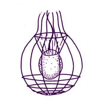
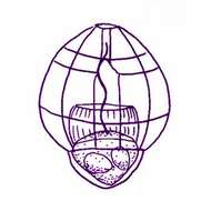
Stephanoeca campanula (Kent, 1880) Boucaud-Camou, 1967. Lorica goblet or bell shaped, scarcely longer than broad, the basal region narrower, conically pointed, the anterior two-thirds expanding abruptly and in a marked manner in comparison with the first-named area, the anterior border widest, but not everted, pedicel equling the length of the lorica, contained protoplast symmetrically ovate, occupying and projecting slightly beyond the conical basal area, the fully expanded collar enclosed entirely within the wider anterior area of the cavity of the lorica, the flagellum extending for about half its length beyond its anterior border, two contractile vacuoles, posteriorly located, nucleus spherical, subcentral. Length of lorica 32 microns