-
Anna Halász, Catherine S. McFadden, Dafna Aharonovich, Robert Toonen, Yehuda Benayahu
Zookeys
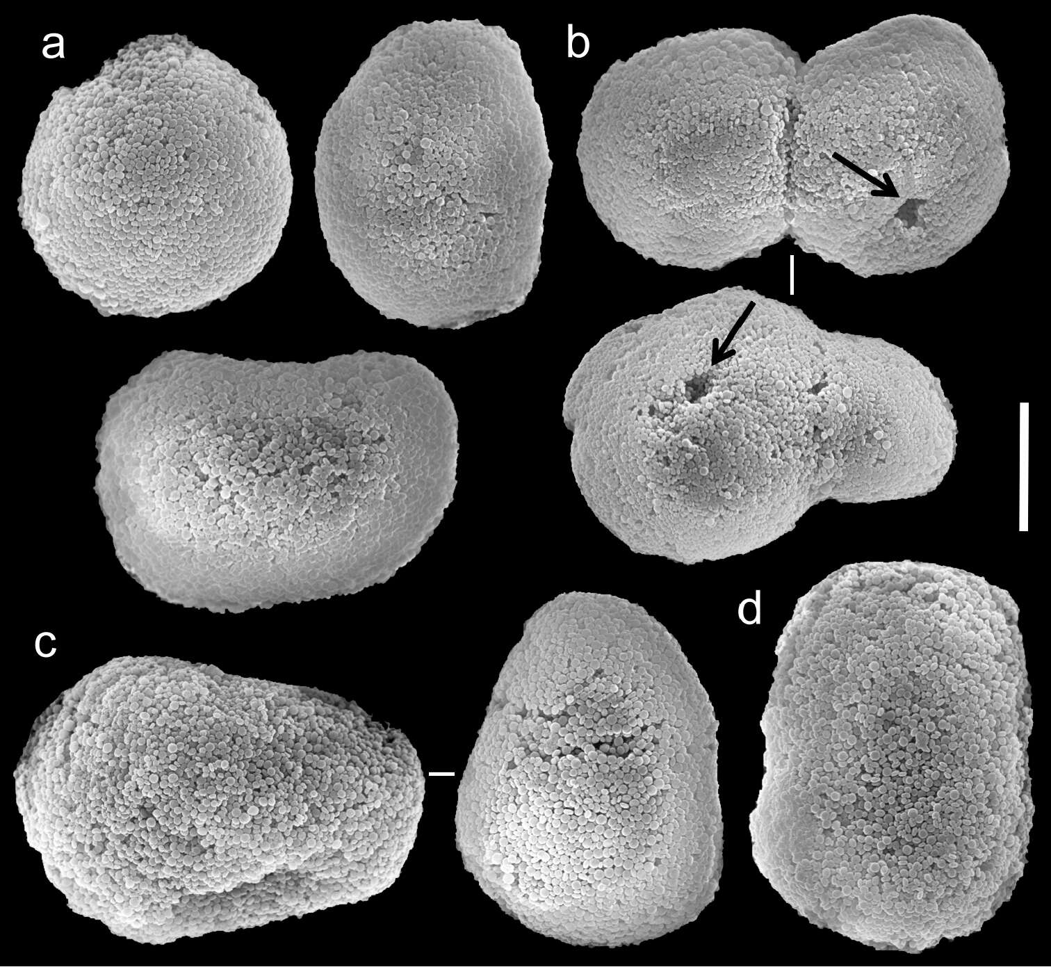
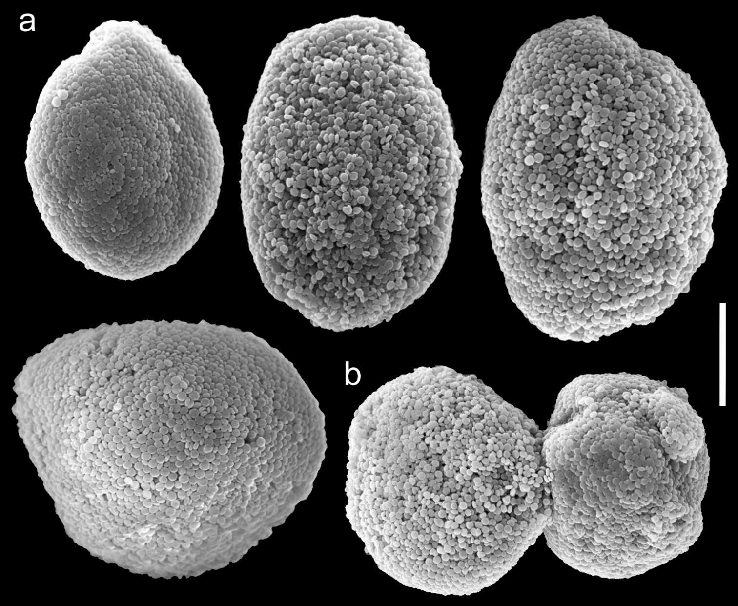
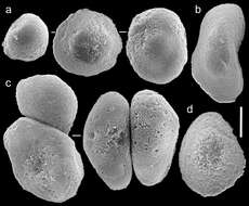
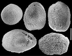
Figure 18.Scanning electron micrographs of polyp sclerites of Ovabunda macrospiculata (Gohar, 1940) paratype (ZMTAU Co 35790). a Regular sclerites b Irregular sclerite c Fused sclerites d Pear-shaped sclerite. Scale bar 10 µm.
-
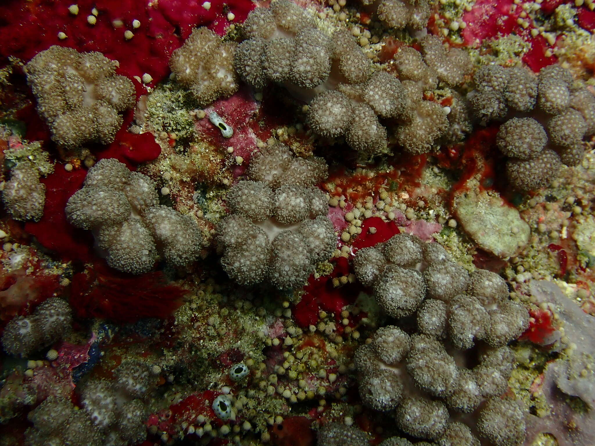
Naisisili, Fiji
-
Anna Halász, Catherine S. McFadden, Dafna Aharonovich, Robert Toonen, Yehuda Benayahu
Zookeys
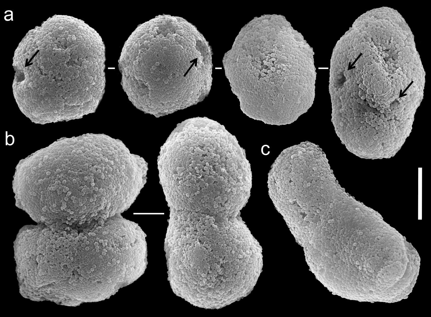
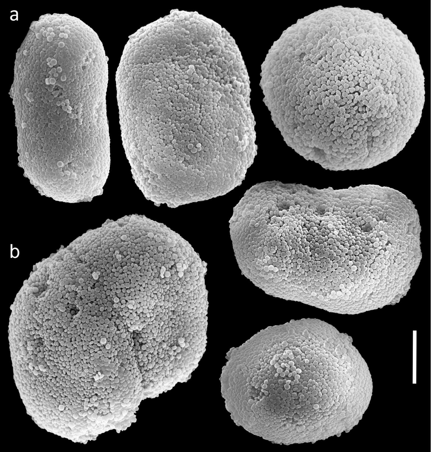
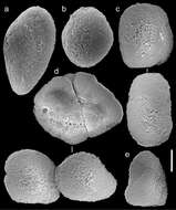
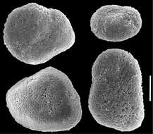
Figure 19.Scanning electron micrographs of polyp sclerites of Ovabunda macrospiculata (Gohar, 1940) paratype (ZMTAU Co 35791). a Regular sclerite b Irregular sclerites c Fused sclerites. Arrows indicate surface dents. Scale bar 10 µm.
-
Anna Halász, Catherine S. McFadden, Dafna Aharonovich, Robert Toonen, Yehuda Benayahu
Zookeys
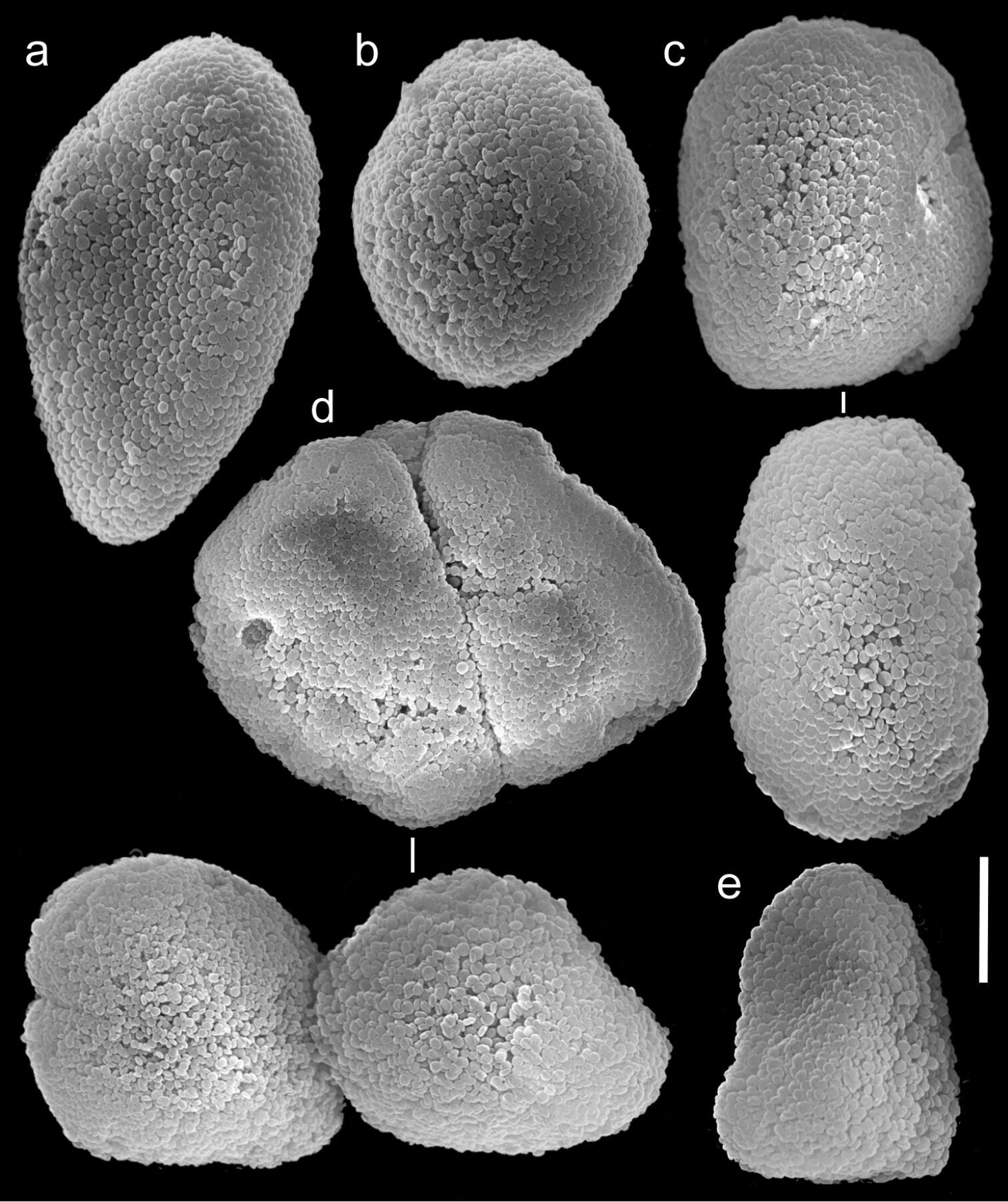
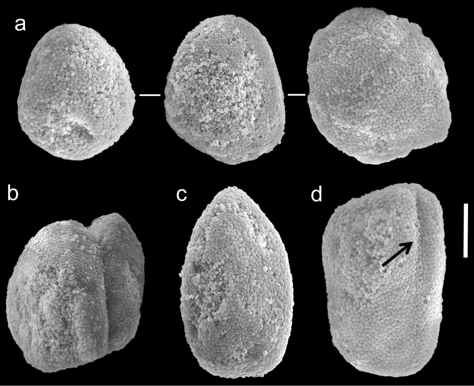
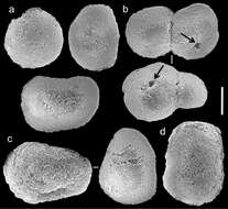
Figure 20.Scanning electron micrographs of polyp sclerites of Ovabunda verseveldti (Benayahu, 1990) holotype (ZMTAU Co 26048). a Egg-shaped sclerite b Regular sclerite c Rectangular sclerites d Fused sclerites e Irregular sclerite. Scale bar 10 µm.
-
2005 California Academy of Sciences
CalPhotos
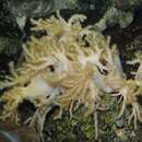
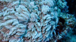
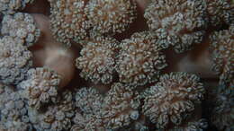
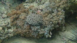
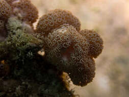
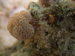
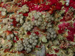
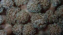
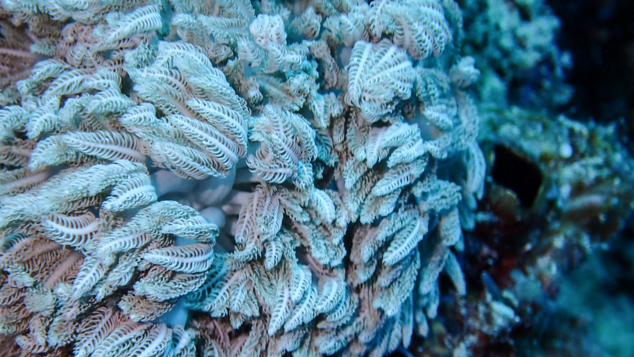
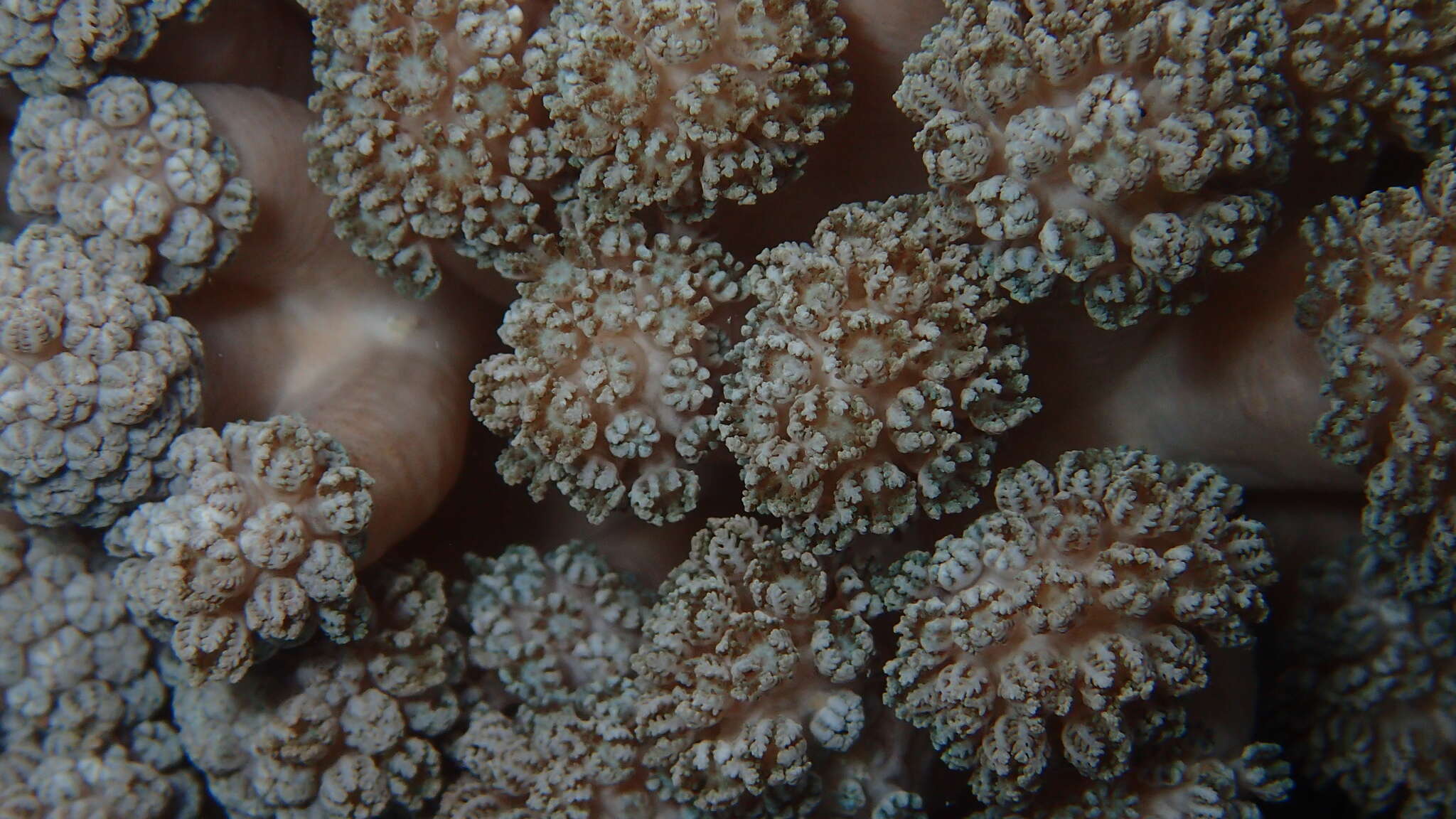
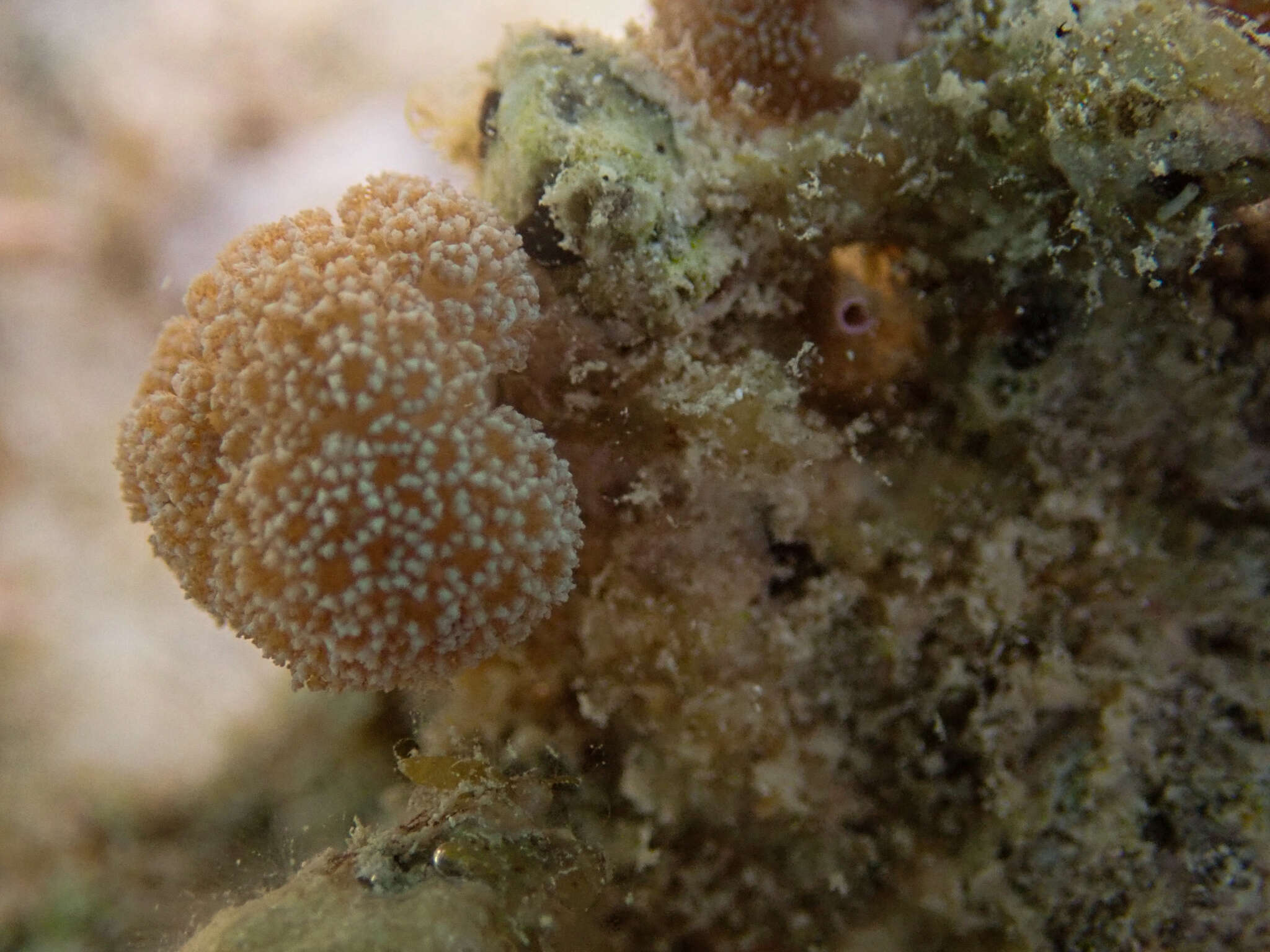
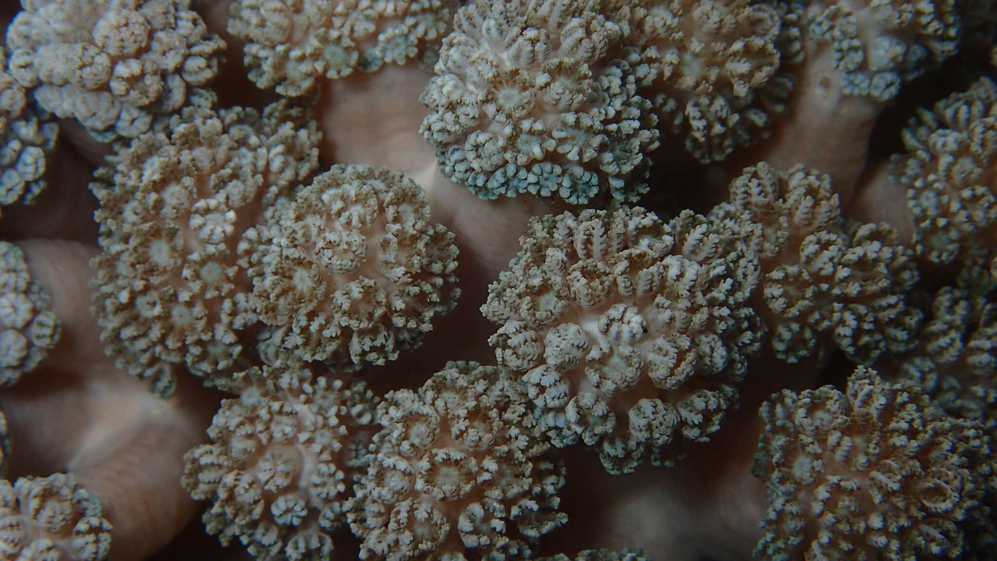
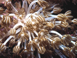
The tentacles of these polyps continuously open and close. Subclass Octocorallia.
-
Anna Halász, Catherine S. McFadden, Dafna Aharonovich, Robert Toonen, Yehuda Benayahu
Zookeys
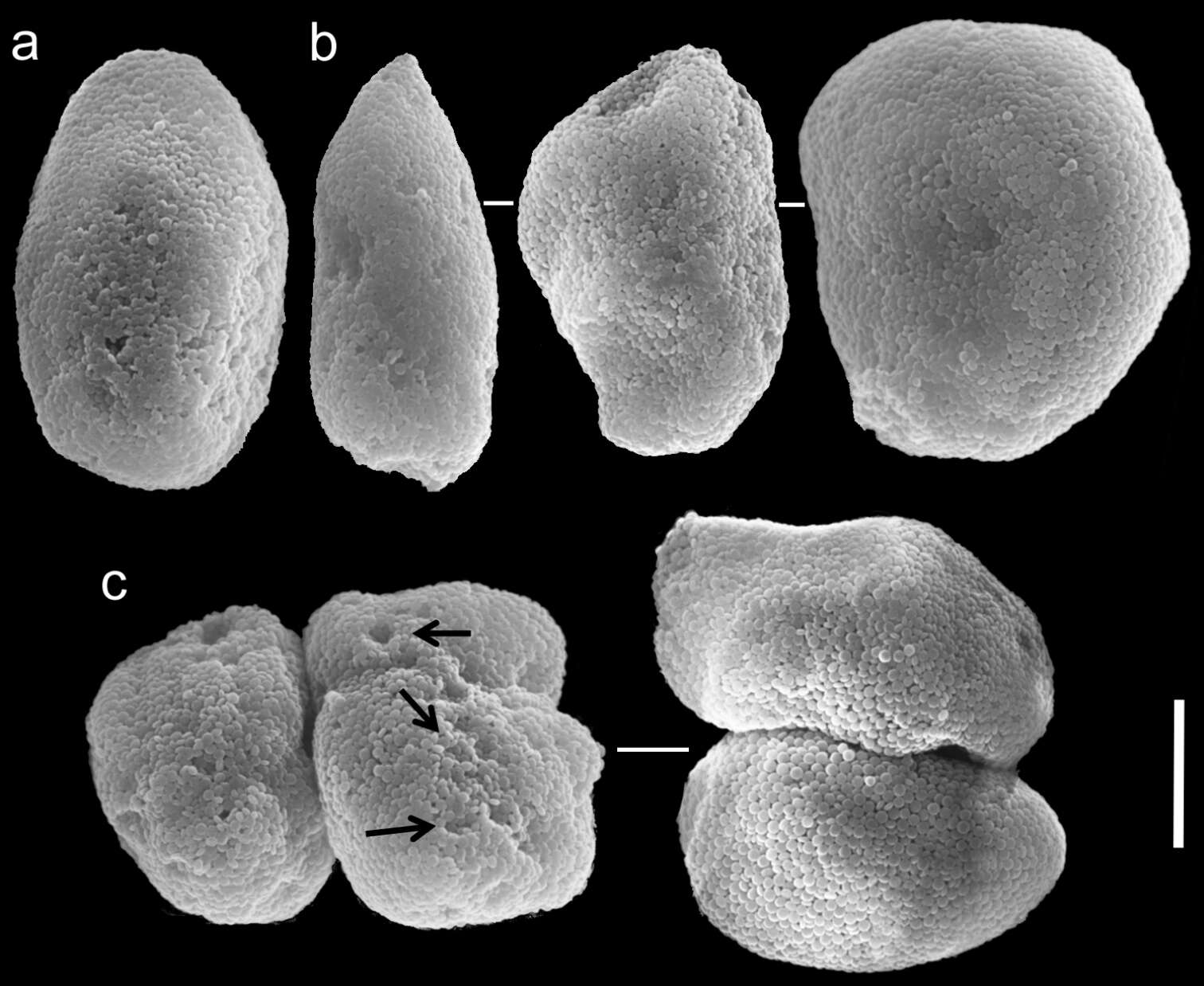
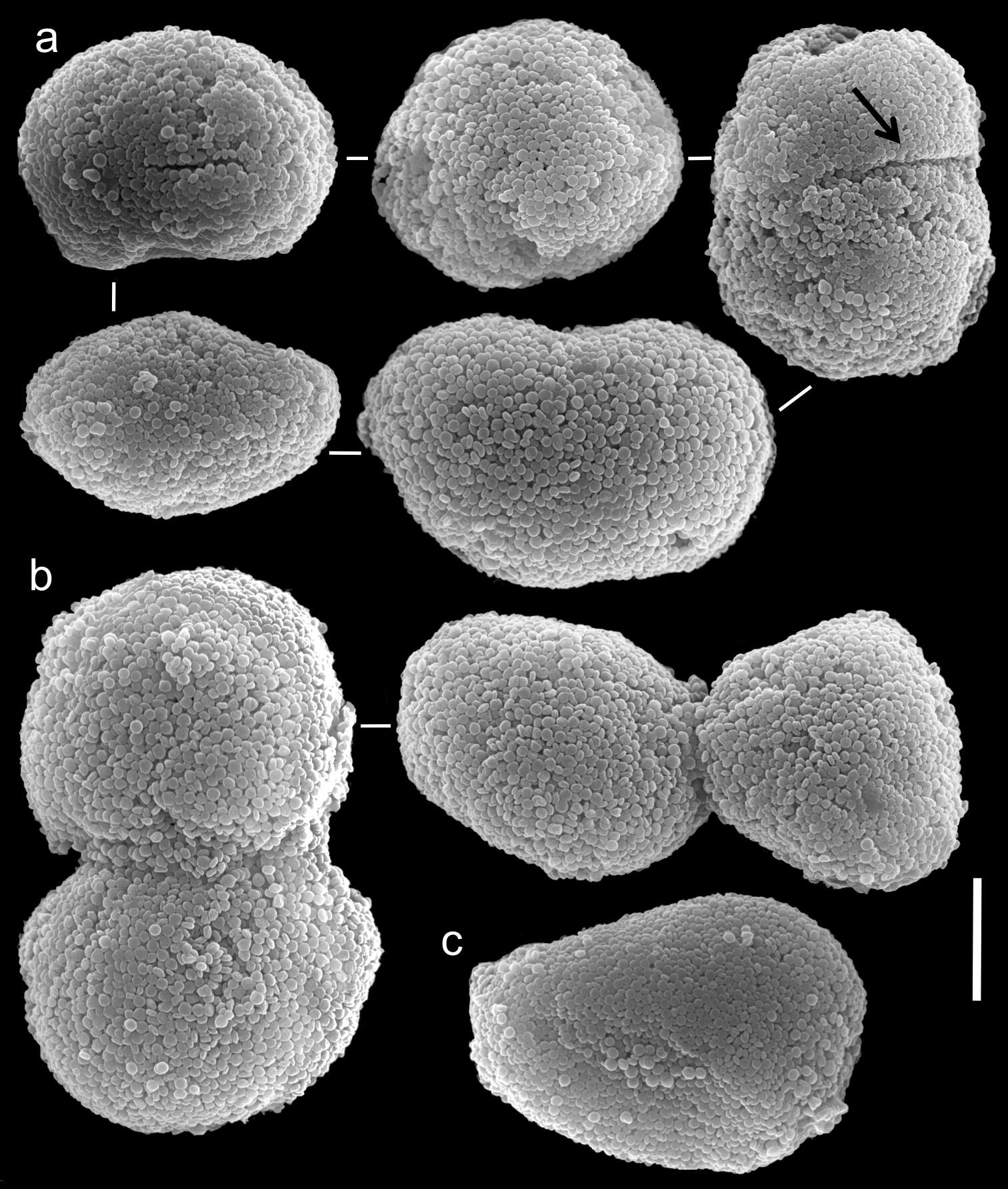
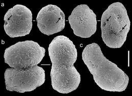
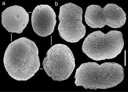
Figure 3.Scanning electron micrographs of polyp sclerites of Ovabunda ainex (Reinicke, 1997) paratype (RMNH Coel. 23540). a Regular sclerites b Fused sclerites c Irregular sclerite. Arrows indicate surface dents. Scale bar: 10 µm.
-
Anna Halász, Catherine S. McFadden, Dafna Aharonovich, Robert Toonen, Yehuda Benayahu
Zookeys
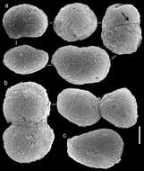
Figure 4.Scanning electron micrographs of polyp sclerites of Ovabunda arabica (Reinicke, 1995) paratype (RMNH Coel. 18675). a Regular sclerites b Fused sclerites c Pear-shaped sclerite. Arrow indicates surface irregularity, might represent the fusion area of two individual sclerites. Scale bar 10 µm.
-
Anna Halász, Catherine S. McFadden, Dafna Aharonovich, Robert Toonen, Yehuda Benayahu
Zookeys
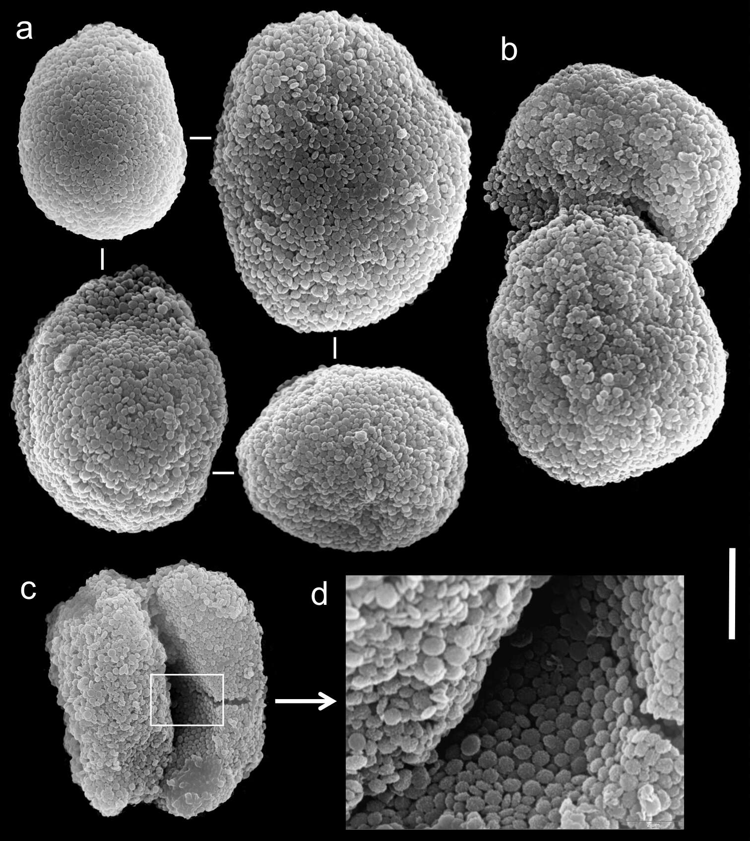
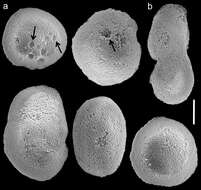
Figure 5.Scanning electron micrographs of polyp sclerites of Xenia crista Reinicke, 1997 holotype (RMNH 18677). a Regular sclerites b–c Fused sclerites d White rectangle in c indicates magnified area. Scale bar 10 µm.
-
Anna Halász, Catherine S. McFadden, Dafna Aharonovich, Robert Toonen, Yehuda Benayahu
Zookeys
Figure 6.Scanning electron micrographs of polyp sclerites of Ovabunda benayahui (Reinicke, 1995) holotype (RMNH Coel. 19664). a Regular sclerites b Fused sclerite. Arrows indicate surface dents. Scale bar 10 µm.
-
Anna Halász, Catherine S. McFadden, Dafna Aharonovich, Robert Toonen, Yehuda Benayahu
Zookeys
Figure 7.Scanning electron micrographs of polyp sclerites of Ovabunda benayahui Reinicke, 1995 (ZMTAU Co 26043). a Regular sclerites b Fused sclerites c Egg-shaped sclerite d Rectangular sclerite. Arrow indicates surface crest. Scale bar 10 µm.
-
Anna Halász, Catherine S. McFadden, Dafna Aharonovich, Robert Toonen, Yehuda Benayahu
Zookeys
Figure 8.Scanning electron micrographs of polyp sclerites of Ovabunda biseriata (Verseveldt & Cohen, 1971) holotype (HUJ I Co. 72). Arrow indicates surface dents. Scale bar 10 µm.
-
Anna Halász, Catherine S. McFadden, Dafna Aharonovich, Robert Toonen, Yehuda Benayahu
Zookeys
Figure 9.Scanning electron micrographs of polyp sclerites of Ovabunda obscuronata (Verseveldt & Cohen, 1971) holotype (HUJ I Co. 120). Scale bar 10 µm.
-
Anna Halász, Catherine S. McFadden, Dafna Aharonovich, Robert Toonen, Yehuda Benayahu
Zookeys
Figure 10.Scanning electron micrographs of polyp sclerites of Ovabunda crenata (Reinicke, 1997) (RMNH Coel. 23517). a Regular sclerites b Fused sclerites c Egg-shaped sclerites d Rectangular sclerite. Arrows indicate surface dents. Scale bar 10 µm.
-
Anna Halász, Catherine S. McFadden, Dafna Aharonovich, Robert Toonen, Yehuda Benayahu
Zookeys
Figure 5.Scanning electron micrographs of polyp sclerites of Xenia crista Reinicke, 1997 holotype (RMNH 18677). a Regular sclerites b–c Fused sclerites d White rectangle in c indicates magnified area. Scale bar 10 µm.
-
Anna Halász, Catherine S. McFadden, Dafna Aharonovich, Robert Toonen, Yehuda Benayahu
Zookeys
Figure 11.Scanning electron micrographs of polyp sclerites of Ovabunda faraunensis (Verseveldt & Cohen, 1971) holotype (HUJ I. Co. 140). a Regular sclerites b Fused sclerite. Scale bar 10 µm.
-
Anna Halász, Catherine S. McFadden, Dafna Aharonovich, Robert Toonen, Yehuda Benayahu
Zookeys
Figure 12.Scanning electron micrographs of polyp sclerites of Ovabunda gohari (Reinicke, 1997) paratype (RMNH Coel. 23436). a Regular sclerites b Fused sclerite. Scale bar 10 µm.
-
Anna Halász, Catherine S. McFadden, Dafna Aharonovich, Robert Toonen, Yehuda Benayahu
Zookeys
Figure 13.Scanning electron micrographs of polyp sclerites of Ovabunda hamsina (Reinicke, 1997) holotype (RMNH Coel. 23904). a Regular sclerites b Fused sclerites. Scale bar 10 µm.
-
-
-
-
-
-
-