-
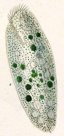
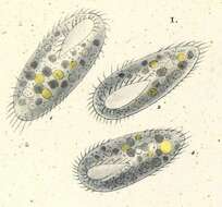
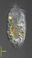
100-300 micron long. Elongate, anterior wider than posterior.
-
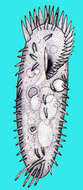
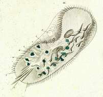
The cell body measures 100-300 micron in lenght and is dorsoventrally flattened, with a rounded anterior end and a slyghtly pointed posterior end. It has two ovoid macronuclei and two spherical micronuclei. The somatic ciliature consists of 8 frontal cirri, 5 trasverse cirri, 3 caudal cirri, 20-25 right marginal cirri and 15 left marginal cirri ( Tuffrau, 1965 ).
-
-
-
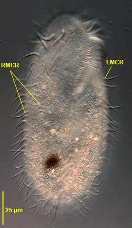
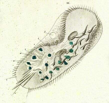
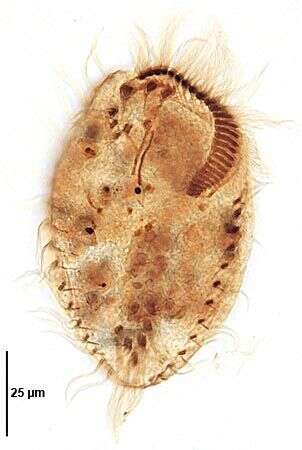
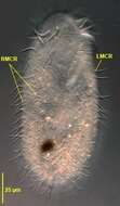
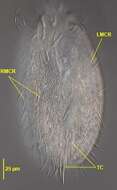
Ventral view of Pleurotricha lanceolata (EHRENBERG,1835) STEIN, 1859. RMCR= two right marginal cirral rows. LMCR=single left marginal cirral row.Collected from a flood-irrigated grass lawn in Boise,Idaho. May 2008. DIC.
-
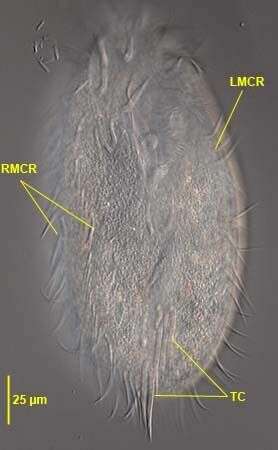
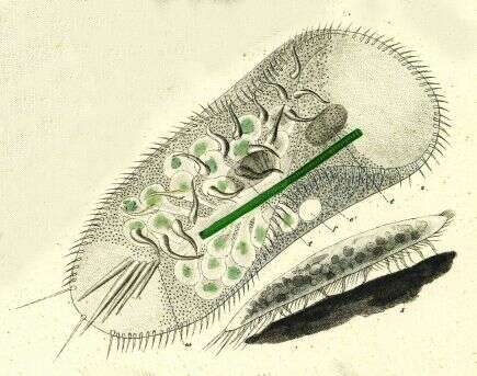
Ventral view of Pleurotricha lanceolata (EHRENBERG,1835) STEIN, 1859. RMCR= two right marginal cirral rows. LMCR=single left marginal cirral row.T=transverse cirri in 2 groups (3+2).Collected from a flood-irrigated grass lawn in Boise,Idaho. May 2008. DIC.
-
Originally described by Ehrenberg under the name Stylonychia lanceolata.
-
Originally described by Ehrenberg under the name Stylonychia lanceolata.
-
Originally described by Ehrenberg under the name Stylonychia lanceolata.
-
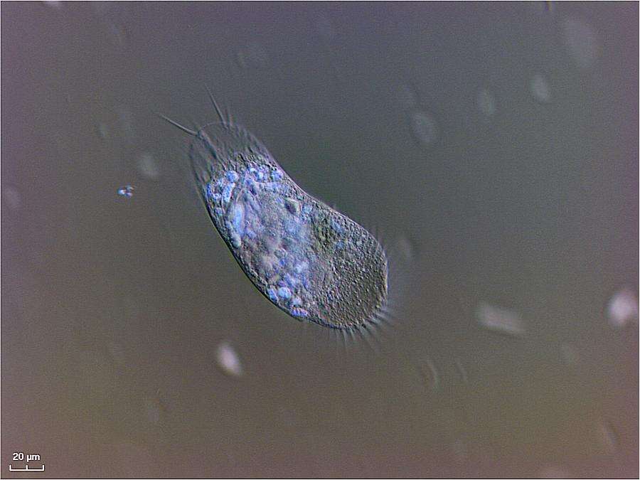
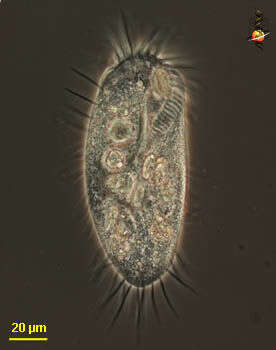
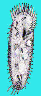
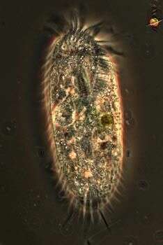
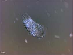
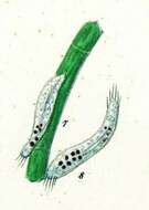
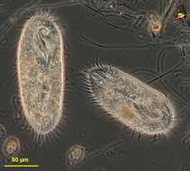
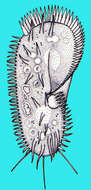
Histriculus (his-trick-you-lus) is a hypotrich ciliate with cirri forming a row all around the margin and including across the back of the cell. Cell not flexible, by which it is distinguished from the very similar Oxytricha, also not with three long caudal cirri, by which it is distinguished from Stylonychia. With adoral zone of membranelles. Phase contrast.
-
Histriculus (his-trick-you-lus) is a hypotrich ciliate with cirri forming a row all around the margin and including across the back of the cell. Cell not flexible, by which it can be distinguished from the very similar Oxytricha, also not with three long caudal cirri, by which it is distinguished from Stylonychia. With adoral zone of membranelles. Phase contrast.
-
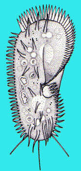
Histriculus (his-trick-you-lus) is a hypotrich ciliate, which can be distinguished by the distribution of the cirri - the aggregates of cilia used in locomotion - on the ventral side. There is an anterior (top of image) array of membranelles (aggregates of cilia) which are used to collect food - typically algae. Differential interference contrast. Material from Nymph Creek and Nymph Lake, thermal sites within Yellowstone National Park, photograph by Kathy Sheehan and David Patterson.
-
-
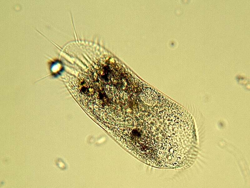
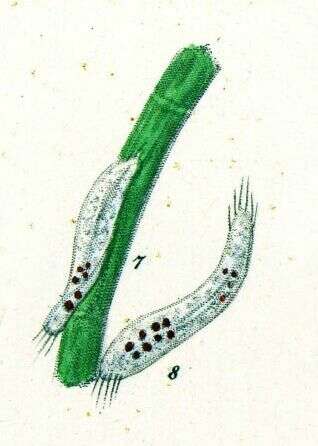
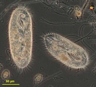
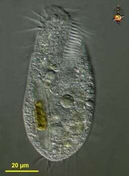
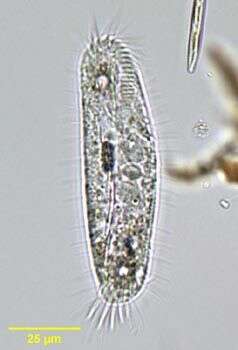
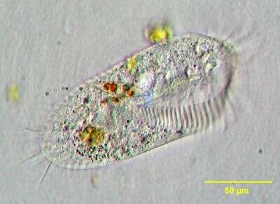
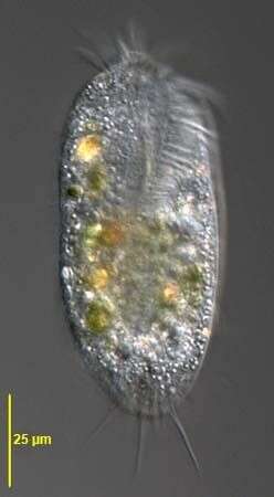
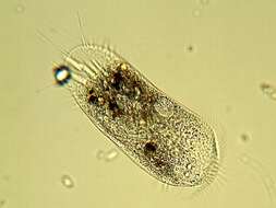
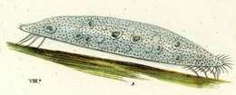
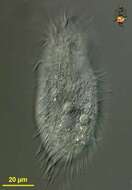
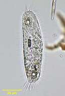
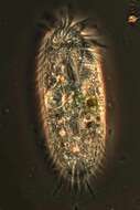
Tachysoma, a hypotrich ciliate. The body is relatively elongate, dorsoventrally flattened and rounded anteriorly and posteriorly. The adoral zone of membranelles is limited to about one quarter of the body length. The three long frontal cirri are seen here. There are five characteristic long transverse cirri. Caudal cirri are absent. The right and left marginal cirral files do not join posteriorly. There are two spherical macronuclei flanking a dense relatively large micronucleus (seen well in this image). Two small refractile lipid globules, considered characteristic (termed Fettkorn by Foissner), are clearly seen in the anterior and posterior quarters of the cell in this image. The contractile vacuole (not seen in this image) is located on the left in the mid portion of the cell. Tachysoma feeds on bacteria, green algae and diatoms. From freshwater pond near Boise, Idaho. Brightfield illumination.
-
Originally described by Ehrenberg under the name Oxytricha eurystoma.
-
-
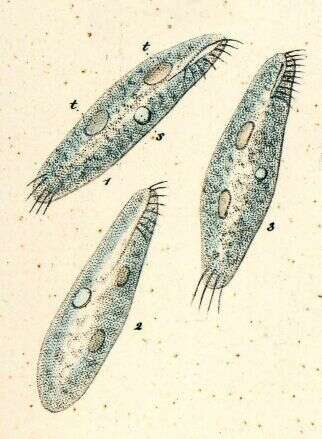
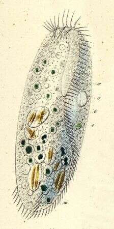
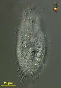
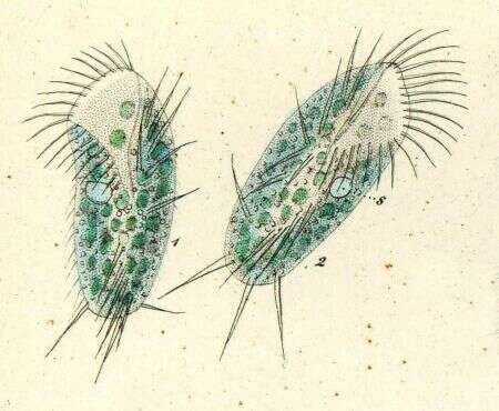
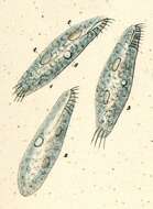
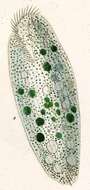
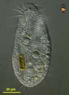
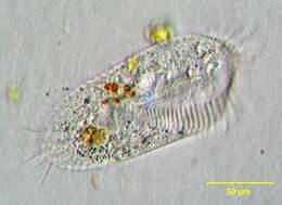
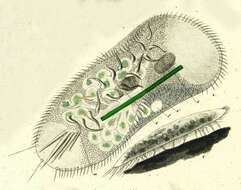
Stylonychia, a widely distributed hypotrich ciliate. The dorsoventrally flattened body is elongate and broadly rounded anteriorly, narrowing posteriorly. The adoral zone of membranelles is strongly developed and rests on an anteriorly protruding collar. The two rows of marginal cirri are slightly out of the focal plane in this image. They do not meet posteriorly. The three characteristic caudal cirri are seen here. There are short dorsal cilia (not seen here). Two ellipsoid macronuclei are visible in this image. One of two small spherical micronuclei is seen at the inferior margin of the posterior macronucleus. This individual has been feeding on diatoms and green algae. From freshwater pond near Boise, Idaho. Brightfield illumination.
-
Inflexible, elongate, oval, dorso-ventrally flattened body with a large and powerful AZM. There are rows of marginal cirri that are not continuous posteriorly. Three long, strong and prominent caudal cirri help to distinguish members of this genus.
-
-
-
Originally described by Ehrenberg under the name Stylonychia silurus
-
Ventral infraciliature of Stylonychia pustulata (MUELLER, 1786) EHRENBERG, 1835. Collected from a eutrophic freshwater pond in Boise, Idaho. June 2008. Protargol A (see Foissner, W. Europ. J. Protistol., 27:313-330;1991).Brightfield.
-
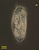
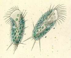
Ventral view of Stylonychia pustulata (MUELLER, 1786) EHRENBERG, 1835. Collected from a eutrophic freshwater pond in Boise, Idaho. June 2008. DIC.