-
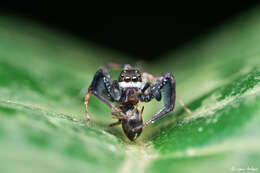
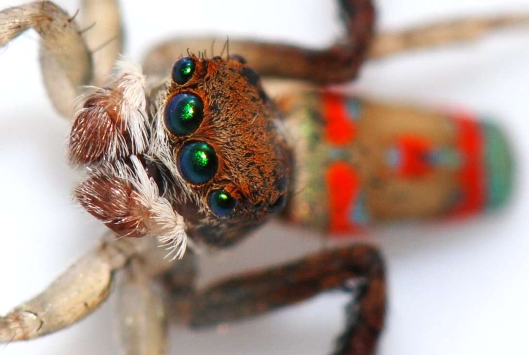
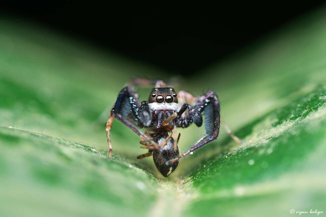
Maratus pavonisI was outside at sunset last night and notice a flash of orange. I was pleased that I had time to come in and add my flash and 20mm extension on the camera before finding that this little spider was still waving his legs and looking for a female.
those big green eyes!!These spiders attract mates by standing and lifting their 3rd legs into the air. They then make a beautiful display by lifting flaps from the side of their abdomen and tilting their abdomen. The display can be seen here:
www.xs4all.nl/~ednieuw/australian/salticidae/Peacock_spid...
-
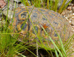
Avne, Languedoc-Roussillon, France
-
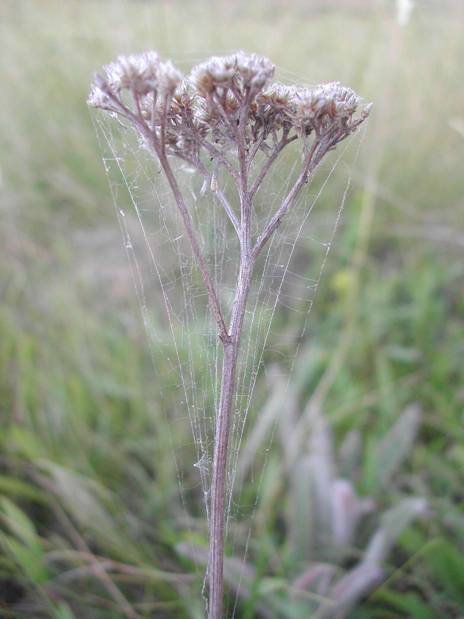
Dictyna web on yarrow plant
-
-
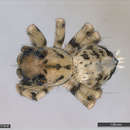
Figures 177–182.Habitats and natural history of some Selenops species. 177 Selenops arikok sp. n. from Aruba guarding egg sac with several spiderlings inside 178 Selenops geraldinae Corronca eating a fly and guarding her egg sac on a bromeliad, Gaspar Grande Island, Trinidad and Tobago 179 Selenops willinki Corronca on a tree trunk, Little Tobago, Trinidad and Tobago. Terminal setal tufts and festoon pattern are visible 180 Selenops micropalpus Muma on the trunk of Bursera simaruba, in a dry forest, St. Lucia. Terminal setal tufts and festoon pattern are visible 181 Selenops mexicanus Keyserling on a large tree trunk outside of Cueva Actun Kan, Guatemala. Note the alternating light and dark leg annulations 182 Egg sacs of Selenops bifurcatus Banks on rocks in a wash in dry forest and thornscrub, Zacatan, Guatemala. Selenops bifurcatus sometimes guards the yellowish egg sacs, and other times does not.
-
Jeremy Miller, Cahyo Rahmadi
Zookeys
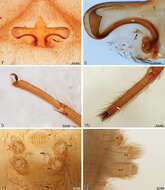
Figures 7–12.Amauropelma matakecil sp. n., female holotype (MZB.Aran.500) 7 Epigynum, ventral view. Note that the right epigynal tooth has broken off leaving a round hole; the tooth itself is lying unattached near the epigastric furrow 8 Vulva, dorsal view, left side, cleared, white arrow indicates fertilization duct 9 Right tarsus, leg I, prolateral view 10 Left tarsus, leg I, dorsal view, arrow indicates tarsal organ 11 Spinnerets, anal tubercle, and tracheal spiracle, posterior view, arrow indicates tracheal spiracle 12 Spinnerets, lateral view. ALS, anterior lateral spinneret; AT, anal tubercle; CD, copulatory duct; ET, epigynal tooth; PLS, posterior lateral spinneret; PMS, posterior median spinneret; S, spermatheca.
-
Mykola M. Kovblyuk, Zoya A. Kastrygina, Mikhail M. Omelko
Zookeys
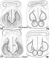
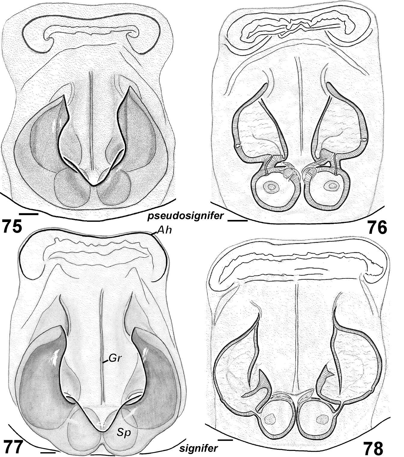
Figures 75–78.Females of Haplodrassus pseudosignifer (75–76 from Crimea) and Haplodrassus signifer (77–78 from Crimea): 75, 77 epigyne, ventral view 76, 78 epigyne, dorsal view. Abbreviations: Ah anterior hood; Gr groove of epigyne; Spspermatheca.
-
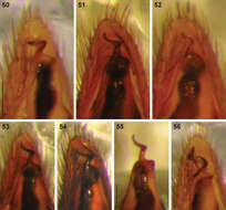
Figures 50–56.Digital microscope photographs of emboli of Afrotropical Cambalida species in ventral view: 50 Cambalida compressa sp. n. 51 Cambalida coriacea Simon, 1909 52 Cambalida deminuta (Simon, 1909) 53 Cambalida dippenaarae sp. n. 54 Cambalida fulvipes (Simon, 1896) 55 Cambalida griswoldi sp. n. 56 Cambalida loricifera (Simon, 1885). Scale bars = 0.1mm.
-
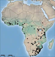
Figure 71.Distribution of Copa flavoplumosa Simon, 1885 in the Afrotropical Region.
-
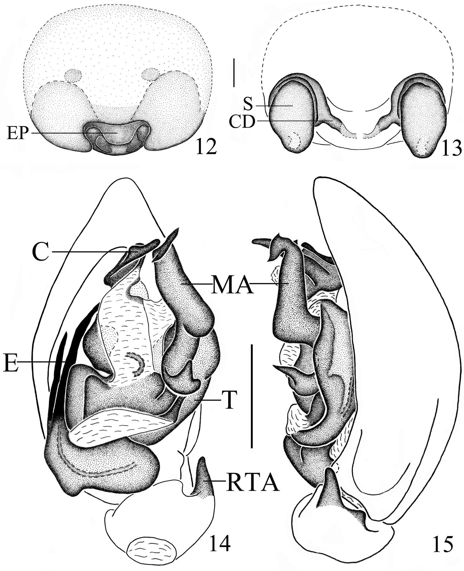
Figures 12–15.Mallinella sphaerica sp. n., 12 epigyne, ventral view 13 vulva 14 left male palp, ventral view 15 same, retrolateral view. Scale bars: 0.2 mm (12–13); 0.5 mm (14–15).
-
Dan Quan, Jian Chen, Jie Liu
Zookeys
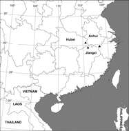
Figure 17.Collection localities of Sinopoda serrata (Wang, 1990) in China.
-
Yuri M. Marusik, Mikhail M. Omelko
Zookeys
Figures 6–12.Male palp of Cryptothele verrucosa (6–11) and Cryptothele alluaudi (12). 6 bulbus, ventral 7 bulbus, ventro-prolateral 8, 10 bulbus, retrolateral 9 bulbus, prolateral 11 palp with removed bulbus, retrolateral 12 palp, ventral (after Marusik and Omelko (2012)). Abbreviations: Co conductor; Ea terminal process of embolus; Eb embolus base; Em embolus; Ep posterior process of embolus; Sd seminal duct; Sp subtegular process; St subtegulum; Te triangle extesion of tegulum; Ts threads of subtegulum.
-
Feng Zhang, Bao-Shi Zhang, Zhi-Sheng Zhang
Zookeys
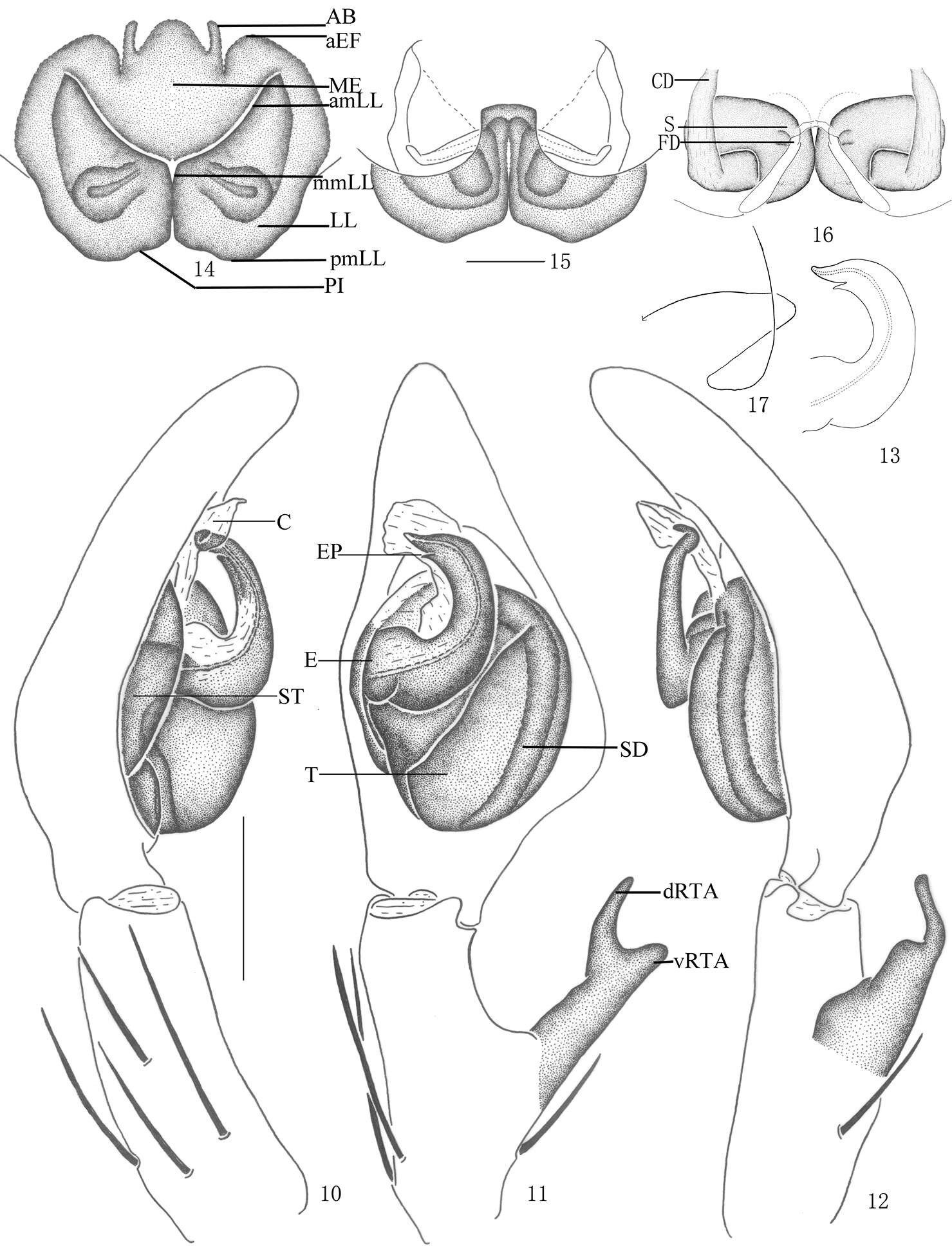
Figures 10–17.Pseudopoda acuminata sp. n., 10–13 Male (SP–SC–03–0050): 10–12 Left palp (10 prolateral 11 ventral 12 retrolateral) 13 embolus (ventral) 14–17 Female (SP–SC–03–0052): 14–16 Epigyne (14 ventral 15 dorsal 16 apical); 17 Schematic course of internal duct system, dorsal. Abbreviations: AB anterior bands; aEF anterior margin of epigynal field; amLL anterior margin of lateral lobes; C conductor; CD copulatory duct; dRTA dorsal branch of retrolateral tibial apophysis; E embolus; EP embolic projection; FD fertilization duct; LL lateral lobes of epigyne; MF median field of epigyne; mmLL median margin of lateral lobes; pmLL posterior margins of lateral lobes; PI posterior incisions; S spermathecae; SD sperm duct; ST subtegulum; T tegulum; vRTA ventral branch of retrolateral tibial apophysis. Scale bars: 0.5 mm.
-
Dmitri V. Logunov, Yuri M. Marusik
Zookeys
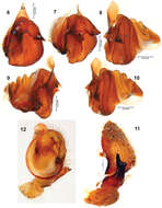
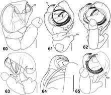
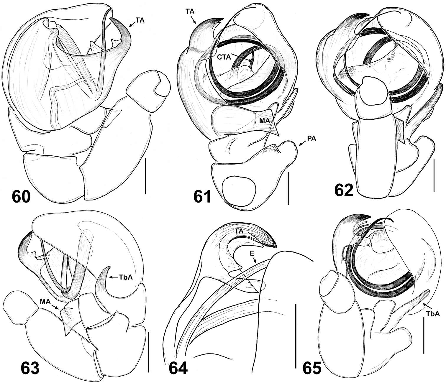
Figures 60–65.Copulatory organs of Eupoa lobli sp. n. (the holotype). 60 male palp, median view 61–62, 65 ditto, ventral view 63 ditto, retrolateral view 64 tegular apophysis, retrolateral view. Abbreviations as explained in ‘Material and methods’. Scale bars: 0.1 mm.
-
Cyril Courtial, Lionel Picard, Frédéric Ysnel, Julien Pétillon
Zookeys
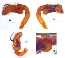
Figure 2.Pictures of the male palp of Eustiromastix guianae. A–C male palp in lateral, retrolateral and ventral views, respectively D detail of the tibial apophysis, ventral view. MA median apophysis, E embolus. Scales: A–C 1 mm, D 0.5 mm.
-
Yuri M. Marusik, Alexander A. Fomichev, Mikhail M. Omelko
Zookeys
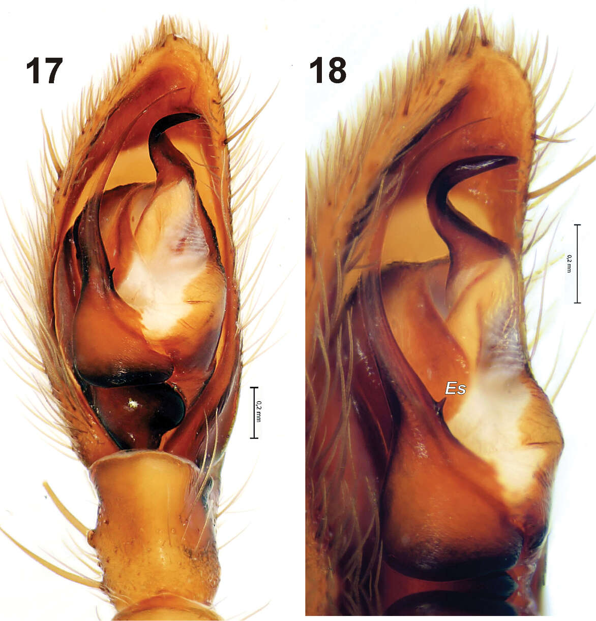
Figures 17–18.Male palp of Gnaphosa ustyuzhanini. 17 ventral 18 prolateral. Scale = 0.2 mm. Es – embolic spine.
-
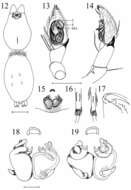
Figures 12–19.Heser vijayanagara sp. n. 12 Female allotype, dorsal view of body 13 Male palp, ventral, with conductor (C), embolus (E), and median apophysis (MA) indicated 14 Male palp, retrolateral 15 Epigyne, ventral 16 Female metatarsi III (left) and IV, with ventral terminal preening comb 17 Female cheliceral teeth 18 Vulva, ventral 19 Vulva, dorsal. Scale bars: 12: 1.0; 13–17: 0.5; 18–19: 0.1.
-
Sarah C. Crews, Mark S. Harvey
Zookeys
Map 5.Northern Australia (inset) showing the distribution of Karaops gen. n. Karaops jenniferae sp. n. (black square), Karaops alanlongbottomi sp. n. (white circle), Karaops keithlongbottomi sp. n. (black circle), Karaops larryoo sp. n. (white square), Karaops dawara sp. n. (white stars).
-
-
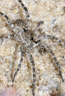
Mpumalanga, South Africa
-
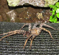
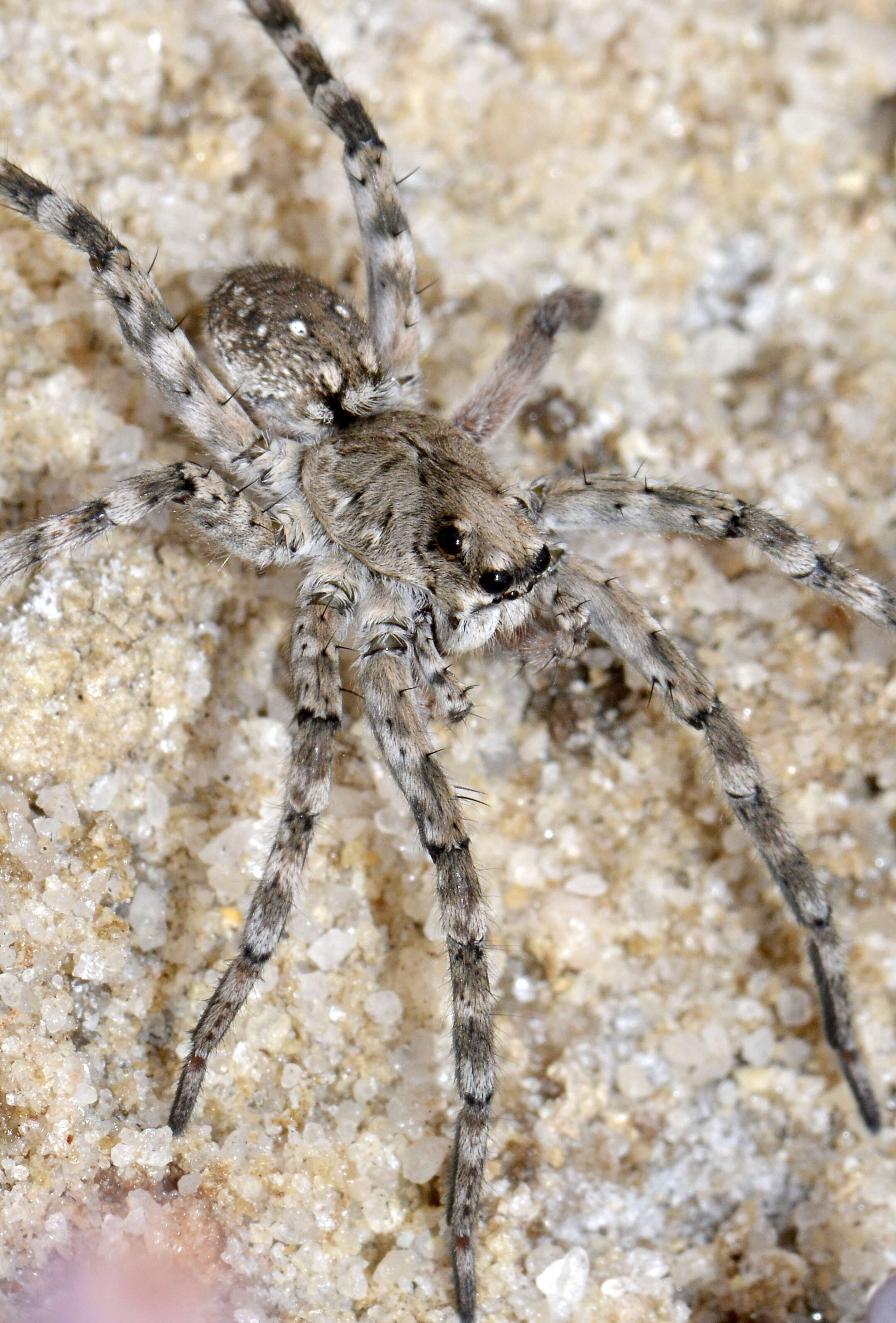
We saw this girl on a salt lake and she could run like the wind. Photo: JeaniD: Artoriopsis Expolita (Polished Wolf Spider) Ethan Yeoman
-
, Northern Territory, Australia
-
Lephalale, Limpopo, South Africa
-
These are well adapted to fishing. The can walk on water, using air trapped in leg hairs. They can dive for extended periods and even weave a web under water. Their range is Costa Rica to Argentina.


