-
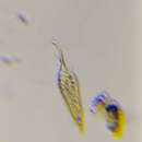
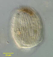
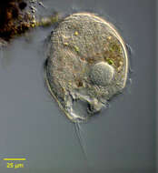
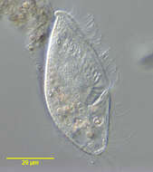
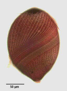
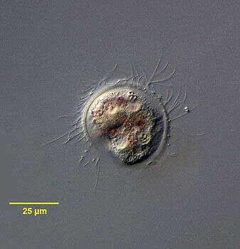
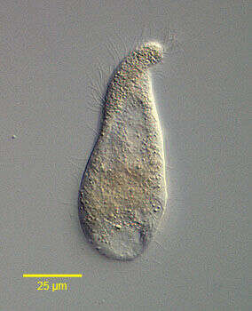
Portrait of Brachonella spiralis. Heterotrich ciliate with twisted anterior end. Blunt posterior with terminal contractile vacuole. Often with longer tuft of caudal cilia. Densely packed dark granules are usually present in the anterior of the cell. This used to be called Metopus spiralis. From stagnant freshwater with decomposing leaves near Boise, Idaho. Brightfield.
-
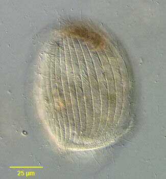
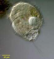
Dorsal surface of the metopid ciliate, Brachonella spiralis. The body is broadly conical anteriorly with a narrower obliquely truncate posterior. The long S-shaped peristome winds around the entire circumference of the cell terminating in the cytostome. This spiraling cytostome distinguishes Brachonella (Jankowski 1964) from similar genus Metopus in which the peristome runs obliquely from anterior to posterior but does not spiral around the long axis. The cytostome is paralleled on the right by a perizonal stripe of kineties and on the left an adoral zone of membranelles. The uniform longitudinal dorsal kineties and the posterior end of the perizonal stripe of kineties are seen well in this image
-
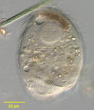
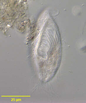
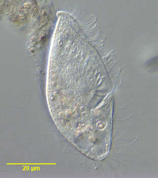
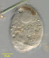
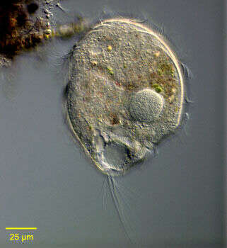
Portrait of the metopid ciliate, Brachonella spiralis. The body is broadly conical anteriorly with a narrower obliquely truncate posterior. The long S-shaped peristome winds around the entire circumference of the cell terminating in the cytostome (the anterior end of the peristome is seen at the right anteriorly and the termination at the right posteriorly in this image). This spiraling cytostome distinguishes Brachonella (Jankowski 1964) from similar genus Metopus in which the peristome runs obliquely from anterior to posterior but does not spiral around the long axis. The cytostome is paralleled on the right by a perizonal stripe of kineties. On the left is an adoral zone of membranelles. There are uniform longitudinal dorsal kineties with a tuft of longer caudal cilia. A distinctive aggregate of brownish refractile granules typical of most metopids is noted anteriorly. There is spherical anterior macronucleus. The micronucleus is not seen here. There is a posterior terminal contractile vacuole. Brachonella is found in sapropelic habitats and contains methanogenic symbionts in the cytoplasm. From stagnant freshwater source with rotting vegetation near Boise, Idaho. DIC optics.
-
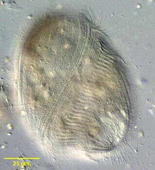
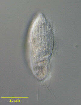
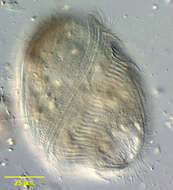
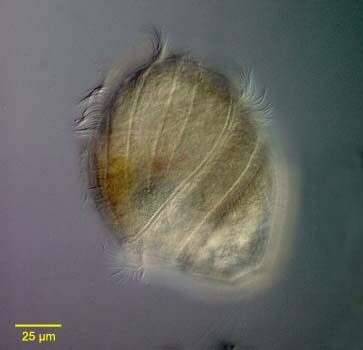
Ventral surface of the metopid ciliate, Brachonella spiralis. The body is broadly conical anteriorly with a narrower obliquely truncate posterior. The long S-shaped peristome winds around the entire circumference of the cell terminating in the cytostome. This spiraling cytostome distinguishes Brachonella (Jankowski 1964) from similar genus Metopus in which the peristome runs obliquely from anterior to posterior but does not spiral around the long axis. The cytostome is paralleled on the right by a perizonal stripe of kineties and on the left an adoral zone of membranelles (both seen well in this image). There are uniform longitudinal dorsal kineties with a tuft of longer caudal cilia. A distinctive aggregate of brownish refractile granules typical of most metopids is noted anteriorly. There is a spherical anterior macronucleus. The micronucleus is not seen here. There is a posterior terminal contractile vacuole. Brachonella is found in sapropelic habitats and contains methanogenic symbionts in the cytoplasm. From stagnant freshwater source with rotting vegetation near Boise, Idaho. DIC optics.
-
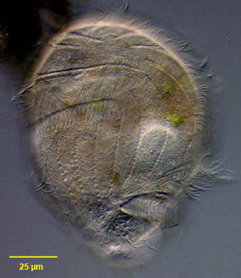
Portrait of the metopid ciliate, Brachonella spiralis. The body is broadly conical anteriorly with a narrower obliquely truncate posterior. The long S-shaped peristome winds around the entire circumference of the cell terminating in the cytostome. This spiraling cytostome distinguishes Brachonella (Jankowski 1964) from similar genus Metopus in which the peristome runs obliquely from anterior to posterior but does not spiral around the long axis. The cytostome is paralleled on the right by a perizonal stripe of kineties. On the left is an adoral zone of membranelles. There are uniform longitudinal dorsal kineties with a tuft of longer caudal cilia. A distinctive aggregate of brownish refractile granules typical of most metopids is noted anteriorly. There is a spherical anterior macronucleus. The micronucleus is not seen here. There is a posterior terminal contractile vacuole. Brachonella is found in sapropelic habitats and contains methanogenic symbionts in the cytoplasm. From stagnant freshwater source with rotting vegetation near Boise, Idaho. DIC optics.
-
Ventral surface of the metopid ciliate, Brachonella spiralis(Smith,1897;Jankowski,1964)stained by a silver carbonate technique(see Foissner, W.Europ. J. Protistol.27,313-330;1991). The body is broadly conical anteriorly with a narrower obliquely truncate posterior. The long S-shaped peristome winds around the entire circumference of the cell terminating in the cytostome. This spiraling cytostome distinguishes Brachonella from the similar genus Metopus in which the peristome runs obliquely from anterior to posterior but does not spiral around the long axis. The cytostome is paralleled on the right by a perizonal stripe of kineties and on the left an adoral zone of membranelles (both seen well in this image). Somatic kineties run obliquely anterior to the cytostome and longitudinally posterior to it. A distinctive aggregate of brownish refractile granules typical of most metopids is noted anteriorly. The spherical anterior macronucleus and micronucleus are not seen here. Brachonella is found in sapropelic habitats and contains methanogenic symbionts in the cytoplasm. From stagnant freshwater source with rotting vegetation near Boise, Idaho. Brightfield.
-
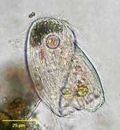
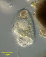
Optical section of the large metopid ciliate, Brachonella caduca (Kahl 1927, Jankowski, 1964). Synonym of Metopus caduca (Kahl, 1927). The cell is pyriform with a broadly domed anterior and tapered truncate posterior end. The long peristome spirals from the dorsum anteriorly around the circumference of the cell to terminate posteriorly at the cytostome (seen here just above contractile vacuole) on the same longitudinal line as the peristome origin. 2 to 3 kinities lie anterior and parallel to the peristome. Approximately 9 longitudinal somatic kineties occur posterior to the peristome. The anterior margin of the peristome is paralleled by a peristomal stripe of kineties, the posterior margin by an adoral zone of membranelles. The somatic kineties of the right and left side terminate anteriorly at a bare suture. There is a long tuft of caudal cilia. A single contractile vacuole is located at the posterior end. There is a single large eccentric spherical macronucleus and adjacent micronucleus. B. caduca is an obligate anaerobe. From stagnant freshwater sapropelic sediment rich in hydrogen sulfide near Boise, Idaho. DIC optics.
-
Ventral view of the large metopid ciliate, Brachonella caduca (Kahl 1927, Jankowski, 1964). Synonym of Metopus caduca (Kahl, 1927). The cell is pyriform with a broadly domed anterior and tapered truncate posterior end. The long peristome spirals from the dorsum anteriorly around the circumference of the cell to terminating posteriorly at the cytostome on the same longitudinal line as the peristome origin. 2 to 3 kinities lie anterior and parallel to the peristome. Approximately 9 longitudinal somatic kineties occur posterior to the peristome. The anterior margin of the peristome is paralleled by a peristomal stripe of kineties, the posterior margin by an adoral zone of membranelles. The somatic kineties of the right and left side terminate anteriorly at a bare suture. There is a long tuft of caudal cilia. A single contractile vacuole is located at the posterior end. There is a single large eccentric spherical macronucleus and adjacent micronucleus. B. caduca is an obligate anaerobe. From stagnant freshwater sapropelic sediment rich in hydrogen sulfide near Boise, Idaho. DIC optics.
-

Dorsolateral view of the large metopid ciliate, Brachonella caduca (Kahl 1927, Jankowski, 1964). Synonym of Metopus caduca (Kahl, 1927). The cell is pyriform with a broadly domed anterior and tapered truncate posterior end. The long peristome spirals from the dorsum anteriorly around the circumference of the cell to terminating posteriorly at the cytostome on the same longitudinal line as the peristome origin (seen well in this image). 2 to 3 kinities lie anterior and parallel to the peristome. Approximately 9 longitudinal somatic kineties occur posterior to the peristome. The anterior margin of the peristome is paralleled by a peristomal stripe of kineties, the posterior margin by an adoral zone of membranelles. The somatic kineties of the right and left side terminate anteriorly at a bare suture. There is a long tuft of caudal cilia. A single contractile vacuole is located at the posterior end. There is a single large eccentric spherical macronucleus and adjacent micronucleus. B. caduca is an obligate anaerobe. From stagnant freshwater sapropelic sediment rich in hydrogen sulfide near Boise, Idaho. DIC optics.
-
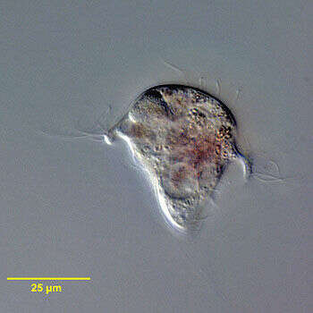
Detail of the well-developed adoral zone of membranelles along the left border of the large buccal cavity of Bothrostoma undulans, a metopid ciliate that is the type species for the genus. From sapropelic sediments in freshwater aquaculture pond near Boise, Idaho. DIC optics.
-
Dorsal view of Bothrostoma undulans, a metopid ciliate that is the type species for the genus. The somatic kineties are longitudinal. There is a long tuft of caudal cilia. From sapropelic sediments in freshwater aquaculture pond near Boise, Idaho. DIC optics
-
Portrait of Bothrostoma undulans, a metopid ciliate that is the type species for the genus. The elongate body is dorsoventrally flattened, narrowing anteriorly to a rounded point, which twists slightly to the left. The large buccal cavity extends 2/3 the body length, bordered on the right by a prominent undulating membrane and on the left by a well-developed adoral zone of membranelles. A perizonal stripe of four longitudinal kineties lies along the right anterior margin parallel to the undulating membrane. The somatic kineties are longitudinal. There is a long tuft of caudal cilia. The central round macronucleus and the overlying micronucleus are seen in this image. Several food vacuoles and the posterior terminal contractile vacuole are seen here. From sapropelic sediments in freshwater aquaculture pond near Boise, Idaho. DIC optics.
-
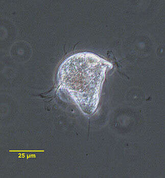
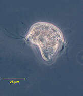
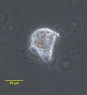
Portrait (coronal section) of the metopid ciliate, Brachonella galeata (Kahl 1927 [Metopus galeatus]; Jankowski, 1964). The anterior half of the cell is broadly helmet-shaped. The posterior is bluntly conical. The peristome encircles the body at the junction of the anterior and posterior halves of the cell, the origin lying just anterior to the termination on the same longitudinal line. The peristome terminates in a posterior cytostome. There is an adoral zone of membranelles on the left margin of the peristome. The somatic cilia are long and sparse. There is a tuft of longer cilia at the posterior end. A single ellipsoid macronucleus is located centrally or anteriorly (not seen well here). There is a single irregularly shaped contractile vacuole at the posterior end. There are purple sulfur bacteria visible in food vacuoles. B. galeata is anaerobic. Collected from anoxic sediment of slow-moving freshwater stream near Boise, Idaho in April 2004. Phase contrast illumination.
-
Portrait (posterior apical view) of the metopid ciliate, Brachonella galeata (Kahl 1927 [Metopus galeatus]; Jankowski, 1964). The anterior half of the cell is broadly helmet-shaped. The margin of the anterior part of the cell is seen well here. The posterior is bluntly conical. The peristome encircles the body at the junction of the anterior and posterior halves of the cell, the origin lying just anterior to the termination on the same longitudinal line. The peristome terminates in a posterior cytostome. There is an adoral zone of membranelles on the left margin of the peristome. The somatic cilia are long and sparse. There is a tuft of longer cilia at the posterior end. A single ellipsoid macronucleus (just to the right of the cell center here) is located centrally or anteriorly. There is a single irregularly shaped contractile vacuole at the posterior end. There are purple sulfur bacteria visible in food vacuoles. B. galeata is anaerobic. Collected from anoxic sediment of slow-moving freshwater stream near Boise, Idaho in April 2004. DIC optics.
-
Portrait of the metopid ciliate, Brachonella galeata (Kahl 1927 [Metopus galeatus]; Jankowski, 1964). The anterior half of the cell is broadly helmet-shaped. The posterior is bluntly conical. The peristome encircles the body at the junction of the anterior and posterior halves of the cell, the origin lying just anterior to the termination on the same longitudinal line. The peristome terminates in a posterior cytostome. There is an adoral zone of membranelles on the left margin of the peristome. The somatic cilia are long and sparse. There is a tuft of longer cilia at the posterior end. A single ellipsoid macronucleus is located centrally or anteriorly. There is a single irregularly shaped contractile vacuole at the posterior end. There are purple sulfur bacteria visible in food vacuoles. B. galeata is anaerobic. Collected from anoxic sediment of slow-moving freshwater stream near Boise, Idaho in April 2004.Phase contrast illumination.
-
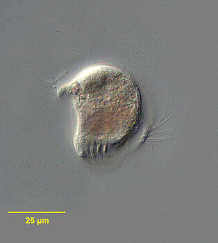
Portrait (anterior oblique view) of the metopid ciliate, Brachonella galeata (Kahl 1927 [Metopus galeatus]; Jankowski, 1964). The anterior half of the cell is broadly helmet-shaped. The posterior is bluntly conical. The peristome encircles the body at the junction of the anterior and posterior halves of the cell, the origin lying just anterior to the termination on the same longitudinal line. The peristome terminates in a posterior cytostome. There is an adoral zone of membranelles on the left margin of the peristome. The posterior part of the AZM is seen in this image. The somatic cilia are long and sparse. There is a tuft of longer cilia at the posterior end. A single ellipsoid macronucleus is located centrally or anteriorly (not seen well here). There is a single irregularly shaped contractile vacuole at the posterior end. There are purple sulfur bacteria visible in food vacuoles. B. galeata is anaerobic. Collected from anoxic sediment of slow-moving freshwater stream near Boise, Idaho in April 2004. DIC optics.
-
Portrait (coronal section) of the metopid ciliate, Brachonella galeata (Kahl 1927 [Metopus galeatus]; Jankowski, 1964). The anterior half of the cell is broadly helmet-shaped. The posterior is bluntly conical. The peristome encircles the body at the junction of the anterior and posterior halves of the cell, the origin lying just anterior to the termination on the same longitudinal line. The peristome terminates in a posterior cytostome. There is an adoral zone of membranelles on the left margin of the peristome. The somatic cilia are long and sparse. There is a tuft of longer cilia at the posterior end. A single ellipsoid macronucleus is located centrally or anteriorly . There is a single irregularly shaped contractile vacuole at the posterior end. There are purple sulfur bacteria visible in food vacuoles. B. galeata is anaerobic. Collected from anoxic sediment of slow-moving freshwater stream near Boise, Idaho in April 2004. DIC optics.
-
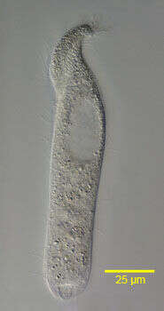
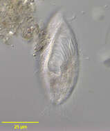
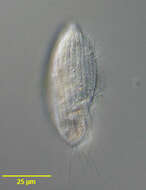
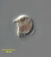
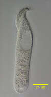
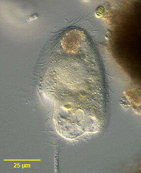
Right side view of Metopus palaeformis (Kahl, 1927).Synonyms probably include Tesnospira alba (Jankowski,1964),M. hyalinus (Kahl,19270 and M. tenuis (Kahl,1927) among others.Morphology is highly variable probably explaining the large number of synonyms. The cell is flask-shaped to elongate (as in this example).The anterior end is twisted to the left resulting in a rounded lip that overhangs the peristome.The spiral peristome is bordered on the left by an adoral zone of membranelles and on the right by five closely spaced kineties,the "perizonal stripe".Just to the right of the posterior termination of the AZM is a short, inconspicuous undulating membrane(usually visible only in silver-stained preparations).The The right somatic kineties parallel the peristome anteriorly and the left somatic kineties terminate at the margin of the peristome.There is no long tuft of caudal cilia. The prominent ellipsoid macronucleus and adjacent micronucleus are in the anterior half. The contractile vacuole is at the posterior end.The cytoplasm contains endosymbiotic methanogenic bacilli (not seen here).There is an aggregate of brown refractile granules at the anterior end typical of the metopid ciliates.Collected from the bottom sediments of an organically enriched rain pool with abundant decaying grass contaminated by Canada goose (Branta canadensis) droppings.Boise, Idaho. January 2006. DIC.
-
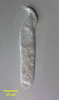
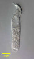
Left lateral view of Metopus palaeformis (Kahl, 1927).Synonyms probably include Tesnospira alba (Jankowski,1964),M. hyalinus (Kahl,19270 and M. tenuis (Kahl,1927) among others.Morphology is highly variable probably explaining the large number of synonyms. The cell is flask-shaped to elongate (as in this example).The anterior end is twisted to the left resulting in a rounded lip that overhangs the peristome.The spiral peristome is bordered on the left by an adoral zone of membranelles and on the right by five closely spaced kineties,the "perizonal stripe".Just to the right of the posterior termination of the AZM is a short, inconspicuous undulating membrane(usually visible only in silver-stained preparations).The The right somatic kineties parallel the peristome anteriorly and the left somatic kineties terminate at the margin of the peristome.There is no long tuft of caudal cilia. The prominent ellipsoid macronucleus and adjacent micronucleus are in the anterior half. The contractile vacuole is at the posterior end.The cytoplasm contains endosymbiotic methanogenic bacilli (not seen here).There is an aggregate of brown refractile granules at the anterior end typical of the metopid ciliates.Collected from the bottom sediments of an organically enriched rain pool with abundant decaying grass contaminated by Canada goose (Branta canadensis) droppings.Boise, Idaho. January 2006. DIC.
-
Right side view of Metopus palaeformis (Kahl, 1927).Synonyms probably include Tesnospira alba (Jankowski,1964),M. hyalinus (Kahl,19270 and M. tenuis (Kahl,1927) among others.Morphology is highly variable probably explaining the large number of synonyms. The cell is flask-shaped (as in this example)to elongate .The anterior end is twisted to the left resulting in a rounded lip that overhangs the peristome.The spiral peristome is bordered on the left by an adoral zone of membranelles and on the right by five closely spaced kineties,the "perizonal stripe".Just to the right of the posterior termination of the AZM is a short, inconspicuous undulating membrane(usually visible only in silver-stained preparations).The The right somatic kineties parallel the peristome anteriorly and the left somatic kineties terminate at the margin of the peristome. The prominent ellipsoid macronucleus and adjacent micronucleus are in the anterior half. The contractile vacuole is at the posterior end.The cytoplasm contains endosymbiotic methanogenic bacilli (not seen here).There is an aggregate of brown refractile granules at the anterior end typical of the metopid ciliates.Collected from the bottom sediments of an organically enriched rain pool with abundant decaying grass contaminated by Canada goose (Branta canadensis) droppings.Boise, Idaho. January 2006. DIC.
-

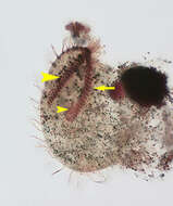
Ventral view of the infraciliature of Metopus palaeformis (Kahl,1927) contracted by fixation and compressed to display details.Synonyms probably include Tesnospira alba (Jankowski,1964),M. hyalinus (Kahl,19270 and M. tenuis (Kahl,1927) among others.Morphology is highly variable probably explaining the large number of synonyms. The cell is flask-shaped (as in this example)to elongate .The anterior end is twisted to the left resulting in a rounded lip that overhangs the peristome.The spiral peristome is bordered on the left by an adoral zone of membranelles (arrow) and on the right by five closely spaced kineties,the "perizonal stripe" (large arrowhead).Just to the right of the posterior termination of the AZM is a short, inconspicuous undulating membrane(small arrowhead).The cytoplasm contains endosymbiotic methanogenic bacilli (not seen here).Collected from the bottom sediments of an organically enriched rain pool with abundant decaying grass contaminated by Canada goose (Branta canadensis) droppings.Boise, Idaho. January 2006.Stained by the silver carbonate technique (see Foissner, W. Europ. J. Protistol., 27:313-330;1991).Brightfield.
-
Ventral view of the infraciliature of Metopus palaeformis (Kahl, 1927) contracted by fixation and compressed to display details...Synonyms probably include Tesnospira alba (Jankowski,1964),M. hyalinus (Kahl,19270 and M. tenuis (Kahl,1927) among others.Morphology is highly variable probably explaining the large number of synonyms. The cell is flask-shaped (as in this example) to elongate .The anterior end is twisted to the left resulting in a rounded lip that overhangs the peristome.The spiral peristome is bordered on the left by an adoral zone of membranelles (large arrowhead) and on the right by five closely spaced kineties,the "perizonal stripe" (small arrowhead).Just to the right of the posterior termination of the AZM is a short, inconspicuous undulating membrane(black arrow).The cytoplasm contains endosymbiotic methanogenic bacilli (not seen here).Collected from the bottom sediments of an organically enriched rain pool with abundant decaying grass contaminated by Canada goose (Branta canadensis) droppings.Boise, Idaho. January 2006.Stained by the silver carbonate technique (see Foissner, W. Europ. J. Protistol., 27:313-330;1991).Brightfield.
-

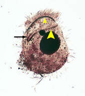
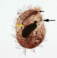
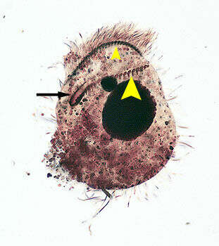
Ventral view of the infraciliature of Metopus palaeformis (Kahl,1927) contracted by fixation and compressed to display details..Synonyms probably include Tesnospira alba (Jankowski,1964),M. hyalinus (Kahl,19270 and M. tenuis (Kahl,1927) among others.Morphology is highly variable probably explaining the large number of synonyms. The cell is flask-shaped (as in this example)to elongate.The anterior end is twisted to the left resulting in a rounded lip that overhangs the peristome. The spiral peristome is bordered on the left by an adoral zone of membranelles (large black arrow) and on the right by five closely spaced kineties,the "perizonal stripe" (small black arrow).Just to the right of the posterior termination of the AZM is a short, inconspicuous undulating membrane(yellow arrow).The cytoplasm contains endosymbiotic methanogenic bacilli (not seen here).Collected from the bottom sediments of an organically enriched rain pool with abundant decaying grass contaminated by Canada goose (Branta canadensis) droppings.Boise, Idaho. January 2006.Stained by the silver carbonate technique (see Foissner, W. Europ. J. Protistol., 27:313-330;1991).Brightfield.