-
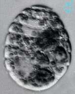
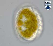
Gyrodinium oblongum cells are oblong from the ventral side, dorso-ventrally flattened posteriorly, length 17 - 25 microns, width 6-10 microns. The epicone and hypocone are approximately equal in size. Cingulum displaced approximately 1/4 - 1/3 of cell length. The sulcus initially narrow but widens at the posterior end . Apical groove not present. Nucleus in the hypocone, approximately 5 microns diameter . Chloroplast single, yellow-brown, forms thin longitudinal strands towards the anterior and posterior ends. Obvious pyrenoid present, 3 - 4 microns diameter, in the centre of the cell.
-
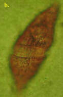
Cells are spindle shaped and asymmetric. The cingulum is narrow and excavated and displaced by more than one third of the body length. The apex is pointed. The antapical part of the cell is slightly bilobed. Chloroplasts are absent but food vacuoles are somteimes visible.
-
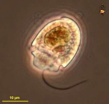
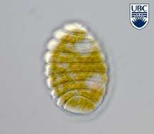
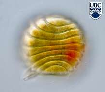
Katodinium (cat-owe-din-ee-um) common coastal marine dinoflagellate. The cingulum (circumferential groove) is located near the posterior of the body so that the epicone (front part) is much bigger than the epicone (back part). This genus is in part distinguished by the groove near the apex of the epicone. This individual is clearly a carnivore. With longitudinal flagellum trailing behind the cell. Phase contrast.
-
Katodinium spec.
-
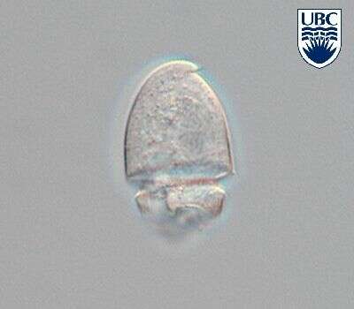
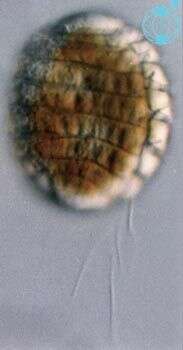
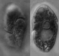
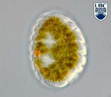
Polykrikos (polly-cry-coz) lebourae Herdman 1923. The image shows a cell in right lateral view. The cell has many cingula. The cell contains no plastids, however a food particle is present.
-
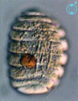
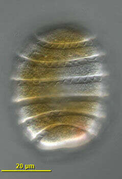
Polykrikos (polly-cry-coz) lebourae Herdman 1923. The image shows a cell in lateral view. The cell has many cingula. There are two nuclei present, one in the posterior of the cell, the other near the anterior end.
-
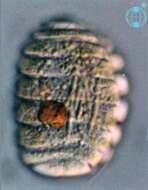
Polykrikos (polly-cry-coz) lebourae Herdman 1923. The image shows a cell in right lateral view. The cell has many cingula.
-
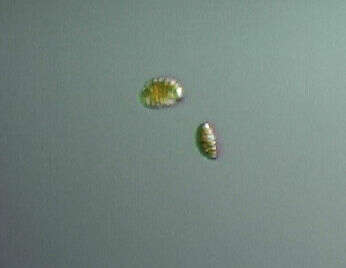
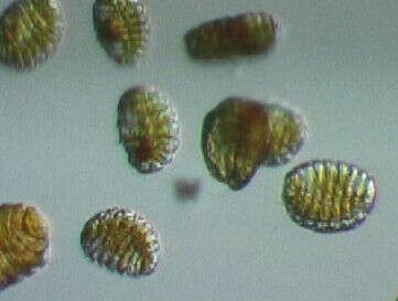
Polykrikos lebourae Herdman 1923
-
Polykrikos lebourae Herdman 1923
-
Polykrikos lebourae Herdman 1923
-
Polykrikos lebourae Herdman 1923
-
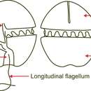
Dorso-lateral view of the numerous transverse flagella lie in the cingular grooves of this phagotrophic gymnodinioid dinoflagellate. Isolated by Bob Moore from Little Sippiwissett marsh near Woods Hole, Massachusetts, USA. Differential interference contrast optics.
-
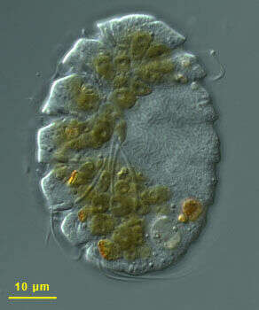
Differential interference contrast image of this phagotrophic dinoflagellate isolated from Chappaquoit beach, Massachusetts. The brown inclusions are partly digested elements from food - this species was observed consuming other dinoflagellates. This species may have more than one nucleus, but in this cell there is a singler nucleus (middle of right hand side of cell). There are also large extrusomes within the cell. Image by all.
-
This guys can be real quick. With so many flagella, one would expect so... Bugs isolated by Bob Moore, identified by Shauna Murray, video by Dan Lahr.
-
A bunch of Polykrikos lebourae moving around. These guys were isolated and picked using a "very hi-tech and complex technique" developed by Bob Moore and Shauna Murray. Video by Dan Lahr.