-
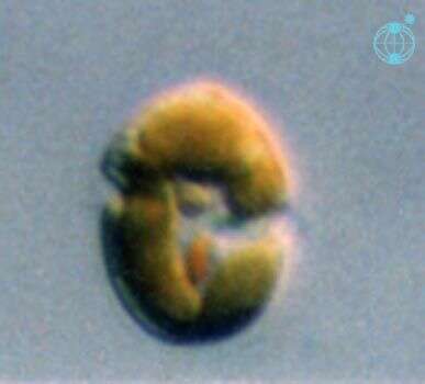
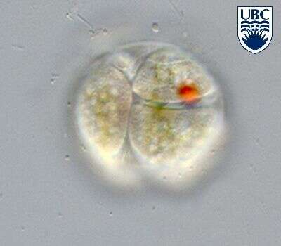
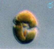
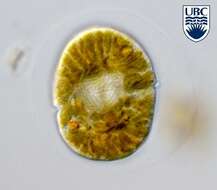
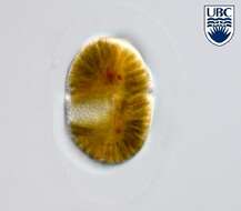
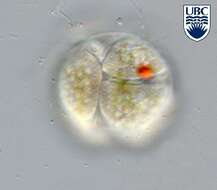
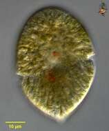
Gymnodinium (jim-no-din-ee-um) danicans Campbell 1973. The image shows a cell in ventral view. The red stigma is visible in the sulcal area. The plastids are yellow-brown and multiple.
-
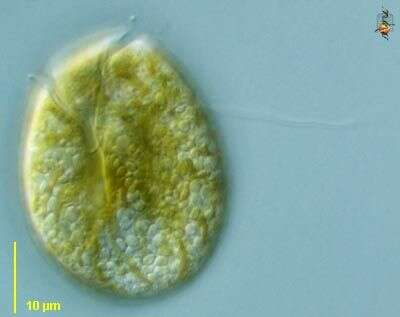
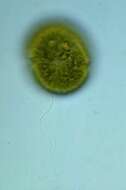
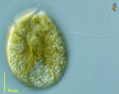
Image showing the off green colour of the chloroplasts of this dinoflagellate, the equatorial groove and the longitudinal (trailing) flagellum.
-
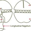
Dinoflagelate with chloroplasts. There are two flagella, one in the groove that runs around the middle of the body and the second lies in the longitudinal groove and extends behind the swimming cell.
-
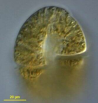
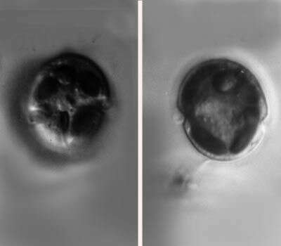
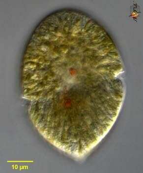
Differential interference contrast micrograph of a living Gymnodinium showing the radiating strands of the chloroplast.
-
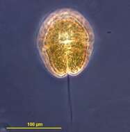
Gymnodinium (Dinoflagellata) is the only genus of naked dinoflagellates found in Lake Kinneret. This genus never forms blooms but occurs quite often in small densities. Like other dinoflagellates, Gymnodinium has 2 unequal flagella, the longitudinal "whiplash" flagellum is seen in this picture, the second, transverse flagellum is hidden in the transverse groove, or cingulum.
-
Gymnodinium spec. in dorsal view. Note the helical transverse flagellum running in the cingulum. The nucleus is lying in the cell centre.
-
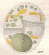
Gymnodinium spec. in mid cell focus showing the central nucleus with nicely visible condensed, rod-shaped chromosomes. The golden-brown chloroplasts are elongated radiating to the cell periphery. Note also the very faint, colorless round cyst around the cell. It is a vegetative division cyst.
-
Gymnodinium spec.
-
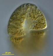
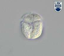
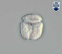
Gymnodinium spec. in ventral view. The sulcus is extending on the episome and the start of the acrobase (apical groove) is visible at the upper cell end.
-
Gymnodinium spec. in ventral view. This heterotrophic species has a sulcus extending onto the episome and a nearly circular cingulum (transverse furrow).
-
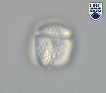
Gymnodinium spec. in dorsal view.
-
Gymnodinium spec. in mid cell focus.
-
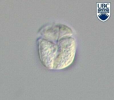
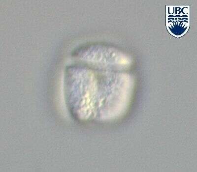
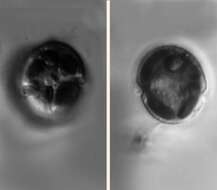
Gymnodinium spec. Two just divided cells in a hyalin division cyst.
-
Gymnodinium spec. Two just divided cells in a hyalin division cyst.
-
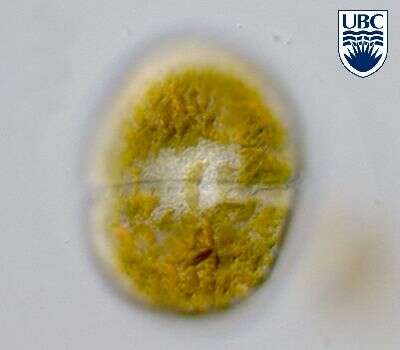
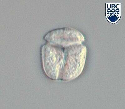
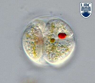
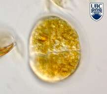
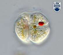
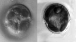
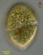
Gymnodinium (jim-no-din-ee-um) danicans Campbell 1973. The image on the right shows a cell in ventral view. The cingulum is in the middle of the cell, and the stigma is in the sulcal area. The image on the right shows a mid-plane focus through a cell, displaying the multiple plastids.
-
Gymnodinium danicans cells are round from the ventral side, dorso-ventrally flattened. Length 13 16 microns, width 12 15 microns, breadth (lateral) approximately 5 microns. Cingulum begins 0.5 0.6 of the way down the cell, relatively wide, 2 - 3 microns, proximal end 1 2 microns lower than distal. Sulcus 2 3 microns wide, with prominent edges. Apical groove not present. A red, usually triangular shaped eyespot, approximately 3 microns long, in or just beside the sulcus. Longitudinal flagellum arising at the anterior end of the sulcus. Nucleus round, central, often to the left of the cell, 3 5 microns diameter. Chloroplasts yellow-green to yellow-brown, peripherally positioned, usually rounded to oval, 3 4 microns diameter. Fast swimming.
-
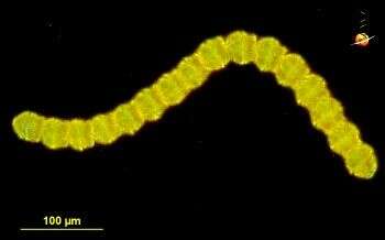
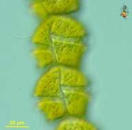
Gymnodinium catenatum (jim-know-din-ee-um) is a chain forming motile species. This chain contains 16 cells, but the chains may be of differing lengths, and other genera may form similar chains. Cells coloured because of presence of chloroplasts. This species produces a toxin that is harmful to humans, and because of the toxicity, correct identification is important and requires expert input which can be obtained from other sites such as
IOC Harmful Algal Bloom Program. Dark ground illumination.
data on this strain.
-
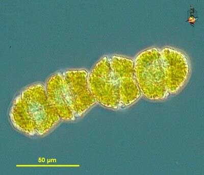
Gymnodinium catenatum (jim-know-din-ee-um) is a chain forming motile species associated with the production of toxins. Girdle (equatorial grooves) visible. Because of the toxicity, correct identification is important and needs expert input, which can be obtained from other sites such as
IOC Harmful Algal Bloom Program. Phase contrast microscopy.
data on this strain.
-
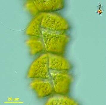
Gymnodinium catenatum (jim-know-din-ee-um) is a chain forming motile species associated with the production of toxins. This image of the ventral surface of several cells shows the equatorial groove or girdle and longitudinal groove or cingulum. Flagella can also be seen. Because of the toxicity, correct identification is important and needs expert input, which can be obtained from other sites such as
IOC Harmful Algal Bloom Program. Differential interference microscopy.
data on this strain.
-
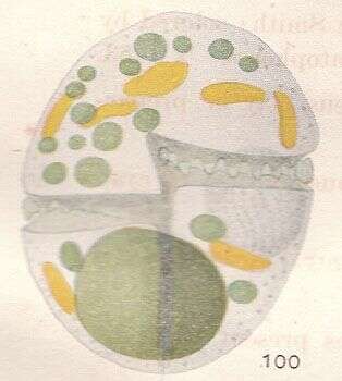
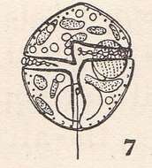
From the original description by Kofoid and Swezy, text is included in the 'large file'.
-
From the original account by Kofoid and Swezy, the full account is included in the large downloadable file.
-
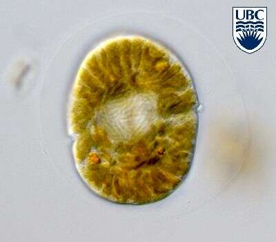
Gymnodinium fuscum,. common freshwater dinoflagellates, with greatly folded radiating plastid, red granules near middle or in posterior half of the cell. With equatorial groove - the cingulum more or less running around the middle of the cell, the longitudinal groove - the sulcus - not strongly developed. This is the type species of the genus Gymnodinium. Differential interference contrast.
-
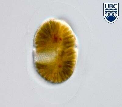
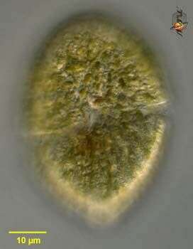
Gymnodinium fuscum,. common freshwater dinoflagellates, with greatly folded radiating plastid, red granules near middle or in posterior half of the cell. This image focusses on the surface to show the equatorial groove - the cingulum, and the longitudinal groove - the sulcus. This is the type species of the genus Gymnodinium. Differential interference contrast.
-
This species of Amphidinium (am-fee-din-ee-um) contains a plastid with chlorophylls a and c (the yellow/brown colour indicates this). All taxa with plastids of this colour have been considered by some to form a group - the Chromophyta or chromists which has at times included some unrelated organisms. It looks as if there is a single radiating plastid. It has a pyrenoid, as another image of the same species shows. Circumferential groove is located near the top of the cell. Differential interference contrast.