-
Description: Français : Certaines espèces de Dinophysis sont responsable de nombreuses intoxication par les coquillages. Date: 14 October 2011. Source: Own work. Author:
Marulio.
-
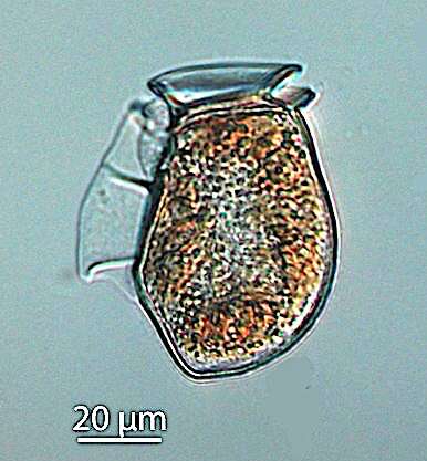
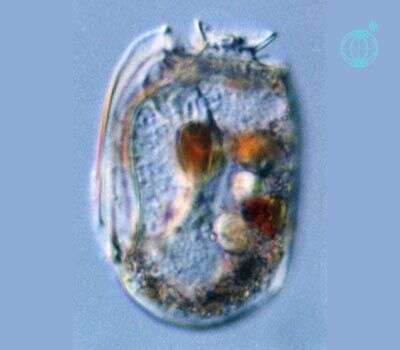
Description: English: Live cell of the thecate dinoflagellate Dinophysis acuta in lateral view. Live sample from net plankton haul (mesh size 20 µm), magnification x400. Date: 29 March 2011. Source: Extracted from
this Commons file. Author: Alexandra Kraberg.
-
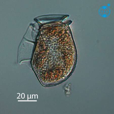
Description: English: Live cell of the thecate dinoflagellate Dinophysis acuta in lateral view. Live sample from net plankton haul (mesh size 20 µm), magnification x400. Date: 29 March 2011. Source:
PlanktonNet. Author: Alexandra Kraberg.
-
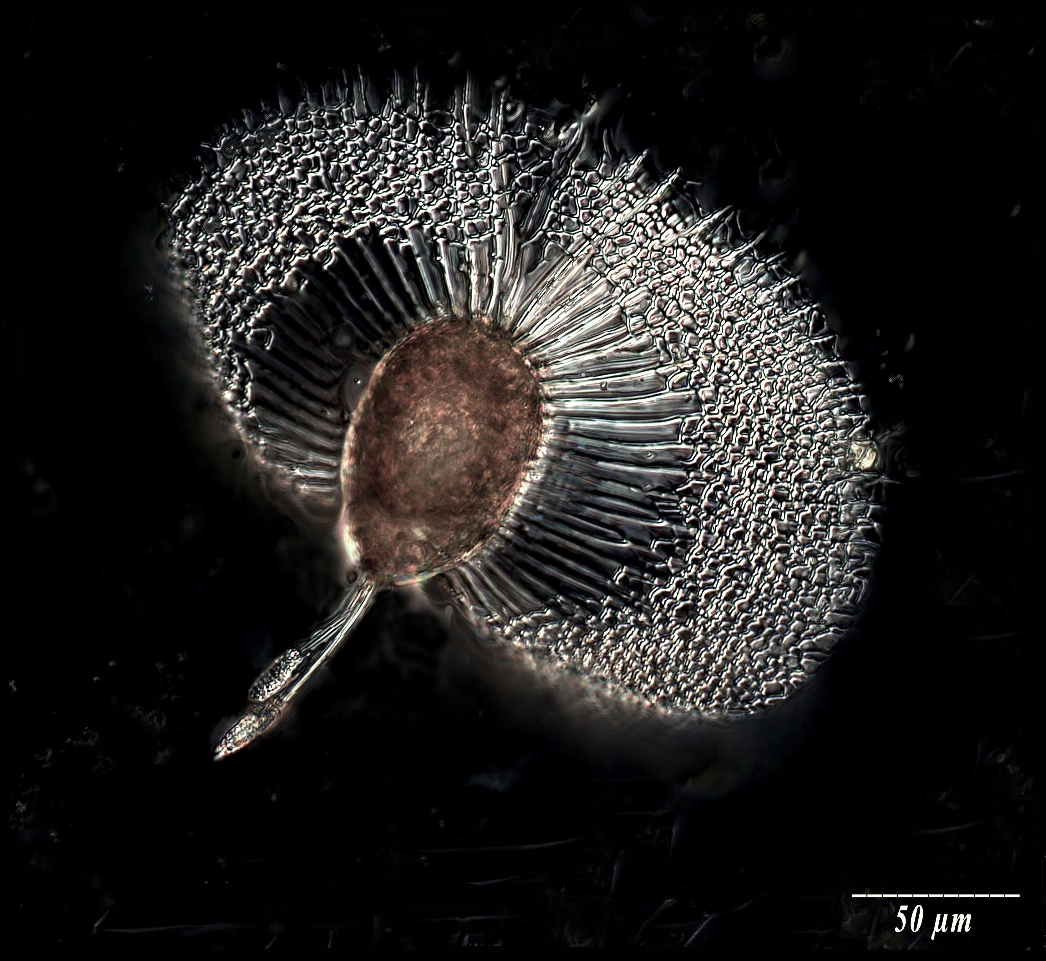
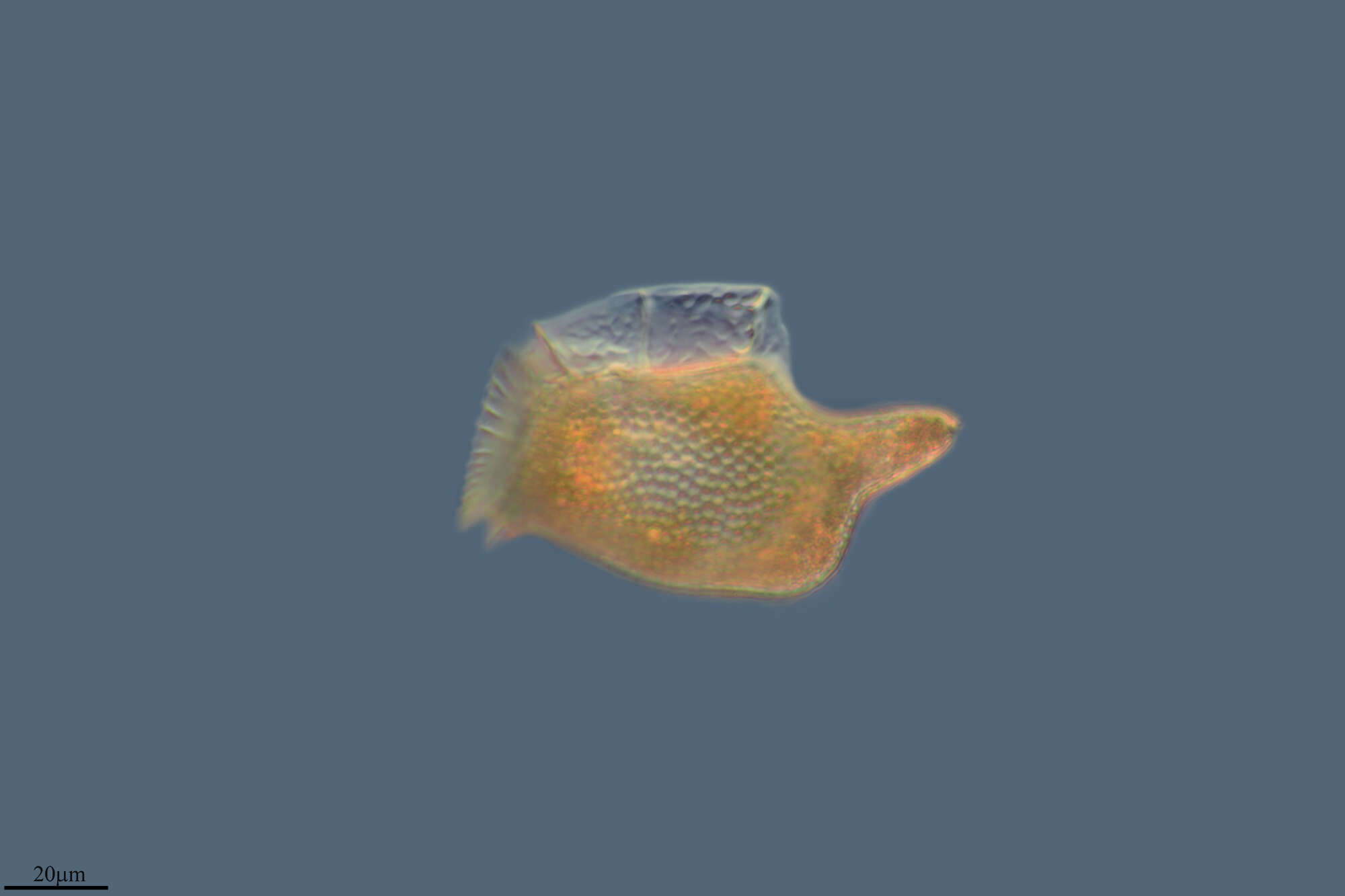
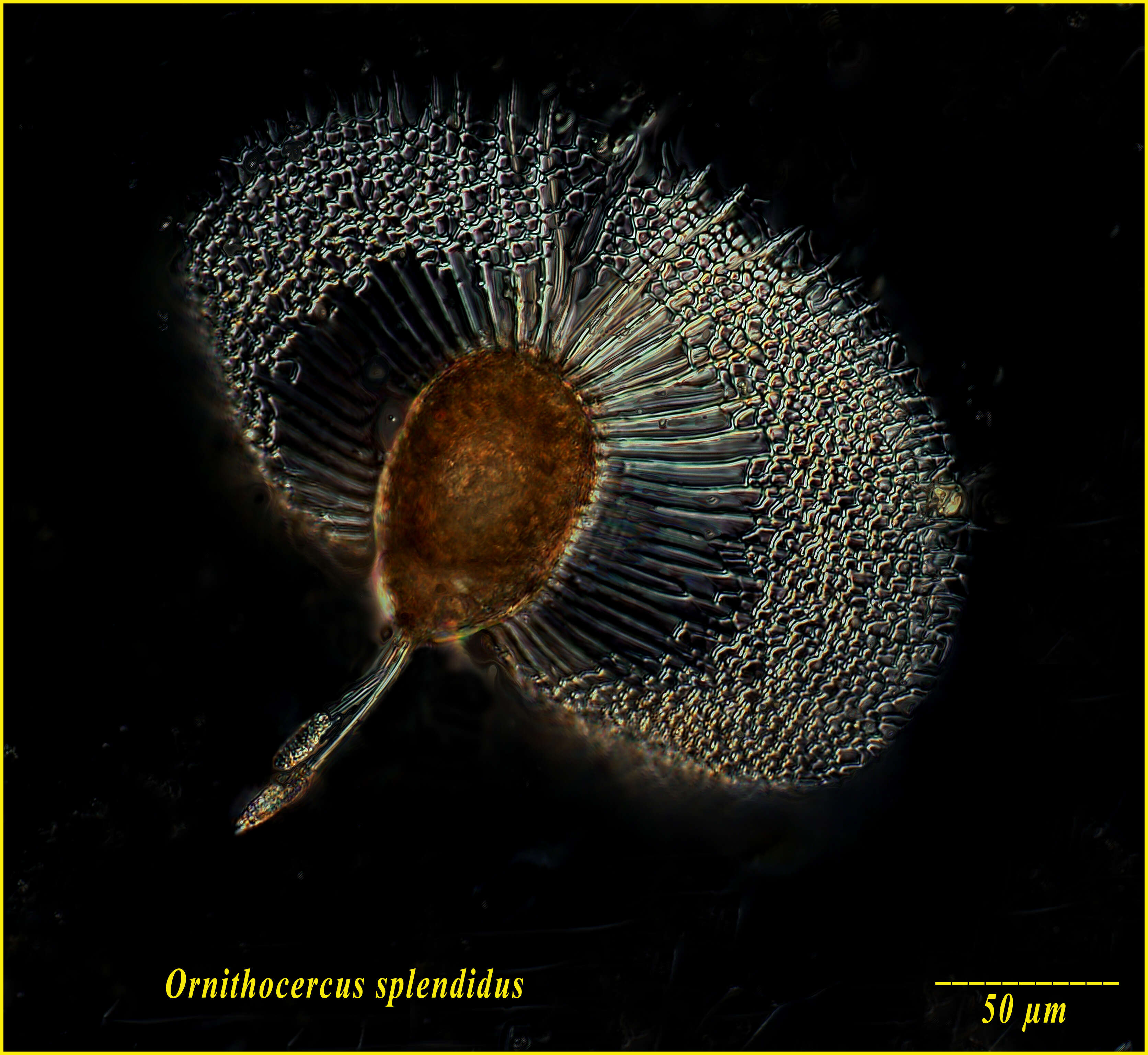
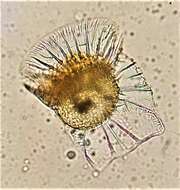
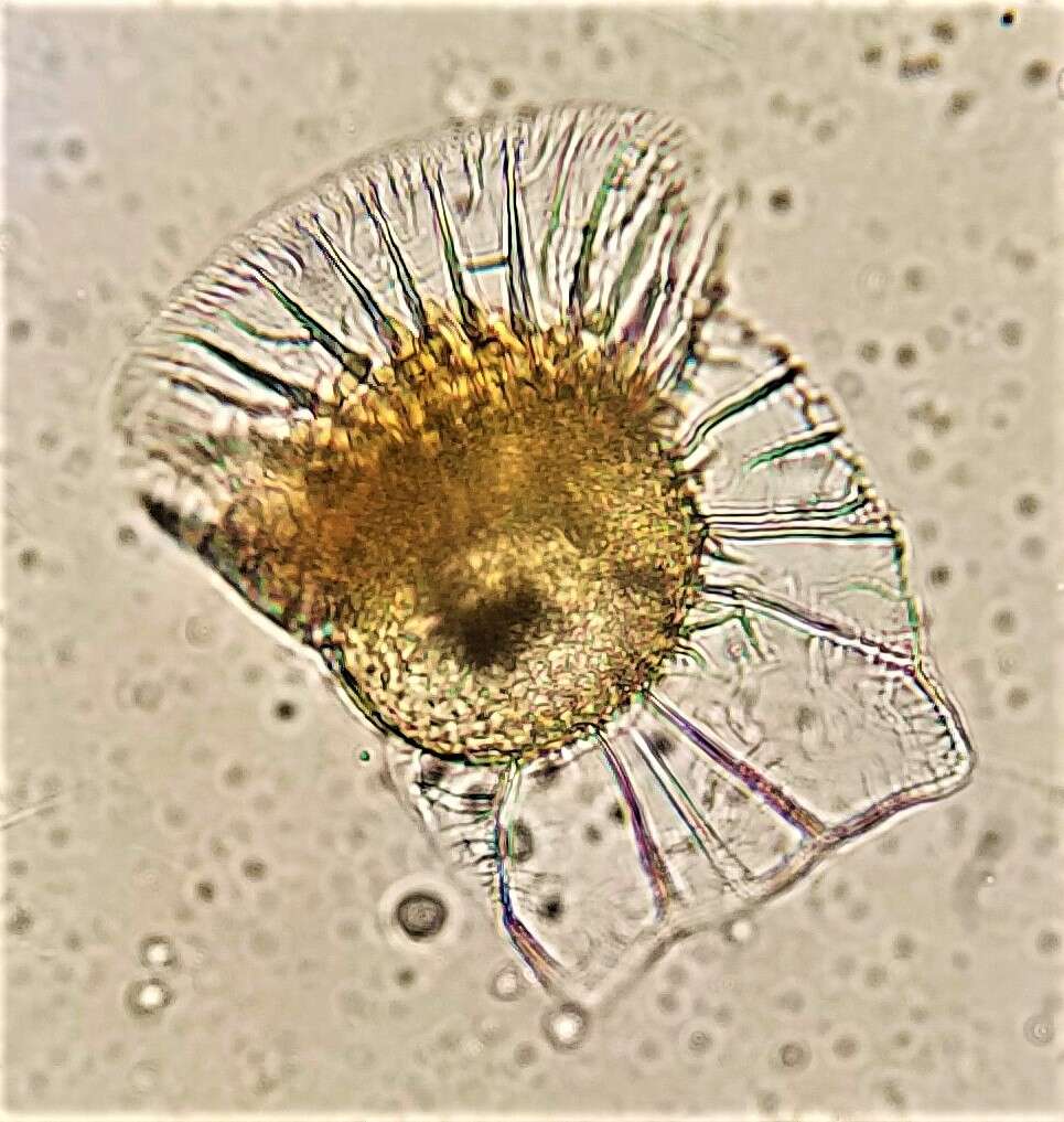
Summary.mw-parser-output table.commons-file-information-table,.mw-parser-output.fileinfotpl-type-information{border:1px solid #a2a9b1;background-color:#f8f9fa;padding:5px;font-size:95%;border-spacing:2px;box-sizing:border-box;margin:0;width:100%}.mw-parser-output table.commons-file-information-table>tbody>tr,.mw-parser-output.fileinfotpl-type-information>tbody>tr{vertical-align:top}.mw-parser-output table.commons-file-information-table>tbody>tr>td,.mw-parser-output table.commons-file-information-table>tbody>tr>th,.mw-parser-output.fileinfotpl-type-information>tbody>tr>td,.mw-parser-output.fileinfotpl-type-information>tbody>tr>th{padding:4px}.mw-parser-output.fileinfo-paramfield{background:#ccf;text-align:right;padding-right:0.4em;width:15%;font-weight:bold}.mw-parser-output.commons-file-information-table+table.commons-file-information-table,.mw-parser-output.commons-file-information-table+div.commons-file-information-table>table{border-top:0;padding-top:0;margin-top:-8px}@media only screen and (max-width:719px){.mw-parser-output table.commons-file-information-table,.mw-parser-output.commons-file-information-table.fileinfotpl-type-information{border-spacing:0;padding:0;word-break:break-word;width:100%!important}.mw-parser-output.commons-file-information-table>tbody,.mw-parser-output.fileinfotpl-type-information>tbody{display:block}.mw-parser-output.commons-file-information-table>tbody>tr>td,.mw-parser-output.commons-file-information-table>tbody>tr>th,.mw-parser-output.fileinfotpl-type-information>tbody>tr>td,.mw-parser-output.fileinfotpl-type-information>tbody>tr>th{padding:0.2em 0.4em;text-align:left;text-align:start}.mw-parser-output.commons-file-information-table>tbody>tr,.mw-parser-output.fileinfotpl-type-information>tbody>tr{display:flex;flex-direction:column}.mw-parser-output.commons-file-information-table+table.commons-file-information-table,.mw-parser-output.commons-file-information-table+div.commons-file-information-table>table{margin-top:-1px}.mw-parser-output.fileinfo-paramfield{box-sizing:border-box;flex:1 0 100%;width:100%}} Description: English: Ornithocercus splendidus Dinoflagellate from the Ionian Sea in September 2010. Found in a sample from 74m depth. Date: 9 December 2010, 18:32:10. Source:
https://www.flickr.com/photos/56879865@N08/5246342627/. Author: tintinnidguy.
-
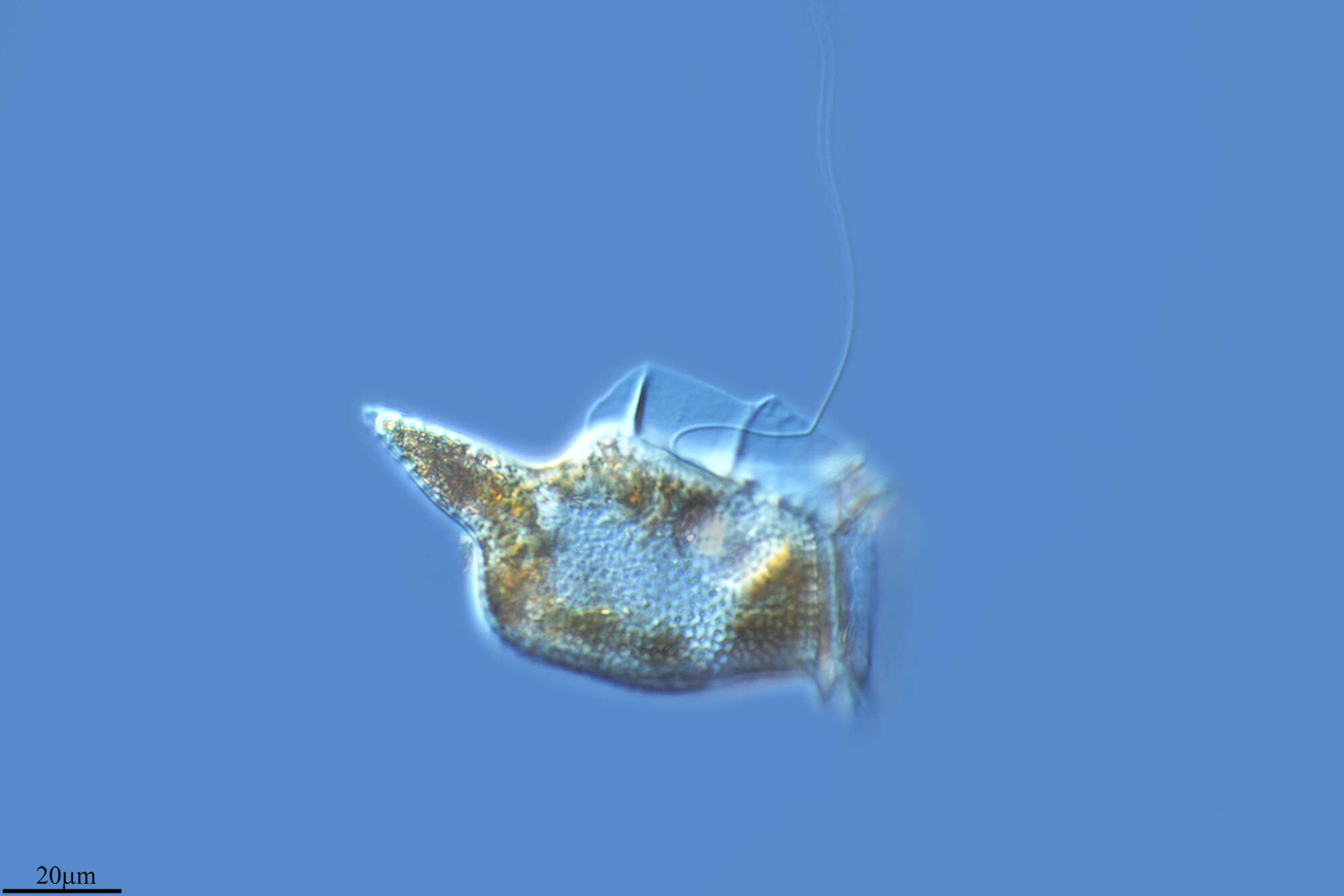
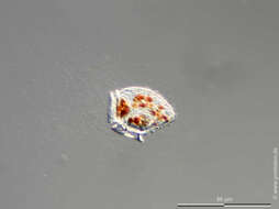
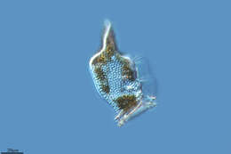
Summary.mw-parser-output table.commons-file-information-table,.mw-parser-output.fileinfotpl-type-information{border:1px solid #a2a9b1;background-color:#f8f9fa;padding:5px;font-size:95%;border-spacing:2px;box-sizing:border-box;margin:0;width:100%}.mw-parser-output table.commons-file-information-table>tbody>tr,.mw-parser-output.fileinfotpl-type-information>tbody>tr{vertical-align:top}.mw-parser-output table.commons-file-information-table>tbody>tr>td,.mw-parser-output table.commons-file-information-table>tbody>tr>th,.mw-parser-output.fileinfotpl-type-information>tbody>tr>td,.mw-parser-output.fileinfotpl-type-information>tbody>tr>th{padding:4px}.mw-parser-output.fileinfo-paramfield{background:#ccf;text-align:right;padding-right:0.4em;width:15%;font-weight:bold}.mw-parser-output.commons-file-information-table+table.commons-file-information-table,.mw-parser-output.commons-file-information-table+div.commons-file-information-table>table{border-top:0;padding-top:0;margin-top:-8px}@media only screen and (max-width:719px){.mw-parser-output table.commons-file-information-table,.mw-parser-output.commons-file-information-table.fileinfotpl-type-information{border-spacing:0;padding:0;word-break:break-word;width:100%!important}.mw-parser-output.commons-file-information-table>tbody,.mw-parser-output.fileinfotpl-type-information>tbody{display:block}.mw-parser-output.commons-file-information-table>tbody>tr>td,.mw-parser-output.commons-file-information-table>tbody>tr>th,.mw-parser-output.fileinfotpl-type-information>tbody>tr>td,.mw-parser-output.fileinfotpl-type-information>tbody>tr>th{padding:0.2em 0.4em;text-align:left;text-align:start}.mw-parser-output.commons-file-information-table>tbody>tr,.mw-parser-output.fileinfotpl-type-information>tbody>tr{display:flex;flex-direction:column}.mw-parser-output.commons-file-information-table+table.commons-file-information-table,.mw-parser-output.commons-file-information-table+div.commons-file-information-table>table{margin-top:-1px}.mw-parser-output.fileinfo-paramfield{box-sizing:border-box;flex:1 0 100%;width:100%}} Description: Français : Ornithocercus quadratus et ses cyanobiontes. Spécimen récolté au filet à plancton WP2 (maille 50 µm) en mars 2021 en rade de Villefranche-sur-Mer (France). Observation réalisée au microscope optique au Laboratoire d'Océanographie de Villefranche. Observation et photographie de J. Millot. English: Ornithocercus quadratus and its cyanobionts. The cells were sampled using a WP2 plankton net (50 µm mesh) in March 2021 in the bay of Villefranche-sur-Mer (France). Observed using a microscope in the Laboratoire d'Océanographie de Villefranche. Observation and photograph by J. Millot. Date: 8 March 2021, 15:55:51. Source: Own work. Author:
Phycoplankton.
-
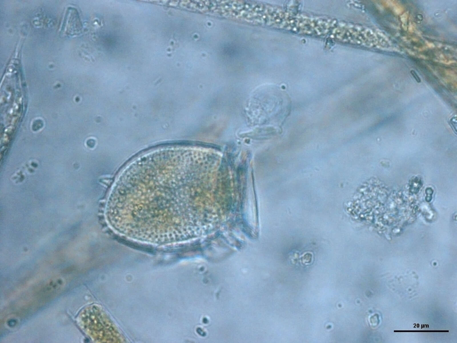
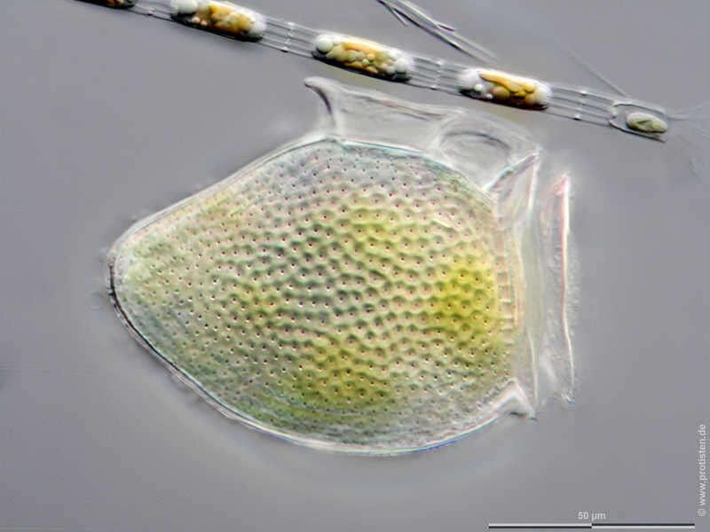
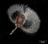
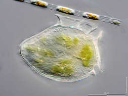
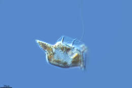
Dinophysis acuta The specimen was gathered in the Kieler Förde (German Baltic Sea). Scale bar indicates 50 µm. The image was built up using several photomicrographic frames with manual stacking technique. Images were taken using Zeiss Axioplan with MFT camera Olympus OM-D E-M5 II.Image under Creative Commons License V 3.0 (CC BY-NC-SA). Place name: Baltic Sea, Kieler Förde, Kiel Fjord (Germany) Latitude: 54.3894126 Longitude: 10.1749055 Multiebenen-Abbildung, manuell gestapelt. Der Messbalken markiert eine Länge von 50 µm. Probe aus der Kieler Förde. Mikrotechnik: Zeiss Axioplan, Kamera: Olympus OM-D E-M5 II. Creative Commons License V 3.0 (CC BY-NC-SA). For permission to use of (high-resolution) images please contact postmaster@protisten.de.
-
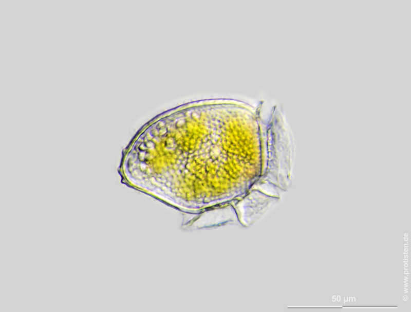
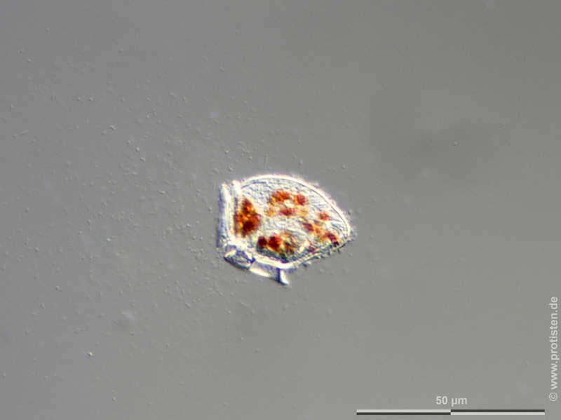
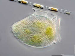
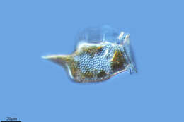
Dinophysis acuta Scale bar indicates 50 µm. The specimen was gathered in the Kieler Förde (Baltic Sea). Sampling date 2/2022. The image was built up using several photomicrographic frames with manual stacking technique. Images were taken using Zeiss Axioplan with Olympus OM-D M5 MKII. Image under Creative Commons License V 3.0 (CC BY-NC-SA). Place name: Baltic Sea, Kieler Förde, Kiel Fjord (Germany) Latitude: 54.3894126 Longitude: 10.1749055 Multiebenen-Abbildung, manuell gestapelt. Der Messbalken markiert eine Länge von 50 µm. Probe aus der Kieler Förde. Datum der Aufsammlung: 2/2022. Mikrotechnik: Zeiss Axioplan, Kamera: Olympus OM-D M5 MKII. Creative Commons License V 3.0 (CC BY-NC-SA). For permission to use of (high-resolution) images please contact postmaster@protisten.de.
-
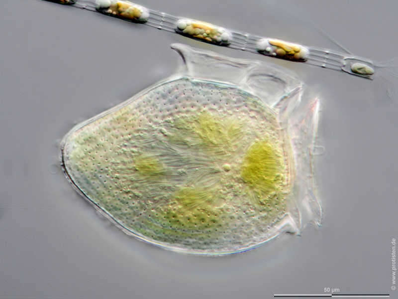
Dinophysis acuta The specimen was gathered in the Kieler Förde (German Baltic Sea). Scale bar indicates 50 µm. The image was built up using several photomicrographic frames with manual stacking technique. Images were taken using Zeiss Axioplan with MFT camera Olympus OM-D E-M5 II.Image under Creative Commons License V 3.0 (CC BY-NC-SA). Place name: Baltic Sea, Kieler Förde, Kiel Fjord (Germany) Latitude: 54.3894126 Longitude: 10.1749055 Multiebenen-Abbildung, manuell gestapelt. Der Messbalken markiert eine Länge von 50 µm. Probe aus der Kieler Förde. Mikrotechnik: Zeiss Axioplan, Kamera: Olympus OM-D E-M5 II. Creative Commons License V 3.0 (CC BY-NC-SA). For permission to use of (high-resolution) images please contact postmaster@protisten.de.
-
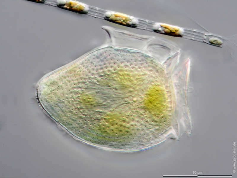
Dinophysis acuta Scale bar indicates 50 µm. The specimen was gathered in the Kieler Förde (Baltic Sea). Sampling date 2/2022. The image was built up using several photomicrographic frames with manual stacking technique. Images were taken using Zeiss Axioplan with Olympus OM-D M5 MKII. Image under Creative Commons License V 3.0 (CC BY-NC-SA). Place name: Baltic Sea, Kieler Förde, Kiel Fjord (Germany) Latitude: 54.3894126 Longitude: 10.1749055 Multiebenen-Abbildung, manuell gestapelt. Der Messbalken markiert eine Länge von 50 µm. Probe aus der Kieler Förde. Datum der Aufsammlung: 2/2022. Mikrotechnik: Zeiss Axioplan, Kamera: Olympus OM-D M5 MKII. Creative Commons License V 3.0 (CC BY-NC-SA). For permission to use of (high-resolution) images please contact postmaster@protisten.de.
-
Grove, O, Galicia, Spain
-
Grove, O, Galicia, Spain
-
Grove, O, Galicia, Spain
-
Grove, O, Galicia, Spain
-
Grove, O, Galicia, Spain
-
Histioneis elongata from the South Pacific, Tara Oceans Expedition station 111. Lugol's-fixed specimen, z-stack of images made using a 40x objective & DIC optics.
-
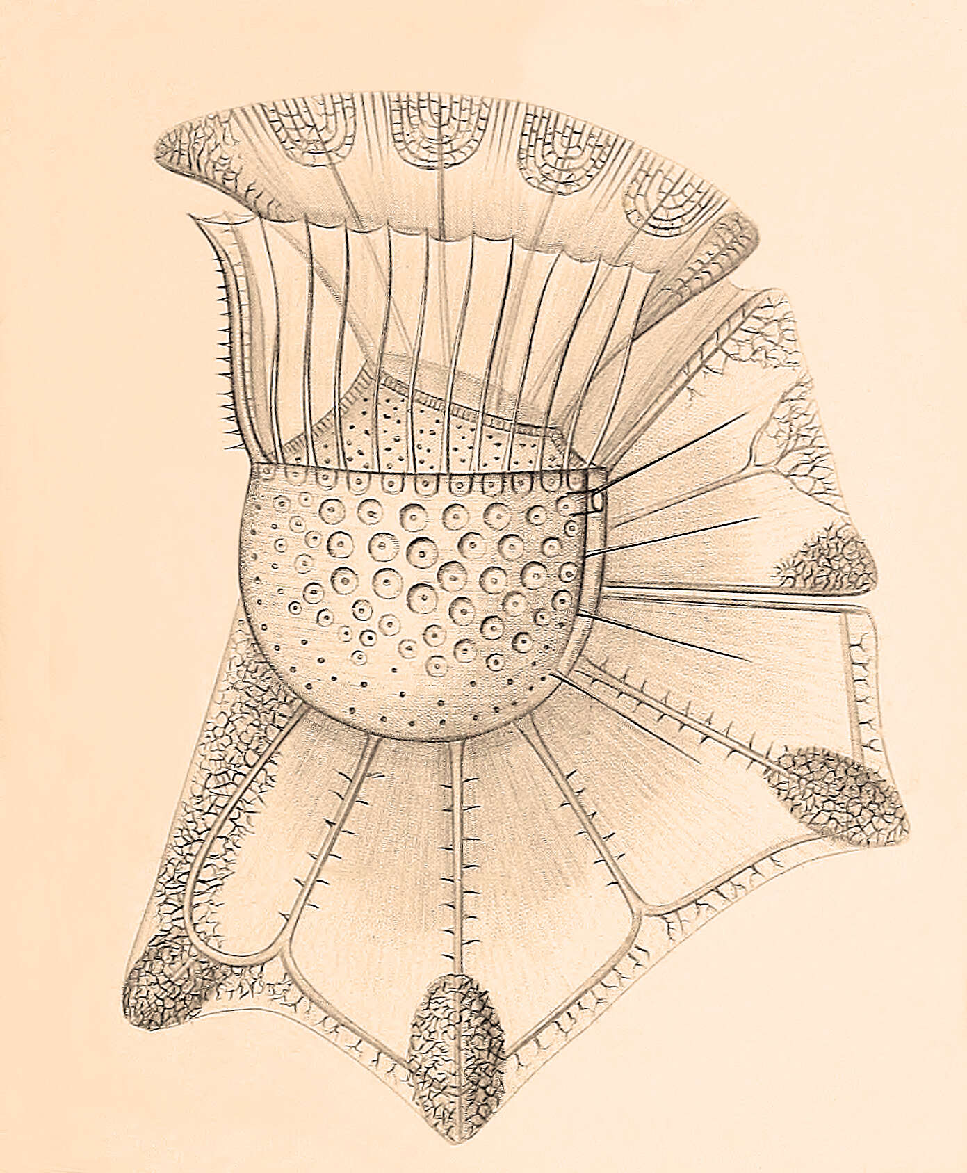
From the original species description: Figure 4 from plate 23 of Stein, F.R. (1883) Der Organismus der Arthrodelen Flagellaten. II. Hlfte. 30 pp & plates.
-
Ornithocercus magnificus from the Bay of Villefranche on Feb 18th 2014. The little orange balls are symbiotic cyanobacteria. Lugol's-fixed specimen, Z-stack of images made using a 40x objective and DIC optics.
-
Dinoflagellate from the Ionian Sea in September 2010. Found in a sample from 74m depth.
-
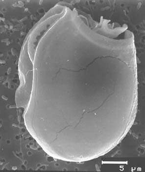
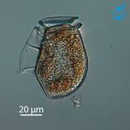
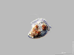
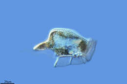
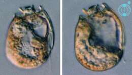
Sinophysis (sine-o-fi/fu-sis) ebriolum (Herdman) Balech 1956. The image on the left shows a cell in right lateral view. The cell is laterally compressed. The epicone is much smaller than the hypocone. The image on the right shows a cell in a mid-focal plane. The cells contain no plastids. The cells are thecate and have cingular lists.
-
Sinophysis ebriolum, scanning electron microscope image. This image was taken by Mona Hoppenrath of a sample from Town Beach, Broome. This work was supported by the Australian Biological Resources Study.
-
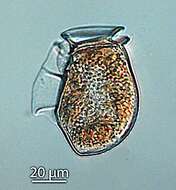
Sinophysis (sine-o-fi/fu-sis) grandis Hoppenrath 2000. The image shows a rectangular cell in left lateral view. The cell is laterally compressed. The epicone is much smaller than the hypocone. The cell contains no plastids, but different coloured food particles are visible. The cell is thecate and has cingular lists.