-
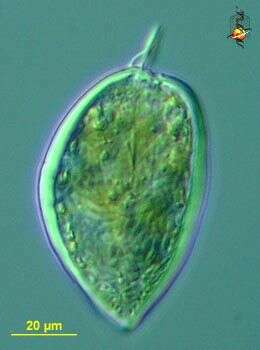
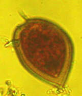
Prorocentrum micans (pro-row-sent-rum my-cans) a marine dinoflagellate, atypical in shape. This genus is associated with toxin production. Differential interference contrast.
-
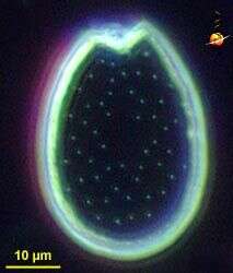
Prorocentrum (pro-row-sen-trum), unusual dinoflagellate with two large valves, often marked within pores which can be used in the identification of species. This image is of a separated plate. Some members of this genus produce toxins. Dark ground illumination.
-
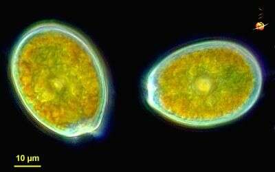
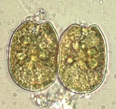
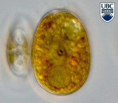
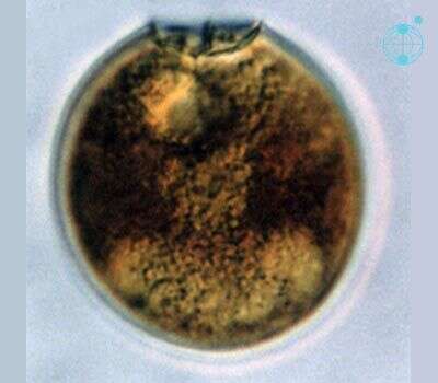
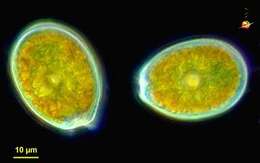
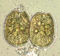
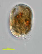
Prorocentrum (pro-row-sen-trum), unusual dinoflagellate with two large valves, often marked with pores which can be used in the identification of species. Two cells, showing the golden colour of the plastids. Dark ground illumination.
-
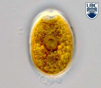
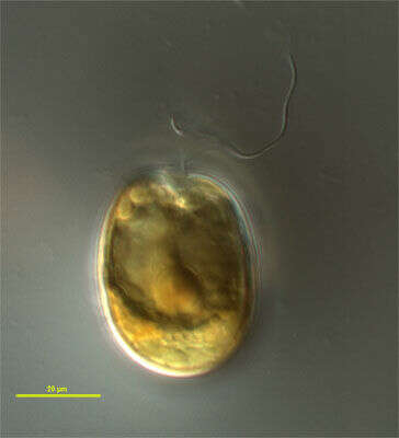
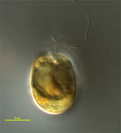
Prorocentrum (pro-row-sen-trum), unusual dinoflagellate with two large valves, which here can seen to be thick skeletal plates. Flagella are located near the apex of the cell. Differential interference microscopy.
-
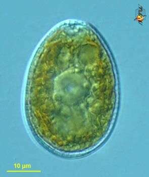
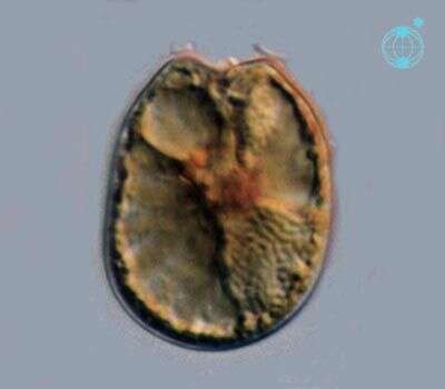
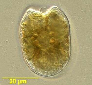
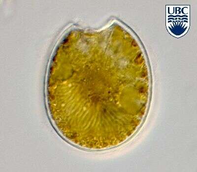
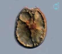
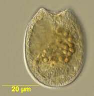
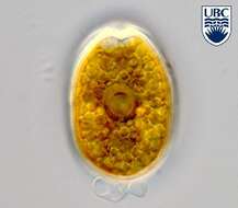
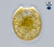
Prorocentrum (pro-row-sent-rum). The image shows one of the two valves of a cell. The cingulum is not visible. The plastid is yellow-brown. There is an apical spine present. The nucleus is in the posterior of the cell.
-
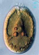
Prorocentrum (pro-row-sent-rum). The image shows one of the two valves of a cell. The cingulum is not visible. The plastid is yellow-brown. There is an apical spine present. The nucleus is in the posterior of the cell.
-
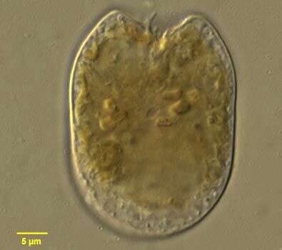
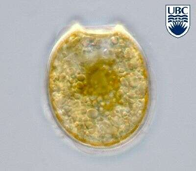
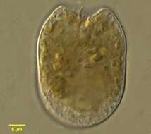
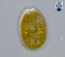
Prorocentrum (pro-row-sent-rum). The image shows one of the two valves of a cell.The nucleus is in the posterior of the cell. The plastids are yellow-brown. The cingulum is not visible.
-
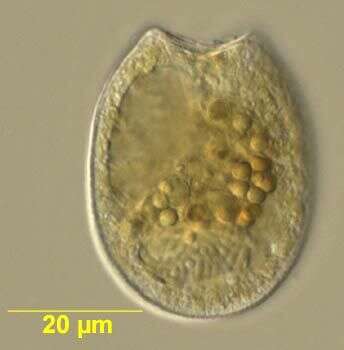
Prorocentrum observed in marine muds and sandy sediments in the vicinity of Broome, Western Australia in September 2003. This image was taken using differential interference contrast optics. This work was supported by the Australian Biological Resources Study.
-
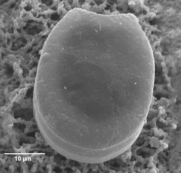
Prorocentrum, observed in marine muds and sandy sediments in the vicinity of Broome, Western Australia in September 2003. This image was taken by scanning electron microscopy. This work was supported by the Australian Biological Resources Study.
-
Prorocentrum observed in marine muds and sandy sediments in the vicinity of Broome, Western Australia in September 2003. This image was taken using differential interference contrast optics. This work was supported by the Australian Biological Resources Study.
-
Prorocentrum observed in marine muds and sandy sediments in the vicinity of Broome, Western Australia in September 2003. This image was taken using differential interference contrast optics. This work was supported by the Australian Biological Resources Study.
-
Prorocentrum observed in marine muds and sandy sediments in the vicinity of Broome, Western Australia in September 2003. This image was taken using differential interference contrast optics. This work was supported by the Australian Biological Resources Study.
-
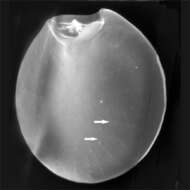
Nomarski interference optics image of one shell of the dinoflagellate.
-
Prorocentrum sp
-
Prorocentrum sp
-
Prorocentrum sp
-
Prorocentrum sp
-
Prorocentrum sp
-
Prorocentrum sp
-
Dinoflagellate from marine sands of Little Sippewisset salt marsh near Woods Hole, MA, USA. Isolated by Laura Wegener Parfrey and Rebecca Zufall.
-
Individual obtained froma bacterial biofilm sample grown out from Sippiwissett Marsh, near Woods Hole Massachusetts. Photo by Bob Moore and Andrew Schurko.
-
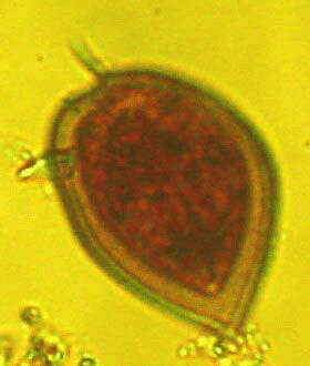
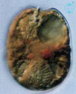
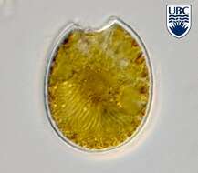
Members of this species have a fusiform body. The apex bears a prominent winged spine. In valve view the cell will have one arched and one convex side.
-
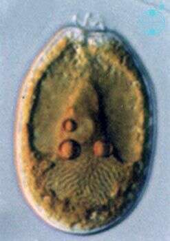
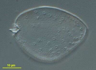
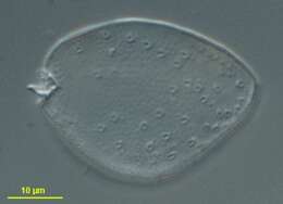
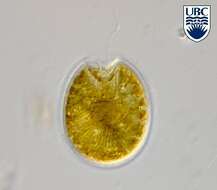
Prorocentrum (pro-row-sent-rum) clipeus Hoppenrath 2000. The image shows one of the two valves of a cell. The nucleus is in the posterior of the cell. The plastids are yellow-brown. The cingulum is not visible. There is an apical spine present.
-
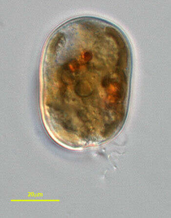
Prorocentrum clipeus cells are round, 37 - 44 long, 35 - 42 microns wide, length to width ratio 1.0-1.1. Apical area a wide rounded indentation, with a small list/collar present to the left side of the apical region. Small spine present, projecting from the apical region. Very small pores, approximately 0.1 microns diameter, present around the periphery of the valve and in short rows radiating towards the centre of the cell. Intercalary region with a horizontal banding pattern. Large yellow-brown plastid fills the cytoplasm. Large extrusomes (12 - 13 microns ) present in the anterior part of the valve, pointing towards the apical area. Nucleus large, approximately 20 microns by 10 microns , in the posterior part of the valve.