-
-
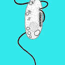
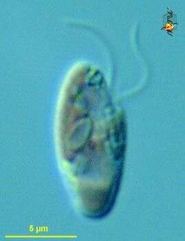
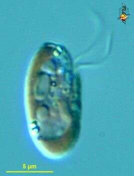
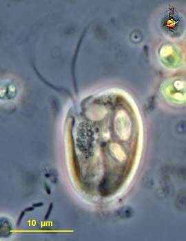
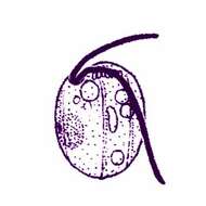
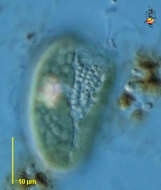
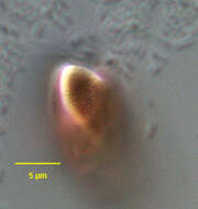
Goniomonas (gop-knee-owe-moan-ass) amphinema. Larsen and Patterson, 1990. Cells are 3 - 9 microns long. Two flagella insert in an anterior lateral pocket, one directed anteriorly, one posteriorly. The two flagella are unequal in length. The long flagellum usually trails over the body and is slightly longer than the cell. The cells are flattened with anterior row of ejectisomes and with or without several longitudinal ridges. The row of ejectisomes is at times difficult to observe. Commonly observed.
-
Goniomonas amphinema Larsen and Patterson, 1990. Cells are 3 - 9 microns long. Two flagella insert in an anterior lateral pocket, one directed anteriorly, one posteriorly. The two flagella are unequal in length. The long flagellum usually trails over the body and is slightly longer than the cell. The cells are flattened with anterior row of ejectisomes and with or without several longitudinal ridges. The row of ejectisomes is at times difficult to observe.
-
-
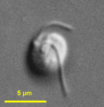
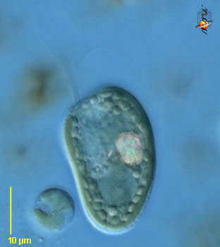
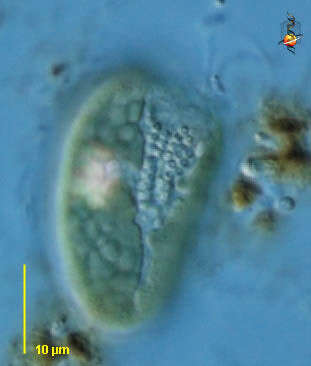
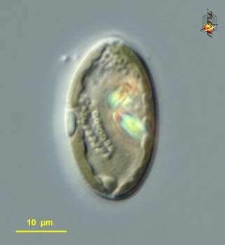
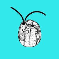
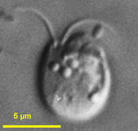
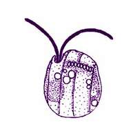
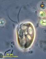
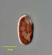
Goniomonas (go-knee-owe-moan-ass) pacifica Larsen and Patterson, 1990. Cells are 4 to 10 microns long and with several distinct longitudinal ridges on both sides of the cell. There is a row of about seven to nine ejectisomes near the anterior end of the cell, which is truncated with the posterior end rounded. Two flagella of similar length emerge from a small anterior depression and are directed anteriorly. When the cells are swimming, two flagella diverge in different directions. Less common than G. amphinema in marine sites.
-
Goniomonas pacifica Larsen and Patterson, 1990. Cells are 4 to 10 microns long and with several distinct longitudinal ridges on both sides of the cell. There is a row of about seven to nine ejectisomes near the anterior end of the cell, which is truncated with the posterior end rounded. Two flagella of similar length emerge from a small anterior depression and are directed anteriorly. When the cells are swimming, the flagella diverge. Less common than G. amphinema. Marine.
-
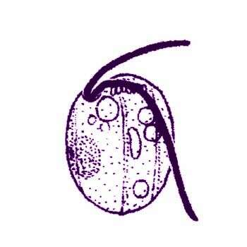
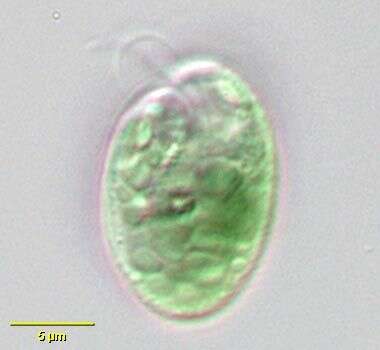
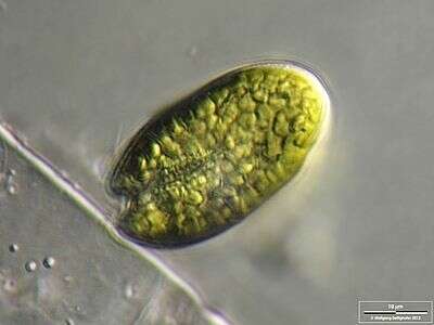
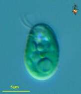
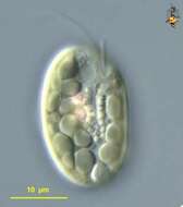
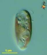
Chroomonas (crow-owe-moan-ass) is one of the cryptomonad flagellates with blue-green plastids. The inclusions are mostly polysaccharide (starch) storage. Two flagella are visible at top (anterior) and the cell surface is irregular because of the small skeletal plates that lie under the cell membrane. With posterior pyrenoid. This may be Chroomonas mesostigmata. Differential Interference Contrast.
data on this strain.
-
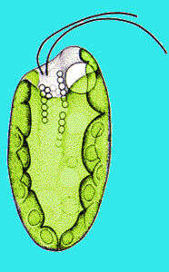
Chroomonas, a blue-green cryptomonad flagellate. Cell is ovoid with two nearly equal-length flagella which emerge from a subapical vestibulum. The flagella can be seen in the invaginated gullet anteriorly. A contractile vacuole is seen adjacent to the gullet on the left. A cup shaped chloroplast contains Cr-phycocyanins, imparting the blue-green color. The periplast of Chroomonas, contains delicate plates sometimes visible on light microscopy as subtle serrations but not seen in this image. Ejectisomes are present along the gullet (small group of refractile granules on the organism's left anteriorly in this image) and beneath the periplast. A stigma is present in some species. Round starch granules are visible peripherally. From temporary rainwater pool near Boise, Idaho. Oblique illumination.
-
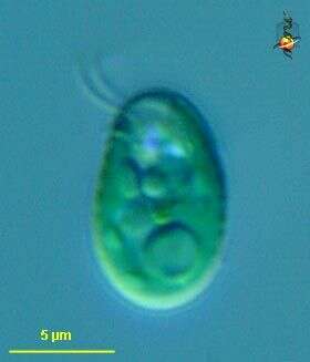
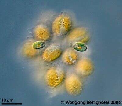
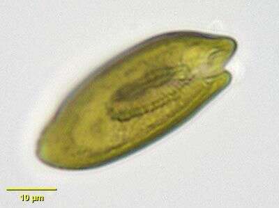
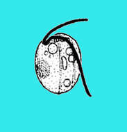
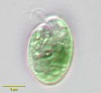
This little cryptomonad was found accompanied by Synura uvella. For details see ZIP archive. Collection from littoral region (stand of Phragmites) of a rain storage reservoir in Kiel (Schleswig-Holstein, Germany). This image was taken using Zeiss Universal with Olympus C7070 CCD camera.
-
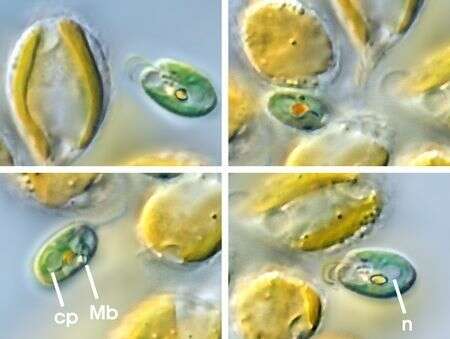
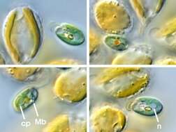
This little cryptomonad was found accompanied by Synura uvella.The picture shows clearly the two flagellae, contractile vacuole, the area of ejectosomes, nucleus (n), stigma, and the pyrenoid covered with amylum (cp). The bright dot represents the Maupas body (Mb). Collection from littoral region (stand of Phragmites) of a rain storage reservoir in Kiel (Schleswig-Holstein, Germany). This image was taken using Zeiss Universal with Olympus C7070 CCD camera.
-
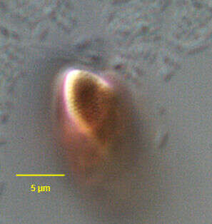
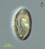
Proteomonas (pro-tea-owe-moan-ass) sulcata, a small cryptophyte = cryptomonad, in which the plastids have a red colour from phycoerythrin pigments. Two flagella emerge from the supapical opening of a broad channel. The channel is underlain with extrusible bodies, ejectisomes. Body with refractile polysaccharide storage materials. Differential interference microscopy.
data on this strain.
-
Cryptomonas (crypt-oh-moan-ass), a common genus of cryptomonad flagellates with chloroplasts. There are two flagella which insert into a groove which opens subapically (the dent near the front of the cell is where is opens). The flagella can be seen upper left. Special extrusomes (ejectisomes) form a little cluster around the flagellar pocket (the pocket can not be seen). Off green chloroplasts, and the orange pink item to the right side is a crystalline inclusion encountered in many cryptomonads. With numerous small polysaccharide inclusions. Differential Interference Contrast.
-
Cryptomonas (crypt-oh-moan-ass), a common genus of cryptomonad flagellates with chloroplasts. There are two flagella which insert into a groove which opens subapically (the dent near the front of the cell is where is opens). The flagella can be seen upper right. Special extrusomes (ejectisomes) form a little cluster around the flagellar pocket (the pocket can not be seen). Two off green chloroplasts, and the orange pink item to the left side is a crystalline inclusion encountered in many cryptomonads. Differential Interference Contrast.
-
Cryptomonas (crypt-oh-moan-ass), a common genus of cryptomonad flagellates with chloroplasts. Special extrusomes (ejectisomes) form a little cluster around the flagellar pocket (the pocket can not be seen). Two off green chloroplasts, and the orange pink item is a crystalline inclusion encountered in many cryptomonads. With numerous small polysaccharide inclusions. Differential Interference Contrast.
-
Cryptomonas (crypt-oh-moan-ass), a common genus of cryptomonad flagellates with chloroplasts. There are two flagella which insert into a groove which opens subapically. Special extrusomes (ejectisomes) form a little cluster around the flagellar pocket . Off green chloroplasts, and the orange pink items are crystalline inclusions encountered in many cryptomonads. With numerous small polysaccharide inclusions. Phase contrast.
-
Cryptomonas (crypt-oh-moan-ass), a common genus of cryptomonad flagellates with chloroplasts. There are two flagella which insert into a groove which opens subapically. Flagella not visible here in this image which emphasises the extrusomes (ejectisomes) that form a little cluster around the flagellar pocket . Off green chloroplasts, and the orange pink items are crystalline inclusions encountered in many cryptomonads. Differential interference contrast.
-
Cryptomonas (crypt-oh-moan-ass), a common genus of cryptomonad flagellates with off green chloroplasts, and the orange pink item out of focus is a crystalline inclusion encountered in many cryptomonads. With numerous small polysaccharide inclusions. Differential interference contrast.
-
-
Cryptomonas, a common cryptomonad flagellate. Prominent extrusomes are seen lining an invaginated canal anteriorly. Two flagella (not well seen in this image) emerge from the canal. The nucleus is seen just posterior to the linear array of extrusomes. This is a relatively large species approximately 45 microns long. From freshwater pond near Boise, Idaho. Brightfield illumination.
-
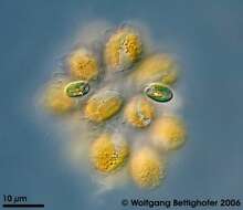
Scale bar indicates 10 µm. Sample from the pond Hegne Moor situated in the vicinity of Lake Constance. The image was built up using several photomicrographic frames with manual stacking technique. Images were taken using Zeiss Universal with Olympus C7070 CCD camera.Image under Creative Commons License V 3.0 (CC BY-NC-SA).
-
Surface detail of the cryptomonad flagellate, Rhodomonas (Karsten,1898). The inner layer of the periplast is composed of overlapping rounded or square proteinaceous organic plates about 0.4 um in diameter. This specimen was collected from a commercial saltwater aquarium in Boise, Idaho, September 2004. DIC.
-
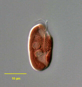
Portrait (lateral view) of the cryptomonad flagellate, Rhodomonas (Karsten,1898). The cells are laterally flattened. The anterior end is obliquely truncate and the posterior rounded. Two subequal flagella insert into a ventral furrow-gullet complex. A single contractile vacuole is seen adjacent to the anterior opening of the ventral furrow. The inner layer of the periplast is composed of overlapping rounded or square proteinaceous organic plates about 0.4 um in diameter. There is a single large boat-shaped chloroplast with a small pyrenoid. Although not always this color, this species is red due to a chloroplast containing Cr-phycoerythrin 545. Like other cryptomonads, ultrastructural studies of Rhodomonas reveal a nucleomorph associated with the plastid. The nucleomorph is thought to represent a nuclear remnant of an ancestral endosymbiotic red alga. The function, if any, of the nucleomorph is unknown. Large ejectosomes are seen here lining the ventral furrow-gullet. Rhodomonas is phototrophic. This genus is found in both freshwater and marine habitats. This specimen was collected from a commercial saltwater aquarium in Boise, Idaho, September 2004. DIC.
-
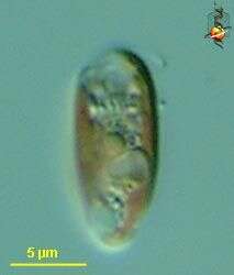
Rhodomonas (row-doe-moan-ass) salina, a cryptomonad / cryptophyte alga, with red coloured plastids, two flagella (not well imaged here) arise in a wide channel which opens near the front of the cell. Differential interference microscopy.
data on this strain.