-
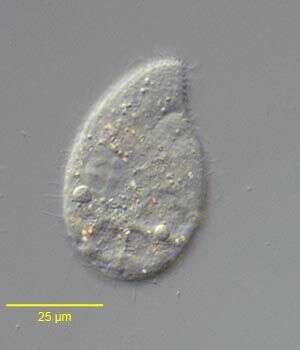
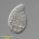
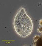
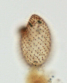
Right lateral view of the colpodid ciliate, Exocolpoda augustini (Foissner, 1987) Foissner, Agatha and Berger, 2002. Foissner erected the family Exocolpodidae based on the life cycle of its members, namely, cell division in free-swimming individuals instead of reproduction in division cysts as seen in the Colpodidae. He felt this life cycle characteristic,the unique boomerang-shaped left oral polykinetid and the unique thick-walled resting cyst of this species warranted its transfer to the new genus, Exocolpoda. The anterior of the cell is cone-shaped and the posterior globular.The small cytostome is in the anterior 1/4 of the cell.There are 25-35 somatic kineties composed of doubly ciliated dikinetids.The right somatic kineties spiral slightly on the long axis to end on the short preoral suture. The left kineties curve more strongly to perpendicularly abut the suture.There are two oral poykinetids. The lekt polykinetid has a unique angulated shape like a boomerang.The macronucleus is spherical.The nucleolus is ribbon-like.There is a single posterior contractile vacuole with a solitary excretory pore.Collected near Boise, Idaho (43°38'21.10"N 116°11'10.78"W elev. 2908 ft.) from an ice-covered temporary puddle containing leaf litter and dead grass.November, 2005.DIC.
-

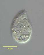
Right lateral view of the colpodid ciliate Exocolpoda augustini (Foissner, 1987) Foissner, Agatha and Berger, 2002.Foissner erected the family Exocolpodidae based on the life cycle of its members, namely, cell division in free-swimming individuals instead of reproduction in division cysts as seen in the Colpodidae. He felt this life cycle characteristic,the unique boomerang-shaped left oral polykinetid and the unique thick-walled resting cyst of this species warranted its transfer to the new genus, exocolpoda. The anterior of the cell is cone-shaped and the posterior globular.The small cytostome is in the anterior 1/4 of the cell.There are 25-35 somatic kineties composed of doubly ciliated dikinetids (the paired cilia are seen well to viewer's right here).The right somatic kineties spiral slightly on the long axis to end on the short preoral suture. The left kineties curve more strongly to perpendicularly abut the suture.There are two oral poykinetids. The left oral polykinetid has a unique angulated shape like a boomerang.The macronucleus is spherical.The nucleolus is ribbon-like.In this specimen the macronucleus has extruded posteriorly during fixation.There is a single posterior contractile vacuole with a solitary excretory pore.Collected near Boise, Idaho (43°38'21.10"N 116°11'10.78"W elev. 2908 ft.) from an ice-covered temporary puddle containing leaf litter and dead grass.November, 2005.Stained by the silver carbonate technique (see Foissner, W. Europ. J. Protistol., 27:313-330;1991).DIC.
-

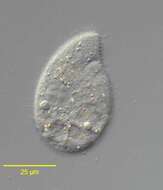
Right lateral view of the colpodid ciliate, Exocolpoda augustini (Foissner, 1987) Foissner, Agatha and Berger, 2002. Foissner erected the family Exocolpodidae based on the life cycle of its members, namely, cell division in free-swimming individuals instead of reproduction in division cysts as seen in the Colpodidae. He felt this life cycle characteristic,the unique boomerang-shaped left oral polykinetid and the unique thick-walled resting cyst of this species warranted its transfer to the new genus, Exocolpoda. The anterior of the cell is cone-shaped and the posterior globular.The small cytostome is in the anterior 1/4 of the cell.There are 25-35 somatic kineties composed of doubly ciliated dikinetids.The right somatic kineties spiral slightly on the long axis to end on the short preoral suture. The left kineties curve more strongly to perpendicularly abut the suture.There are two oral poykinetids. The lekt polykinetid has a unique angulated shape like a boomerang.The macronucleus is spherical.The nucleolus is ribbon-like.There is a single posterior contractile vacuole with a solitary excretory pore.Collected near Boise, Idaho (43°38'21.10"N 116°11'10.78"W elev. 2908 ft.) from an ice-covered temporary puddle containing leaf litter and dead grass.November, 2005.Phase contrast.
-

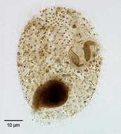
Ventral view of the colpodid ciliate, Exocolpoda augustini (Foissner, 1987) Foissner, Agatha and Berger, 2002.Foissner erected the family Exocolpodidae based on the life cycle of its members, namely, cell division in free-swimming individuals instead of reproduction in division cysts as seen in the Colpodidae. He felt this life cycle characteristic,the unique boomerang-shaped left oral polykinetid and the unique thick-walled resting cyst of this species warranted its transfer to the new genus, Exocolpoda. The anterior of the cell is cone-shaped and the posterior globular.The small cytostome is in the anterior 1/4 of the cell.There are 25-35 somatic kineties composed of doubly ciliated dikinetids (the paired cilia are seen well to viewer's right here).The right somatic kineties spiral slightly on the long axis to end on the short preoral suture. The left kineties curve more strongly to perpendicularly abut the suture.There are two oral poykinetids. The left oral polykinetid has a unique angulated shape like a boomerang.The macronucleus is spherical.The nucleolus is ribbon-like.In this specimen the macronucleus has extruded posteriorly during fixation.There is a single posterior contractile vacuole with a solitary excretory pore.Collected near Boise, Idaho (43°38'21.10"N 116°11'10.78"W elev. 2908 ft.) from an ice-covered temporary puddle containing leaf litter and dead grass.November, 2005.Stained by the silver carbonate technique (see Foissner, W. Europ. J. Protistol., 27:313-330;1991).Brightfield.
-
Ventral view of the colpodid ciliate, Exocolpoda augustini (Foissner, 1987) Foissner, Agatha and Berger, 2002.Foissner erected the family Exocolpodidae based on the life cycle members, namely, cell division in free-swimming individuals instead of reproduction in division cysts as seen in the Colpodidae. He felt this life cycle characteristic,the unique boomerang-shaped left oral polykinetid and the unique thick-walled resting cyst of this species warrented its transfer to the new genus, Exocolpoda. The anterior of the cell is cone-shaped and the posterior globular.The small cytostome is in the anterior 1/4 of the cell.There are 25-35 somatic kineties composed of doubly ciliated dikinetids (the paired cilia are seen well to viewer's right here).The right somatic kineties spiral slightly on the long axis to end on the short preoral suture. The left kineties curve more strongly to perpendicularly abut the suture.There are two oral poykinetids. The left oral polykinetid has a unique angulated shape like a boomerang.The macronucleus is spherical.The nucleolus is ribbon-like.In this specimen the macronucleus has extruded posteriorly during fixation.There is a single posterior contractile vacuole with a solitary excretory pore.Collected near Boise, Idaho (43°38'21.10"N 116°11'10.78"W elev. 2908 ft.) from an ice-covered temporary puddle containing leaf litter and dead grass.November, 2005.Stained by the silver carbonate technique (see Foissner, W. Europ. J. Protistol., 27:313-330;1991).Brightfield.
-
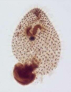
Ventral view of the colpodid ciliate, Exocolpoda augustini (Foissner, 1987) Foissner, Agatha and Berger, 2002.Foissner erected the family Exocolpodidae based on the life cycle of its members, namely, cell division in free-swimming individuals instead of reproduction in division cysts as seen in the Colpodidae. He felt this life cycle characteristic,the unique boomerang-shaped left oral polykinetid and the unique thick-walled resting cyst of this species warranted its transfer to the new genus, Exocolpoda. The anterior of the cell is cone-shaped and the posterior globular.The small cytostome is in the anterior 1/4 of the cell.There are 25-35 somatic kineties composed of doubly ciliated dikinetids (the paired cilia are seen well to viewer's right here).The right somatic kineties spiral slightly on the long axis to end on the short preoral suture. The left kineties curve more strongly to perpendicularly abut the suture.There are two oral poykinetids. The left oral polykinetid has a unique angulated shape like a boomerang.The macronucleus is spherical.The nucleolus is ribbon-like.In this specimen the macronucleus has extruded posteriorly during fixation.There is a single posterior contractile vacuole with a solitary excretory pore.Collected near Boise, Idaho (43°38'21.10"N 116°11'10.78"W elev. 2908 ft.) from an ice-covered temporary puddle containing leaf litter and dead grass.November, 2005.Stained by the silver carbonate technique (see Foissner, W. Europ. J. Protistol., 27:313-330;1991).Brightfield.
-
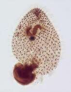
Dorsal infraciliature of the colpodid ciliate, Exocolpoda augustini (Foissner, 1987) Foissner, Agatha and Berger, 2002.Foissner erected the family Exocolpodidae based on the life cycle of its members, namely, cell division in free-swimming individuals instead of reproduction in division cysts as seen in the Colpodidae. He felt this life cycle characteristic,the unique boomerang-shaped left oral polykinetid and the unique thick-walled resting cyst of this species warranted its transfer to the new genus, Exocolpoda. The anterior of the cell is cone-shaped and the posterior globular.The small cytostome is in the anterior 1/4 of the cell.There are 25-35 somatic kineties composed of doubly ciliated dikinetids (the paired cilia are seen well to viewer's right here).The right somatic kineties spiral slightly on the long axis to end on the short preoral suture. The left kineties curve more strongly to perpendicularly abut the suture.There are two oral poykinetids. The left oral polykinetid has a unique angulated shape like a boomerang.The macronucleus is spherical.The nucleolus is ribbon-like.In this specimen the macronucleus has extruded posteriorly during fixation.There is a single posterior contractile vacuole with a solitary excretory pore.Collected near Boise, Idaho (43°38'21.10"N 116°11'10.78"W elev. 2908 ft.) from an ice-covered temporary puddle containing leaf litter and dead grass.November, 2005.Stained by the silver carbonate technique (see Foissner, W. Europ. J. Protistol., 27:313-330;1991).Brightfield.
-
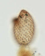
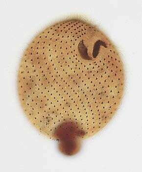
Dorsal silverline system of of the colpodid ciliate, Exocolpoda augustini (Foissner, 1987) Foissner, Agatha and Berger, 2002. Foissner erected the family Exocolpodidae based on the life cycle of its members, namely, cell division in free-swimming individuals instead of reproduction in division cysts as seen in the Colpodidae. He felt this life cycle characteristic,the unique boomerang-shaped left oral polykinetid and the unique thick-walled resting cyst of this species warranted its transfer to the new genus, Exocolpoda. The silverline system is of the "cucullus" type.Collected near Boise, Idaho (43°38'21.10"N 116°11'10.78"W elev. 2908 ft.) from aan ice-covered temporary puddle containing leaf litter and dead grass.November, 2005.Stained by the dry silver nitrate technique (see Foissner, W. Europ. J. Protistol., 27:313-330;1991).Brightfield.
-
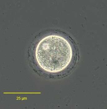
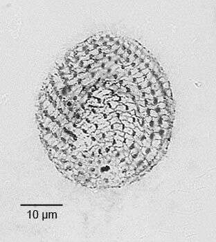
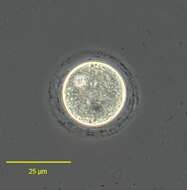
Resting cyst of the colpodid ciliate, Exocolpoda augustini (Foissner, 1987) Foissner, Agatha and Berger, 2002. Foissner erected the family Exocolpodidae based on the life cycle of its members, namely, cell division in free-swimming individuals instead of reproduction in division cysts as seen in the Colpodidae. He felt this life cycle characteristic,the unique boomerang-shaped left oral polykinetid and the unique thick-walled resting cyst of this species warranted its transfer to the new genus, Exocolpoda. The cyst is of the "cucullus" type the wall of which consists of closely spaced membranes.The cyst wall is further differentiated into a thin meso- and endocyst layer and the much thicker ectocyst layer. Dark granular material is scattered between the membranes of the ectocyst. Foissner believes the thick walled cyst may be an adaptation to the desert and semi-desert habitats where this species is most often found.Collected near Boise, Idaho (43°38'21.10"N 116°11'10.78"W elev. 2908 ft.) from aan ice-covered temporary puddle containing leaf litter and dead grass.November, 2005.Phase contrast.