-
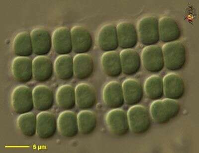
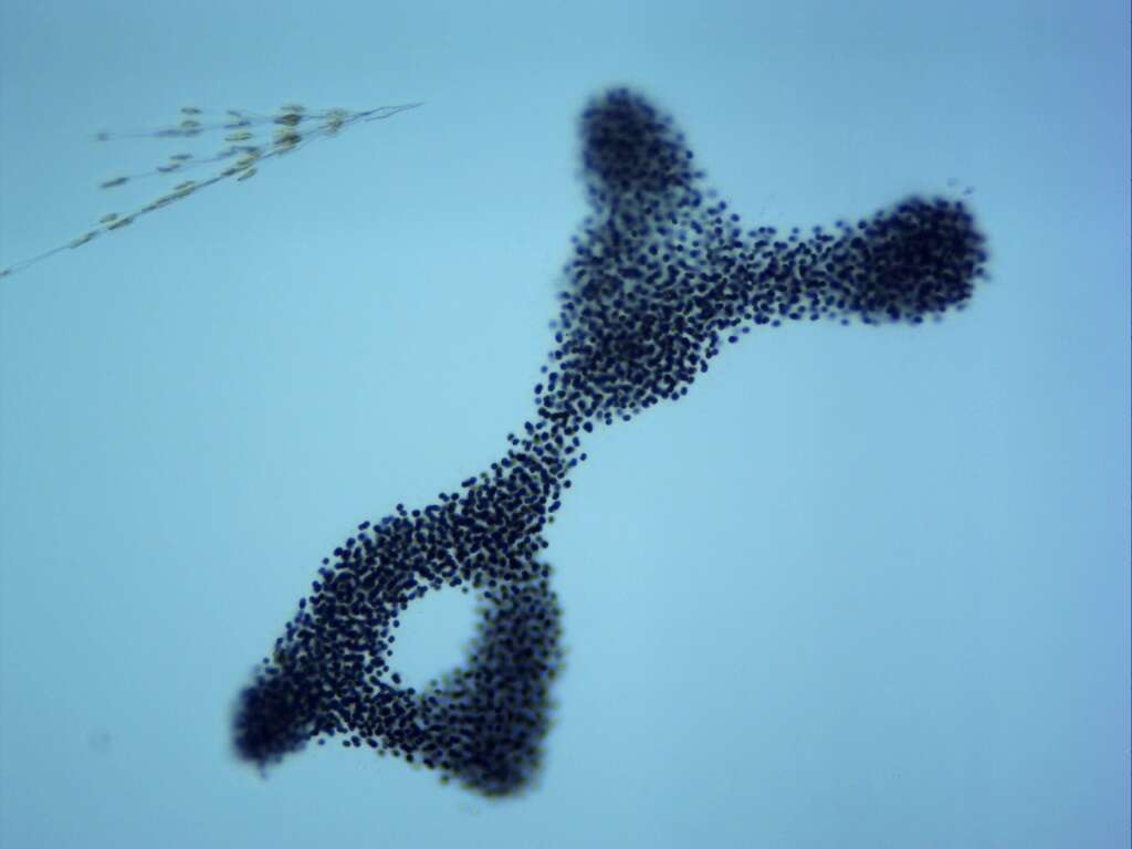
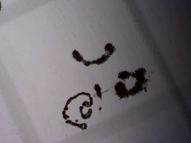
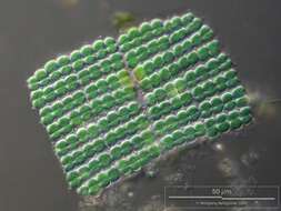
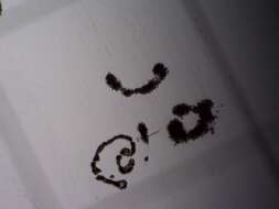
Merismopedia (mer-is-mo-pea-dee-a) is a blue-green alga or cyanobacterium. The genus is distinguished by the square-packed pattern of the coccoid cells. It is common in sediments, but the number of cells which occur in a colony may vary from only a few to thousands. As a cyanobacterium, the photosynthetic pigments are located throughout the cytoplasm. Differential interference contrast
-
Merismopedia. Colony observed in sandy and muddy marine sediments in the vicinity of Broome, Western Australia in September 2003. This image was taken using differential interference contrast optics. This work was supported by the Australian Biological Resources Study.
-
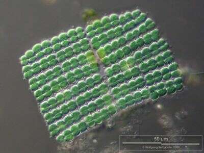
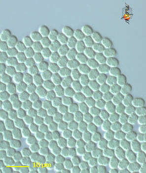
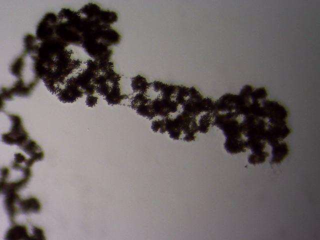
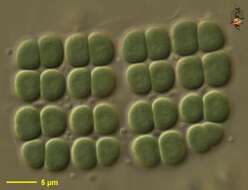
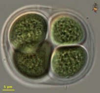
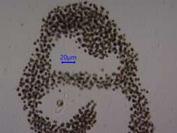
The cells of the bluegreen alga (or cyanobacteria) Merismopedia are arranged in a square plate lying in a mucilaginous envelope. Collected from Bodden, the brackish waters lying between the isles of Hiddensee and Ruegen (German Baltic Sea). This image was taken using Zeiss Universal with Olympus C7070 CCD camera.
-
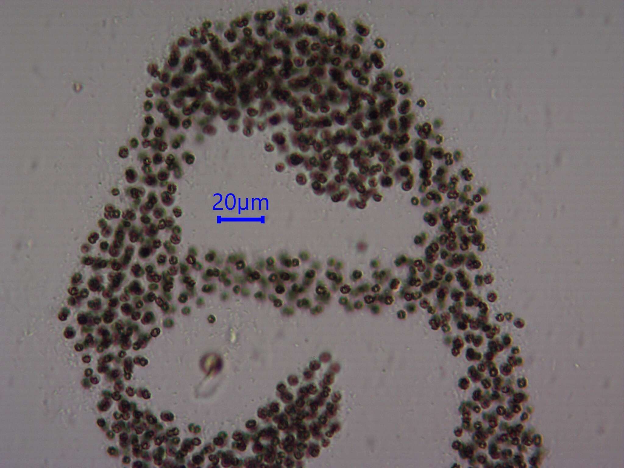
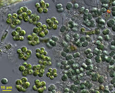
Scale bar indicates 50 µm. Sample from a wetland at the Pillersee (Tyrol, Austria). The image was built up using several photomicrographic frames with manual stacking technique. Images were taken using Zeiss Universal with Olympus C7070 CCD camera.Image under Creative Commons License V 3.0 (CC BY-NC-SA).
-
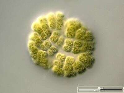
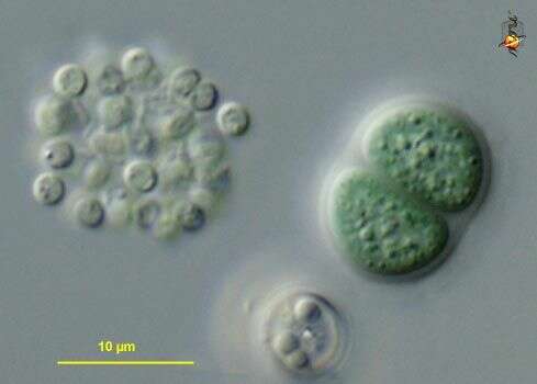
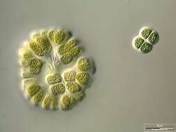
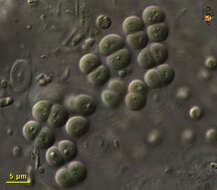
Colony accompanied by Chroococcus turgidus. Scale bar indicates 25 µm. Sample from a wetland at the Pillersee (Tyrol, Austria). The image was built up using several photomicrographic frames with manual stacking technique. Images were taken using Zeiss Universal with Olympus C7070 CCD camera.Image under Creative Commons License V 3.0 (CC BY-NC-SA).
-
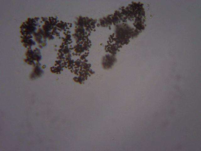
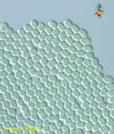
Scale bar indicates 25 µm. Sample from sphagnum pond situated in the northern alpine region of Austria near Salzburg. Images were taken using Zeiss Universal with Olympus C7070 CCD camera.
-
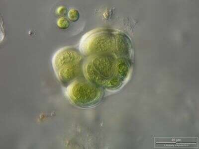
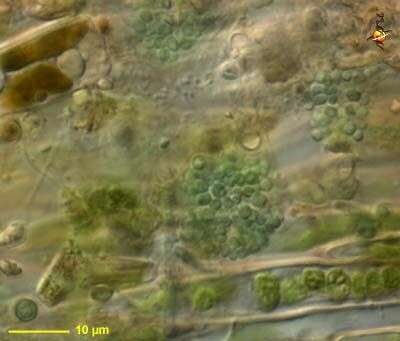
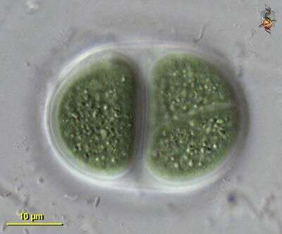
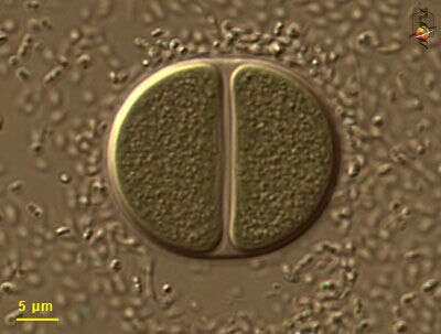
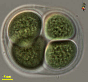
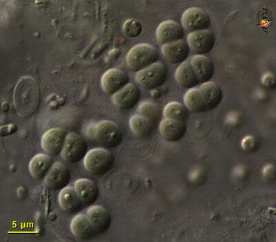
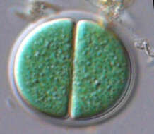
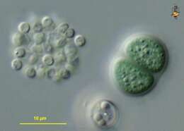
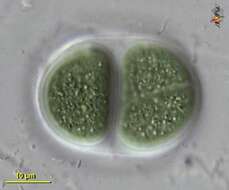
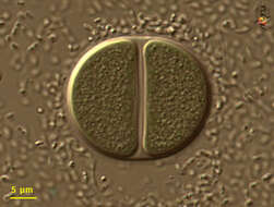
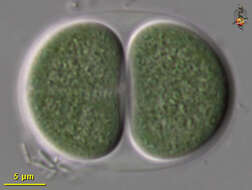
Chroococcus (crow-o-cock-us), large cyanobacterium, typically two (but sometimes one) cells enclosed within a mucus sheath. Photosynthetic pigment distributed through cytoplasm, which may have a granular texture, but does not have subcompartments (organelles). Differential interference contrast.
-
Chroococcus (crow-owe-cock-us) coccoid cyanobacteria, adhering to each other to form extensive flat sheets, no evident mucus sheath or heterocysts. Differential Interference Contrast.
-
Chroococcus (crow-o-cock-us), large cyanobacterium, typically two (but sometimes one) cells enclosed within a mucus sheath. Photosynthetic pigment distributed through cytoplasm, which may have a granular texture, but does not have subcompartments (organelles). Differential interference contrast.
-
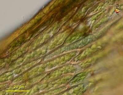
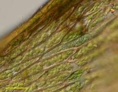
Chroococcus (crow-owe-cock-us) tentative identification. Coccoid blue green algal cells. Found as one of several cyanobacterial epibionts on the leaves of the moss Hygrohypnum, a site which seems to be a focus for nitrogen fixation. In this case the cyanobacterial cells have occupied one of the cortical cells of the plant. Differential interference contrast.
-
Chroococcus (crow-owe-cock-us) tentative identification. Coccoid blue green algal cells. Found as one of several cyanobacterial epibionts on the leaves of the moss Hygrohypnum, a site which seems to be a focus for nitrogen fixation. Differential interference contrast.
-
Chroomonas. Cell observed in freshwater sediments in the vicinity of Broome, Western Australia in September 2003. This image was taken using differential interference contrast optics. Â Â This work was supported by the Australian Biological Resources Study.
-
Variously sized individuals - some species have been reported with at least a five fold size range - so these may be of a single species. Nomarksi, diferential interference contrast optics.
-
Large blue-green algal (cyanobacterial) cells in mucus sheath. Differential interference contrast optics.
-
Blue green algae in a mucus sheath. Differential interference contrast optics.
-
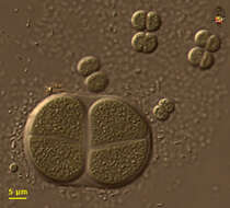
Most Chroococcus cells are quite large. Some empty capsules can be seen to the right of the living cells. Differential interefence contrast optics.
-
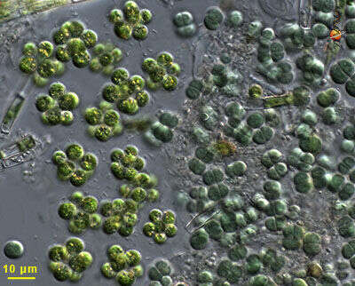
The chroococcus cells are to the right, and the cells of a similar size but a brighter green colour to the left are cells of an unidentified chlorophycean green alga (a eukaryote). This image illustrates the blue-green colour from which the blue-green algae get their name. Differential interference contrast optics.
-
Typical arrangement with paired cells inside a mucus sheath. Differential interference contrast optics.
-
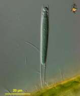
Chamaesiphon (ca-may-sigh-phon) (also known as Entophysalis) elongate cyanobacterium with spores forming at the distal end, basal part with mucus sheath. Found as one of several cyanobacterial epibionts on the leaves of the moss Hygrohypnum, a site which seems to be a focus for nitrogen fixation. Differential interference contrast.
-
-
-
-
-