-
Paraquadrula irregularis The specimen was gathered in a well at Wendelinusplatz in 63820 Elsenfeld (Germany). Copyright Winfried Hölz, Hausen, Germany.Images were taken using Zeiss Axioskop with DSLR.Image under Creative Commons License V 3.0 (CC BY-NC-SA). Place name: Well at Wendelinusplatz in 63820 Elsenfeld (Germany) Latitude: 49.8469263 Longitude: 9.1611486 Probe aus einem Brunnen am Wendelinusplatz in63820 Elsenfeld.Copyright Winfried Hölz, Hausen. Mikrotechnik: Zeiss Axioskop, Kamera: DSLR. Creative Commons License V 3.0 (CC BY-NC-SA). For permission to use of (high-resolution) images please contact postmaster@protisten.de.
-
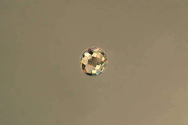
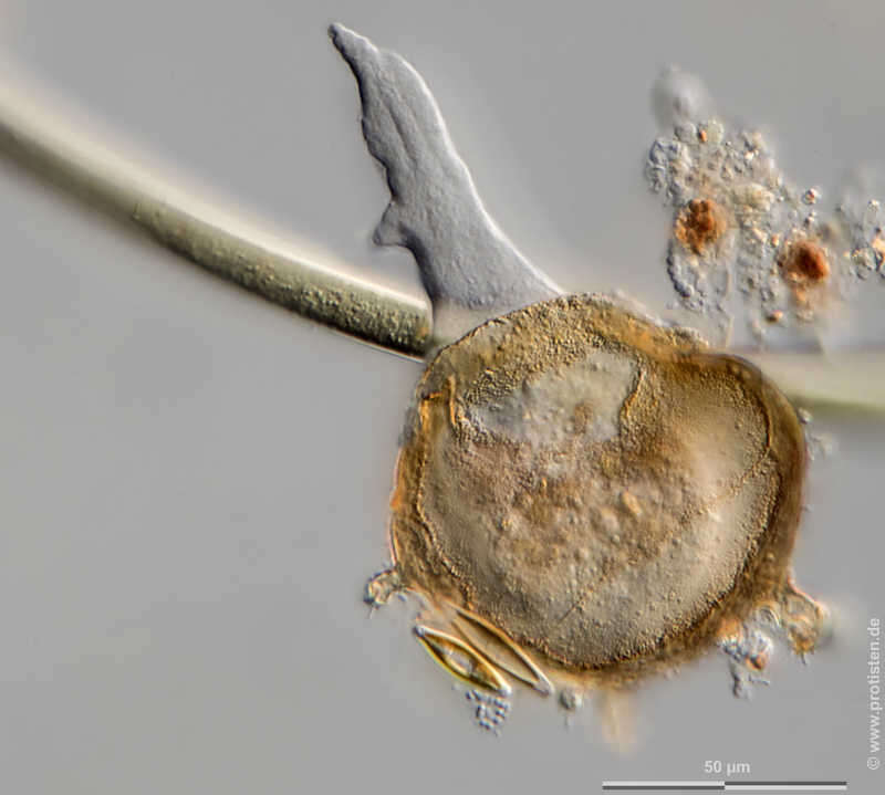

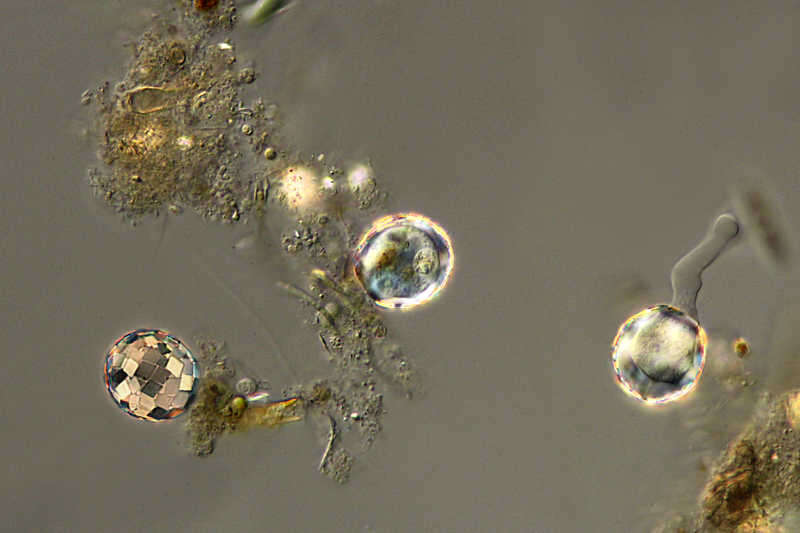
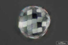
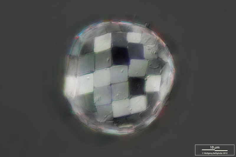
Paraquadrula irregularis Paraquadrula builds its scales from calcite, which is birefringent. This is the reason why in DIC (polarized light) they show such different magnitudes. Scale bar indicates 10 µm. Sample from a bog near Reith/Pillersee (Tyrol, Austria). The image was built up using several photomicrographic frames with manual stacking technique. Images were taken using Zeiss Axioplan with Canon 600D CCD camera.Image under Creative Commons License V 3.0 (CC BY-NC-SA). Place name: Bog near Reith/Pillersee (Tyrol, Austria) Latitude: 47.477767 Longitude: 12.337294 Paraquadrula baut seine Schuppen aus Calcit, welches doppelbrechend ist. Dies ist der Grund, warum sie im DIC (polarisiertes Licht) solche unterschiedlichen Helligkeiten haben. Multiebenen-Abbildung, manuell gestapelt. Der Messbalken markiert eine Länge von 10 µm. Probe aus einem Niedermoor bei Reith/Pillersee in Tirol. Mikrotechnik: Zeiss Axioplan, Kamera: Canon 600D. Creative Commons License V 3.0 (CC BY-NC-SA). For permission to use of (high-resolution) images please contact postmaster@protisten.de.
-
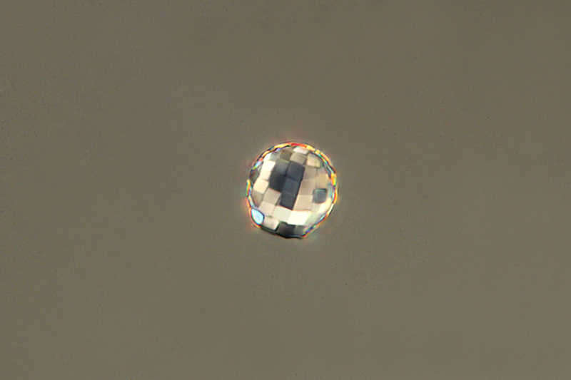
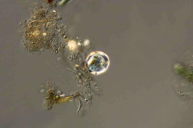
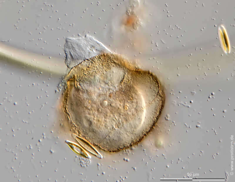
Paraquadrula irregularis The specimen was gathered in a well at Wendelinusplatz in 63820 Elsenfeld (Germany). Copyright Winfried Hölz, Hausen, Germany.Images were taken using Zeiss Axioskop with DSLR.Image under Creative Commons License V 3.0 (CC BY-NC-SA). Place name: Well at Wendelinusplatz in 63820 Elsenfeld (Germany) Latitude: 49.8469263 Longitude: 9.1611486 Probe aus einem Brunnen am Wendelinusplatz in63820 Elsenfeld.Copyright Winfried Hölz, Hausen. Mikrotechnik: Zeiss Axioskop, Kamera: DSLR. Creative Commons License V 3.0 (CC BY-NC-SA). For permission to use of (high-resolution) images please contact postmaster@protisten.de.
-
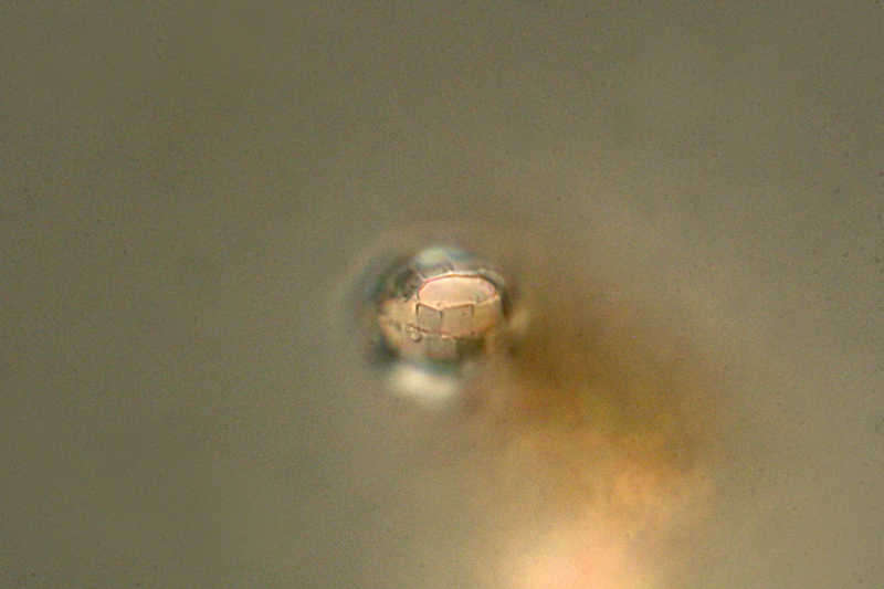
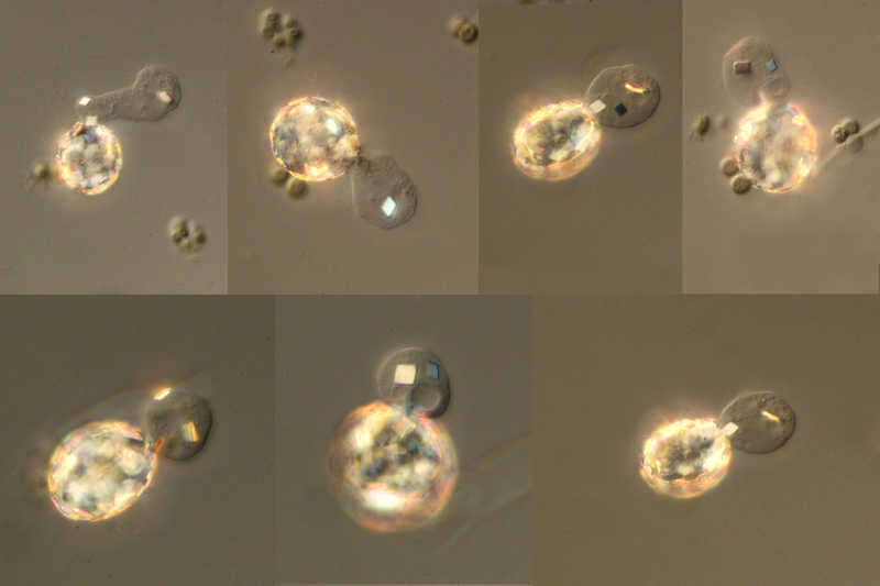
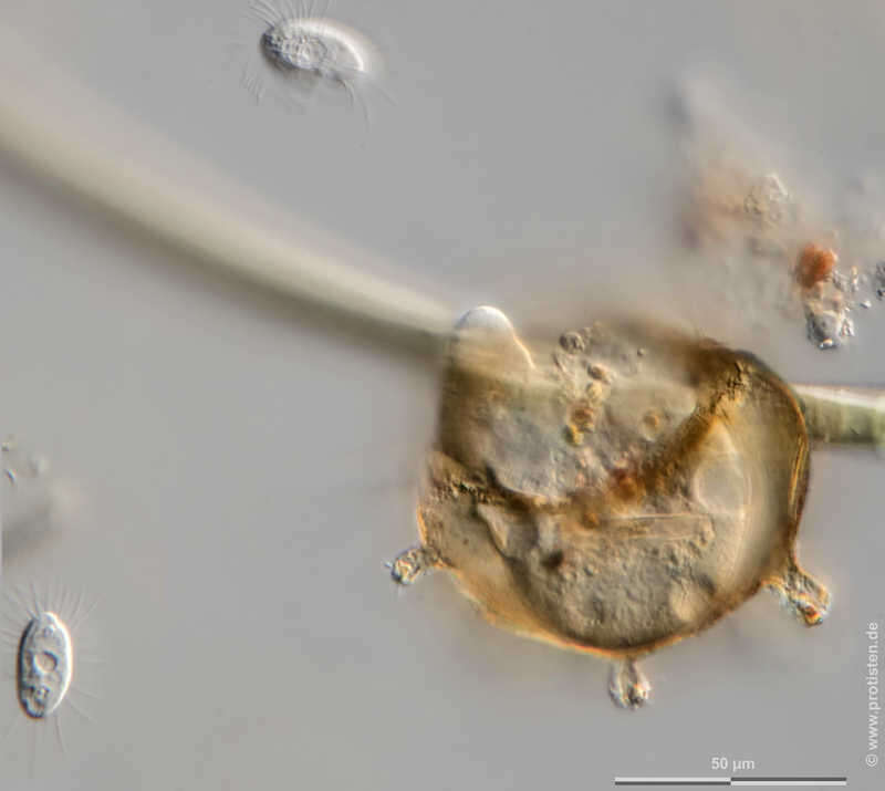
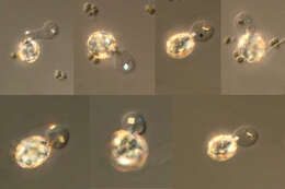
Paraquadrula irregularis Process of binary fission of Paraquadrula irregularis. The specimen was gathered in a well at Wendelinusplatz in 63820 Elsenfeld (Germany). Copyright Winfried Hölz, Hausen, Germany.Images were taken using Zeiss Axioskop with DSLR.Image under Creative Commons License V 3.0 (CC BY-NC-SA). Place name: Well at Wendelinusplatz in 63820 Elsenfeld (Germany) Latitude: 49.8469263 Longitude: 9.1611486 Teilungsvorgang bei Paraquadrula irregularis. Probe aus einem Brunnen am Wendelinusplatz in63820 Elsenfeld.Copyright Winfried Hölz, Hausen. Mikrotechnik: Zeiss Axioskop, Kamera: DSLR. Creative Commons License V 3.0 (CC BY-NC-SA). For permission to use of (high-resolution) images please contact postmaster@protisten.de.
-
Paraquadrula irregularis Paraquadrula builds its scales from calcite, which is birefringent. This is the reason why in DIC (polarized light) they show such different magnitudes. Scale bar indicates 10 µm. Sample from a bog near Reith/Pillersee (Tyrol, Austria). The image was built up using several photomicrographic frames with manual stacking technique. Images were taken using Zeiss Axioplan with Canon 600D CCD camera.Image under Creative Commons License V 3.0 (CC BY-NC-SA). Place name: Bog near Reith/Pillersee (Tyrol, Austria) Latitude: 47.477767 Longitude: 12.337294 Paraquadrula baut seine Schuppen aus Calcit, welches doppelbrechend ist. Dies ist der Grund, warum sie im DIC (polarisiertes Licht) solche unterschiedlichen Helligkeiten haben. Multiebenen-Abbildung, manuell gestapelt. Der Messbalken markiert eine Länge von 10 µm. Probe aus einem Niedermoor bei Reith/Pillersee in Tirol. Mikrotechnik: Zeiss Axioplan, Kamera: Canon 600D. Creative Commons License V 3.0 (CC BY-NC-SA). For permission to use of (high-resolution) images please contact postmaster@protisten.de.
-
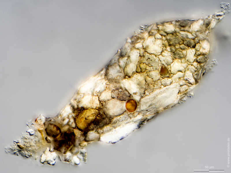
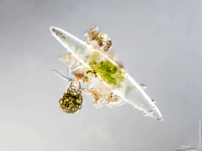
Cylindrifflugia acuminata Synonym: Difflugia acuminata.Scale bar indicates 50 µm. Sample from a pond near Großostheim, Germany. Sampling date 04/2021. The image was built up using several photomicrographic frames with manual stacking technique. Images were taken using Zeiss Axioplan with Olympus OM-D M5 MKII. Image under Creative Commons License V 3.0 (CC BY-NC-SA). Place name: Pond near Großostheim (Germany) Latitude: 49.88482168 Longitude: 9.09980822 Synonym: Difflugia acuminata.Multiebenen-Abbildung, manuell gestapelt. Der Messbalken markiert eine Länge von 50 µm. Probe aus einem Waldteich bei Großostheim. Datum der Aufsammlung: 04/2021. Mikrotechnik: Zeiss Axioplan, Kamera: Olympus OM-D M5 MKII. Creative Commons License V 3.0 (CC BY-NC-SA). For permission to use of (high-resolution) images please contact postmaster@protisten.de.
-
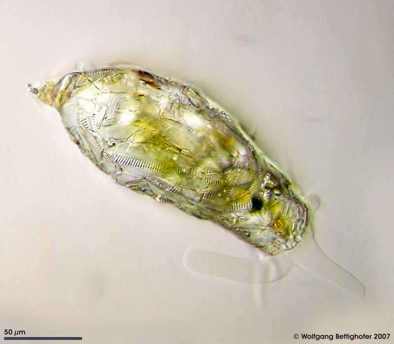
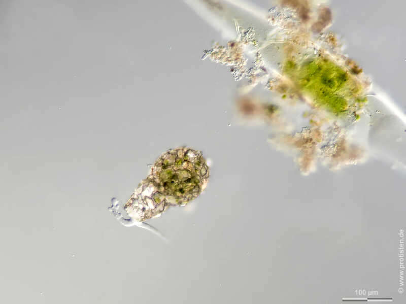
Cylindrifflugia acuminata Synonym: Difflugia acuminata.The xenosoms in the test of Cylindrifflugia acuminata , which originate from pennate diatoms, are clearly visible. The picture was built up using 13 brightfield frames with a manual stacking technique. The specimen was gathered in a tiny freshwater pond at the island of Hiddensee (Baltic Sea, Germany) which shows a fascinating biodiversity of naked and testate amoebae. Images were taken using Zeiss Standard with Olympus C7070 CCD camera.Image under Creative Commons License V 3.0 (CC BY-NC-SA). Place name: Pond Suploch, Hiddensee (Germany) Latitude: 54.538638 Longitude: 13.097802 Synonym: Difflugia acuminata.Die Xenosomen (Fremdkörper) in der Schale von Cylindrifflugia acuminata, die von pennaten Diatomeen stammen, sind deutlich sichtbar. Tiefenschärfe durch Multiebenenabbildung aus 13 Bildebenen, manuell gestapelt. Probe aus einem kleinen Süßwasserteich auf der Insel Hiddensee, welcher eine faszinierende Vielfalt von nackten und beschalten Amöben beherbergt. Mikrotechnik: Zeiss Universal, Kamera: Olympus C7070. Creative Commons License V 3.0 (CC BY-NC-SA). For permission to use of (high-resolution) images please contact postmaster@protisten.de.
-
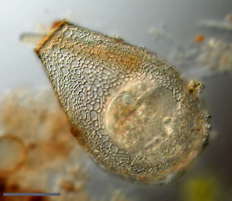
Nebela collaris This picture shows details of the Nebela test. In order to build the test, Nebela collects silicious scales from the stocks of the ingested smaller testate amoebae. The picture was developed using 14 high resolution DIC frames with manual stacking technique. The scale bar indicates 50 µm. Sample from spagnum pond situated in the northern alpine region of Austria near Salzburg. Images were taken using Zeiss Universal with Olympus C7070 CCD camera.Image under Creative Commons License V 3.0 (CC BY-NC-SA). Place name: Bogs near Salzburg (Austria) Latitude: 48.068516 Longitude: 12.954134 Dieses Bild zeigt Details der Nebela-Schale. Um die Schale aufzubauen, sammelt Nebela Kieselschuppen aus den Beständen der aufgenommenen kleineren Schalenamöben. Tiefenschärfe durch Multiebenenabbildung aus 14 Bildebenen, manuell gestapelt. Der Messbalken markiert eine Länge von 25 µm. Probe aus einem Moor in den nördlichen Kalkalpen von Österreich in der Nähe von Salzburg. Mikrotechnik: Zeiss Universal, Kamera: Olympus C7070. Creative Commons License V 3.0 (CC BY-NC-SA). For permission to use of (high-resolution) images please contact postmaster@protisten.de.
-
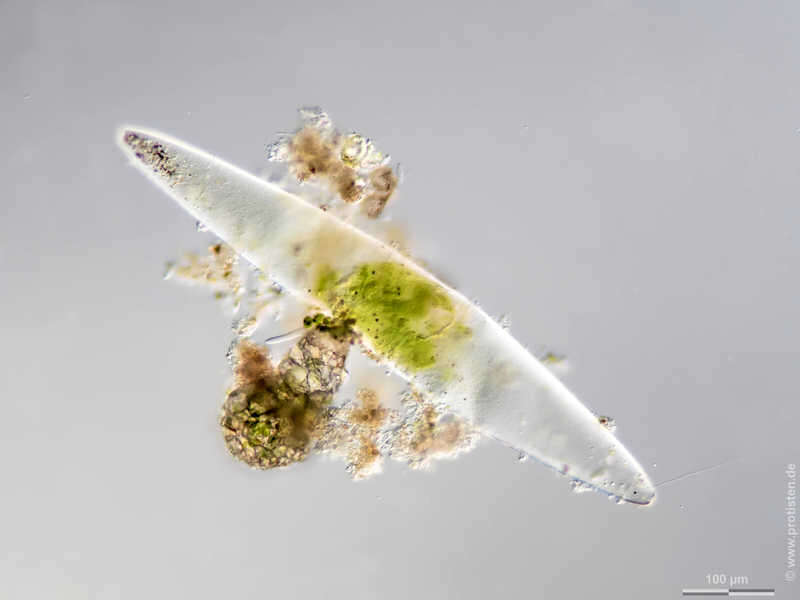
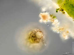
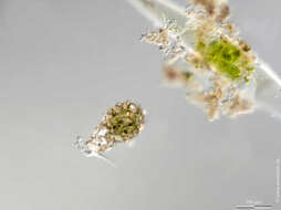
Zivkovicia spectabilis Scale bar indicates 100 µm. Testate ameba feeding on Closterium. Zivkovicia actively penetrates the cell wall of Closterium.The specimen was gathered in a little creek near Rossbach (Lower Franconia, Germany). Sampling date 7/2020 The image was built up using several photomicrographic frames with manual stacking technique. Images were taken using Zeiss Axioplan with Olympus OM-D M5 MKII. Image under Creative Commons License V 3.0 (CC BY-NC-SA). Place name: Creek in 63649 Rossbach (Lower Franconia, Germany) Latitude: 49.87747709 Longitude: 9.23824668 Schalenamöbe frisst Zieralge. Zivkovicia dringt aktiv in die Zellwand von Closterium ein.Multiebenen-Abbildung, manuell gestapelt. Der Messbalken markiert eine Länge von 100/50 µm. Probe aus einem kleinen Bach bei Rossbach (Unterfranken). Datum der Aufsammlung: 7/2020. Mikrotechnik: Zeiss Axioplan, Kamera: Olympus OM-D M5 MKII. Creative Commons License V 3.0 (CC BY-NC-SA). For permission to use of (high-resolution) images please contact postmaster@protisten.de.
-
Zivkovicia spectabilis Scale bar indicates 50 µm. The specimen was gathered in a little creek near Rossbach (Lower Franconia, Germany). Sampling date 7/2020 The image was built up using several photomicrographic frames with manual stacking technique. Images were taken using Zeiss Axioplan with Olympus OM-D M5 MKII. Image under Creative Commons License V 3.0 (CC BY-NC-SA). Place name: Creek in 63649 Rossbach (Lower Franconia, Germany) Latitude: 49.87747709 Longitude: 9.23824668 Multiebenen-Abbildung, manuell gestapelt. Der Messbalken markiert eine Länge von 50 µm. Probe aus einem kleinen Bach bei Rossbach (Unterfranken). Datum der Aufsammlung: 7/2020. Mikrotechnik: Zeiss Axioplan, Kamera: Olympus OM-D M5 MKII. Creative Commons License V 3.0 (CC BY-NC-SA). For permission to use of (high-resolution) images please contact postmaster@protisten.de.
-
Zivkovicia spectabilis Scale bar indicates 100 µm. The specimen was gathered in a little creek near Rossbach (Lower Franconia, Germany). Sampling date 7/2020 The image was built up using several photomicrographic frames with manual stacking technique. Images were taken using Zeiss Axioplan with Olympus OM-D M5 MKII. Image under Creative Commons License V 3.0 (CC BY-NC-SA). Place name: Creek in 63649 Rossbach (Lower Franconia, Germany) Latitude: 49.87747709 Longitude: 9.23824668 Multiebenen-Abbildung, manuell gestapelt. Der Messbalken markiert eine Länge von 100 µm. Probe aus einem kleinen Bach bei Rossbach (Unterfranken). Datum der Aufsammlung: 7/2020. Mikrotechnik: Zeiss Axioplan, Kamera: Olympus OM-D M5 MKII. Creative Commons License V 3.0 (CC BY-NC-SA). For permission to use of (high-resolution) images please contact postmaster@protisten.de.
-
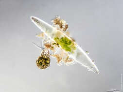
Zivkovicia spectabilis Scale bar indicates 100 µm. Testate ameba feeding on Closterium. Zivkovicia actively penetrates the cell wall of Closterium.The specimen was gathered in a little creek near Rossbach (Lower Franconia, Germany). Sampling date 7/2020 The image was built up using several photomicrographic frames with manual stacking technique. Images were taken using Zeiss Axioplan with Olympus OM-D M5 MKII. Image under Creative Commons License V 3.0 (CC BY-NC-SA). Place name: Creek in 63649 Rossbach (Lower Franconia, Germany) Latitude: 49.87747709 Longitude: 9.23824668 Schalenamöbe frisst Zieralge. Zivkovicia dringt aktiv in die Zellwand von Closterium ein.Multiebenen-Abbildung, manuell gestapelt. Der Messbalken markiert eine Länge von 100/50 µm. Probe aus einem kleinen Bach bei Rossbach (Unterfranken). Datum der Aufsammlung: 7/2020. Mikrotechnik: Zeiss Axioplan, Kamera: Olympus OM-D M5 MKII. Creative Commons License V 3.0 (CC BY-NC-SA). For permission to use of (high-resolution) images please contact postmaster@protisten.de.
-
Centropyxis spinosa Scale bar indicates 50 µm. Sample from a tropical freshwater aquarium. The image was built up using several photomicrographic frames with manual stacking technique. Images were taken using Zeiss Axioplan with Olympus OM-D M5 MKII.Image under Creative Commons License V 3.0 (CC BY-NC-SA). Place name: Tropical freshwater aquarium Latitude: 54.3018013 Longitude: 10.07120132 Multiebenen-Abbildung, manuell gestapelt. Der Messbalken markiert eine Länge von 50 µm. Probe aus einem Süßwasseraquarium für tropische Fische. Mikrotechnik: Zeiss Axioplan, Kamera: Olympus OM-D M5 MKII. Creative Commons License V 3.0 (CC BY-NC-SA). For permission to use of (high-resolution) images please contact postmaster@protisten.de.
-
Centropyxis spinosa The scale bar indicates 50 µm. Sample from a tropical freshwater aquarium. The image was built up using several photomicrographic frames with manual stacking technique. The images were taken using Zeiss Axioplan with Canon EOS 70D.Image under Creative Commons License V 3.0 (CC BY-NC-SA). Place name: Tropical freshwater aquarium Latitude: 54.3018013 Longitude: 10.07120132 Multiebenen-Abbildung, manuell gestapelt. Der Messbalken markiert eine Länge von 50 µm. Probe aus einem Süßwasseraquarium für tropische Fische. Mikrotechnik: Zeiss Axioplan, Kamera: Canon EOS 70D. Creative Commons License V 3.0 (CC BY-NC-SA). For permission to use of (high-resolution) images please contact postmaster@protisten.de.
-
Centropyxis spinosa Scale bar indicates 50 µm. Sample from a tropical freshwater aquarium. The image was built up using several photomicrographic frames with manual stacking technique. Images were taken using Zeiss Axioplan with Olympus OM-D M5 MKII.Image under Creative Commons License V 3.0 (CC BY-NC-SA). Place name: Tropical freshwater aquarium Latitude: 54.3018013 Longitude: 10.07120132 Multiebenen-Abbildung, manuell gestapelt. Der Messbalken markiert eine Länge von 50 µm. Probe aus einem Süßwasseraquarium für tropische Fische. Mikrotechnik: Zeiss Axioplan, Kamera: Olympus OM-D M5 MKII. Creative Commons License V 3.0 (CC BY-NC-SA). For permission to use of (high-resolution) images please contact postmaster@protisten.de.
-

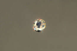
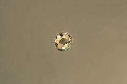
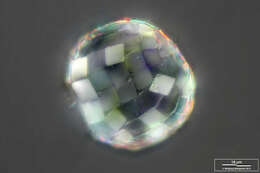
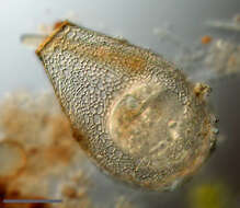
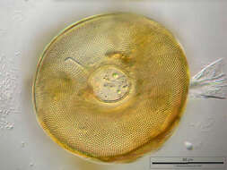
Arcella gibbosa This testate amoeba constructs a proteinaceous shell with numerous tiny alveoli reinforcing the test. Apertural view. This picture was built up using 35 DIC frames with manual stacking technique. The scale bar indicates 50 µm. Sample from a freshwater pond on the island of Hiddensee (Baltic Sea, Germany). This image was taken using Zeiss Universal with Olympus C7070 CCD camera.Image under Creative Commons License V 3.0 (CC BY-NC-SA). Place name: Pond Suploch, Hiddensee (Germany) Latitude: 54.538638 Longitude: 13.097802 Diese Schalenamöbe bildet eine proteinöse Schale mit zahlreichen kleinen, die Schale verstärkenden Hohlräumen (Areolen). Blick auf die Schalenöffnung (Apertur, Pseudostom). Tiefenschärfe durch Multiebenenabbildung aus 35 Bildebenen, manuell gestapelt. Der Messbalken zeigt 50 µm an. Probe aus einem kleinen Süßwasserteich auf der Insel Hiddensee, der eine faszinierende Vielfalt von nackten und beschalten Amöben beherbergt. Mikrotechnik: Zeiss Universal, Kamera: Olympus C7070. Creative Commons License V 3.0 (CC BY-NC-SA). For permission to use of (high-resolution) images please contact postmaster@protisten.de.
-
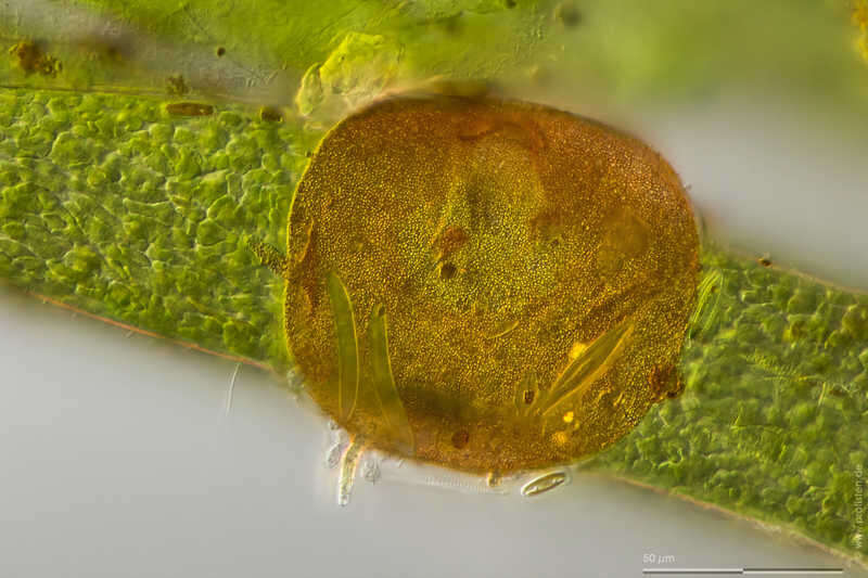
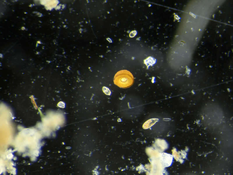
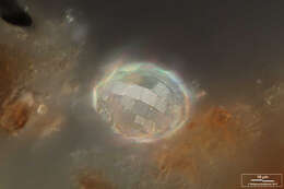
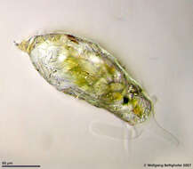
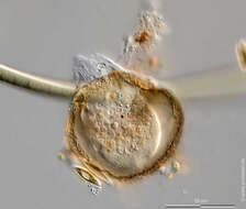
Arcella gibbosa Sample from a pond near Großostheim, Germany. Sampling date 04/2021. Images were taken using Olympus stereomicroscope SZX16 with Olympus OM-D M5 MKII. Image under Creative Commons License V 3.0 (CC BY-NC-SA). Place name: Pond near Großostheim (Germany) Latitude: 49.88482168 Longitude: 9.09980822 Probe aus einem Waldteich bei Großostheim. Datum der Aufsammlung: 04/2021. Mikrotechnik: Olympus Stereomikroskop SZX16, Kamera: Olympus OM-D M5 MKII. Creative Commons License V 3.0 (CC BY-NC-SA). For permission to use of (high-resolution) images please contact postmaster@protisten.de.
-
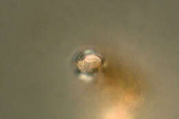
Arcella gibbosa Scale bar indicates 50 µm. Sample from a pond near Großostheim, Germany. Sampling date 04/2021. The image was built up using several photomicrographic frames with manual stacking technique. Images were taken using Zeiss Axioplan with Olympus OM-D M5 MKII. Image under Creative Commons License V 3.0 (CC BY-NC-SA). Place name: Pond near Großostheim (Germany) Latitude: 49.88482168 Longitude: 9.09980822 Multiebenen-Abbildung, manuell gestapelt. Der Messbalken markiert eine Länge von 50 µm. Probe aus einem Waldteich bei Großostheim. Datum der Aufsammlung: 04/2021. Mikrotechnik: Zeiss Axioplan, Kamera: Olympus OM-D M5 MKII. Creative Commons License V 3.0 (CC BY-NC-SA). For permission to use of (high-resolution) images please contact postmaster@protisten.de.