-
Video showing how this amoebae collected at Cedar Swamps in Woods Hole moves around with a single anterior protruding pseudopode. This video made by Dan Lahr under a Zeiss Discovery V12, believe it or not, a dissecting scope.
-
Suserup Skov, Midtsjælland
-
Allindelille Fredskov
-
Fensmark skov
-
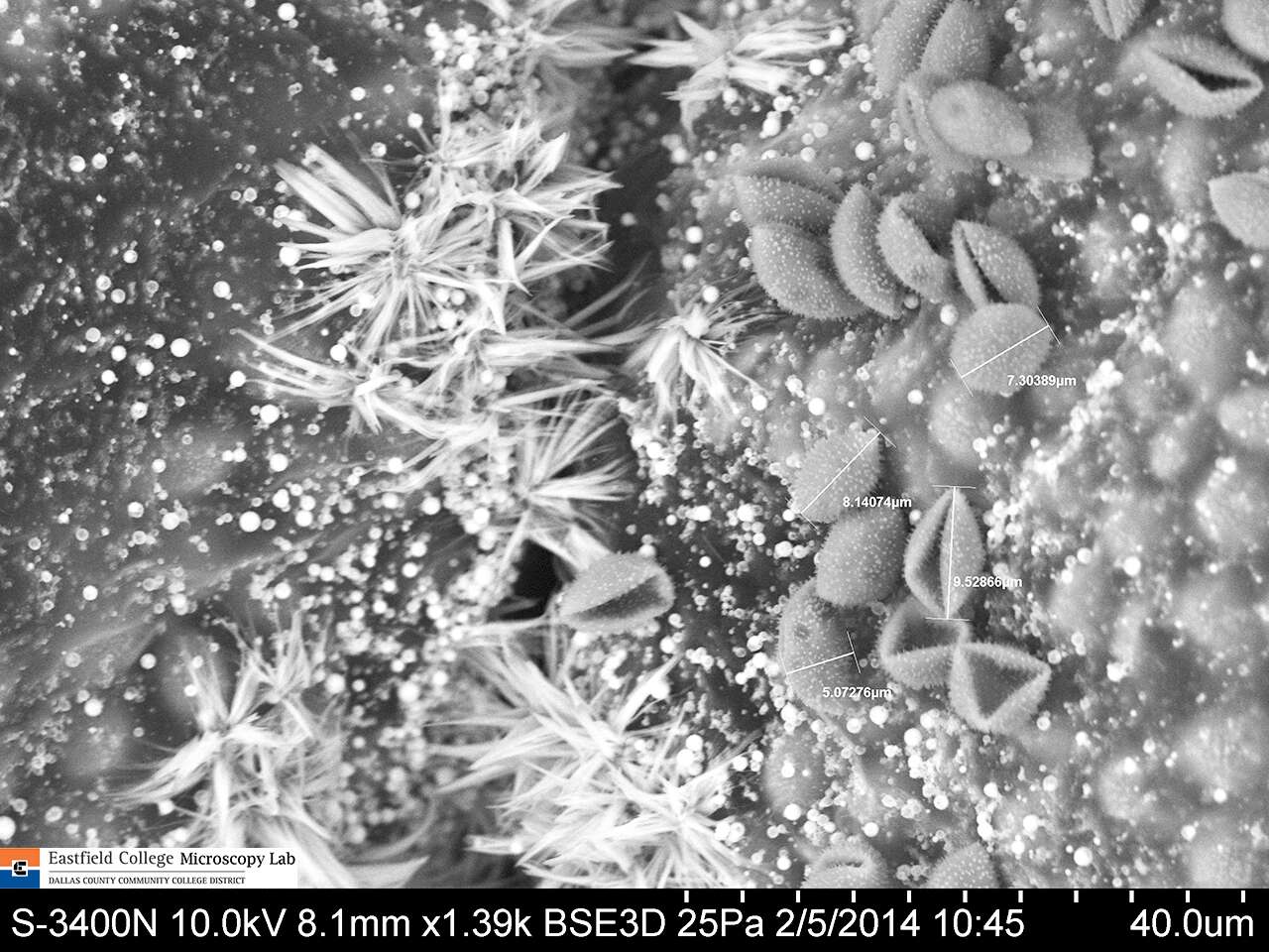
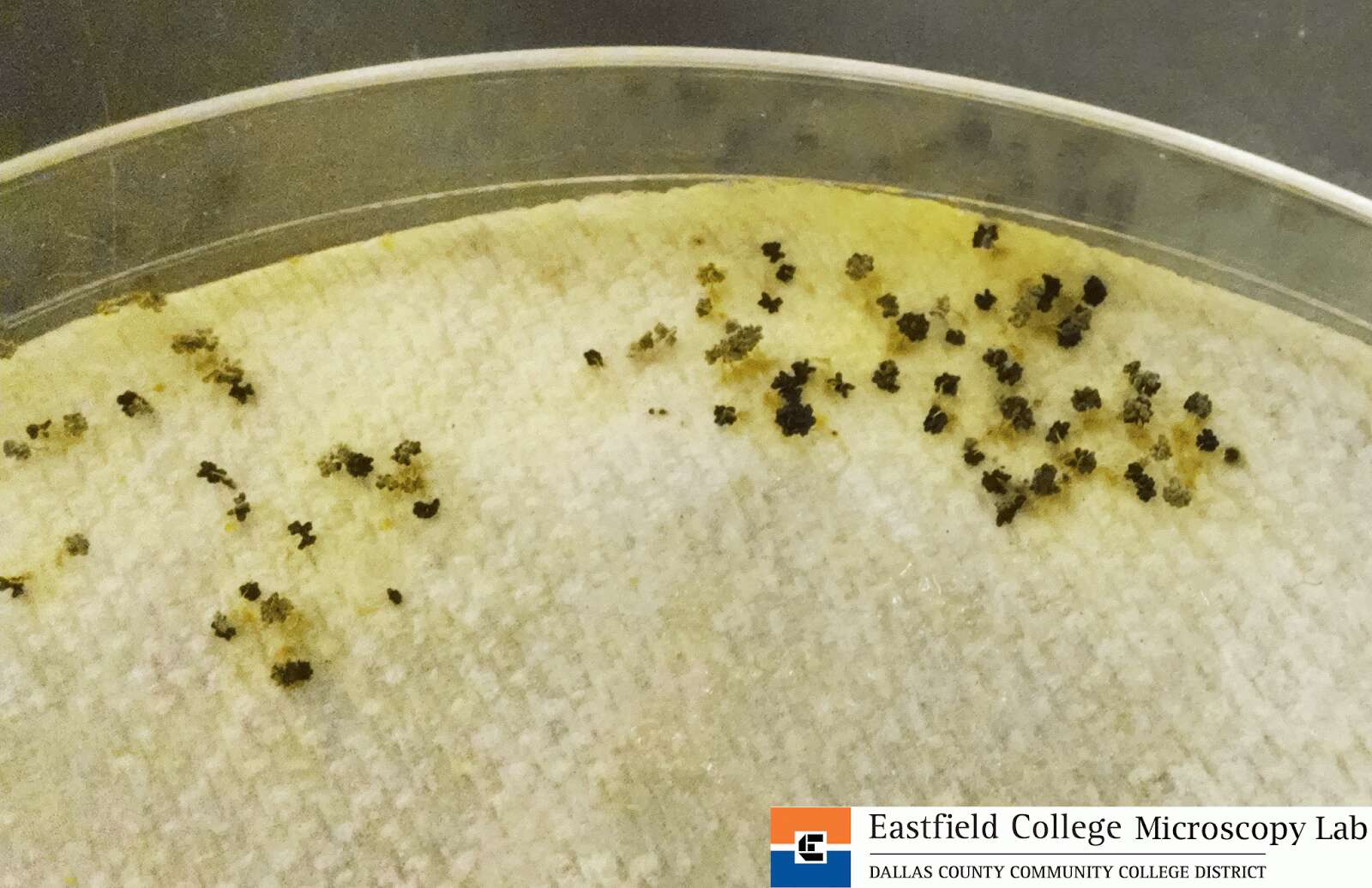
Eastfield College, Mesquite, TX
-
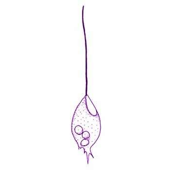
Mastigamoeba punctachora Bernard et al., 2000. With flagellated and non-flagellated cells, 6 - 90 microns long. Cysts were occasionally observed. The outline of the cell is highly variable. The flagellum, when present, is normally 50-80 microns, but may be considerably shorter. The basal region of the flagellum is often slightly thickened and stiffened. Flagellated cells have typically one, conical nucleus, with the flagellum inserting at the point, although the nucleus was occasionally observed further away from the apparent flagellar insertion site. The nucleus contains a conspicuous nucleolus. Nuclei often ontain a granule, usually refractile and usually located to the ab-flagellar side of the nucleolus. Non-flagellated cells with up to eight nuclei, but most usually with one. Pseudopodia are generally rounded and broad, and may emerge as hyaline eruptive structures near the base of the flagellum. Fine conical or branching pseudopodia may also be formed, most commonly from the posterior end. Microtubules and endoplasmic reticulum present, usually around the nucleus. The cytoplasm often contains granules and food vacuoles. Generally one contractile vacuole, but several may occur, especially in the non-flagellated and multinucleate forms. The contractile vacuoles form by fusion of small vesicles and are usually located posteriorly. Occasionally particles adhere to the surface of the cell. Swimming cells are usually globular, less commonly elongate, and swim slowly with the flagellum directed anteriorly. Flagellated and non-flagellated cells may glide on the substrate.
-
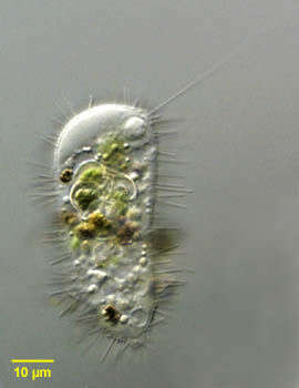
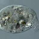
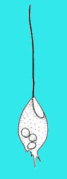
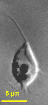
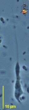
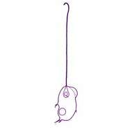
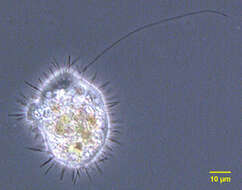
Portrait of Mastigina setosa a flagellated pelobiont. The ameboid monopodial cells have radiating "setae" which are 6-10 micron long filaments each of which has a basal granule (seen in this image). There is a single, long, lazily beating flagellum, which is closely associated with the nucleus (seen at the 12 o clock position adjacent to the cell membrane in this image). Electron microscopy shows the base of the flagellum connected to the nucleus by a cone of microtubules. The flagellum has a variable arrangement of microtubules. The nucleus contains a large endosome. Ingested algae are seen in food vacuoles. There is vigorous cytoplasmic streaming resulting in ameboid locomotion. The flagellum appears to contribute little to motility. Mastigina and the other pelobionts lack mitochondria and dictyosomes. Obvious ameboid locomotion and cytoplasmic streaming may help differentiate Mastigina from Mastigamoeba, a similar pelobiont. From slow-moving organically enriched freshwater runoff stream near Boise, Idaho. Differential interference contrast. Differential interference contrast optics.
-
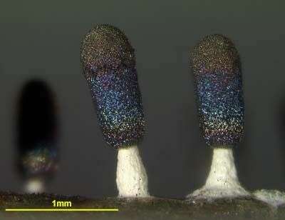
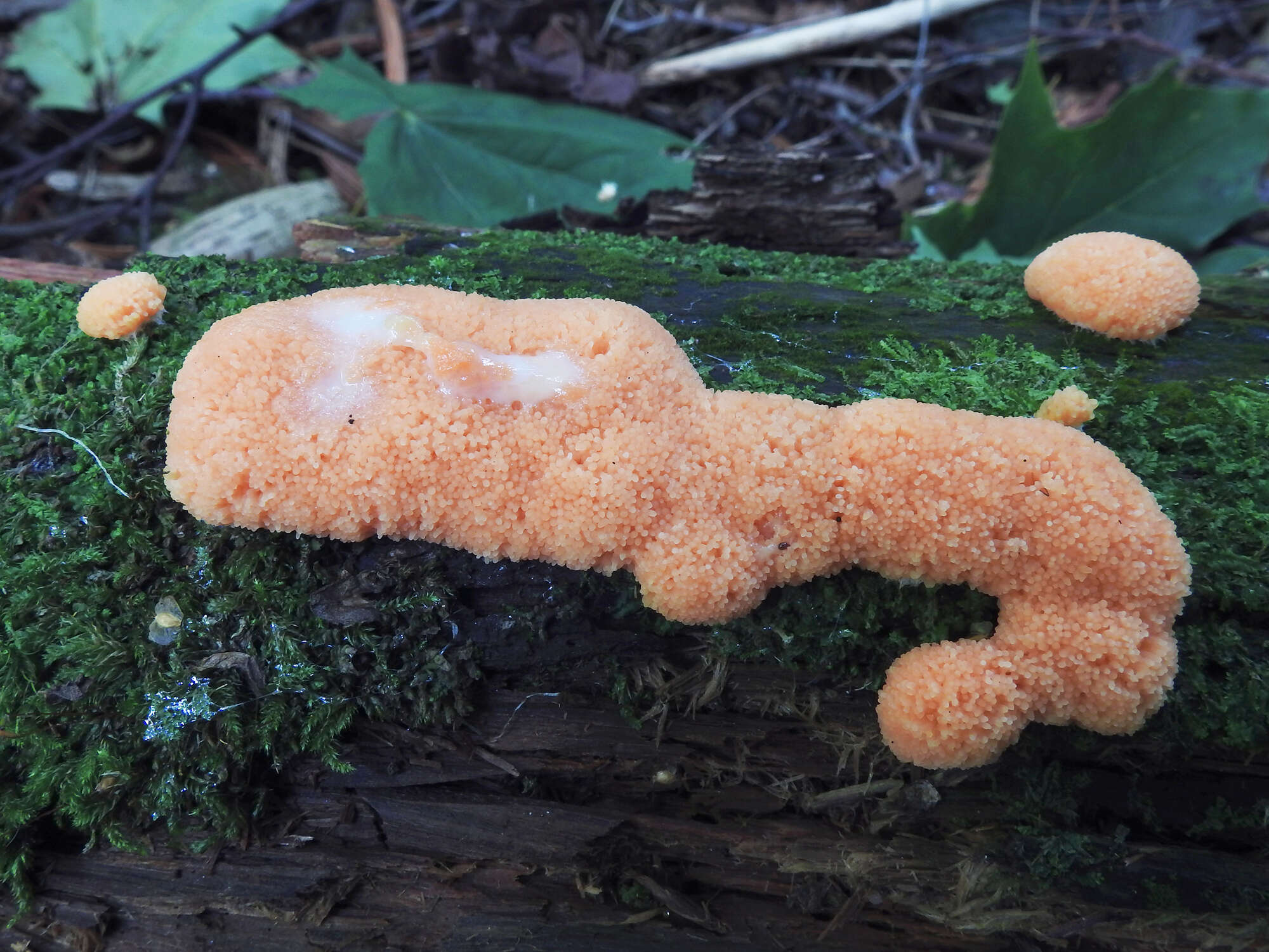
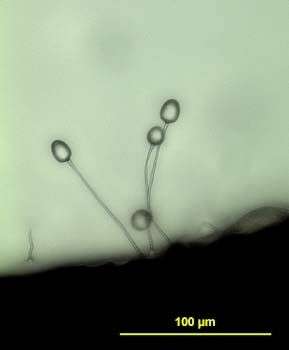
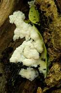
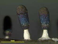
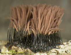
Two mature sporocarps
-
Allindelille Fredskov
-
Midtsjælland, Denmark
-
Eastfield College, Mesquite, TX
-
Helsinki, Uusimaa, Finland
-
-
Portrait of Mastigina setosa a flagellated pelobiont. The ameboid monopodial cells have radiating "setae" which are 6-10 micron long filaments. There is a single, long, lazily beating flagellum, which is closely associated with the nucleus (seen at the 1 o clock position adjacent to the cell membrane in this image). Electron microscopy shows the base of the flagellum connected to the nucleus by a cone of microtubules. The flagellum has a variable arrangement of microtubules. The nucleus contains a large nucleolus. Ingested algae are seen in food vacuoles. There is vigorous cytoplasmic streaming resulting in ameboid locomotion. The flagellum appears to contribute little to motility. Mastigina and the other pelobionts lack mitochondria and dictyosomes. Obvious ameboid locomotion and cytoplasmic streaming may help differentiate Mastigina from Mastigamoeba, a similar pelobiont. From slow-moving organically enriched freshwater runoff stream near Boise, Idaho. Phase contrast.
-
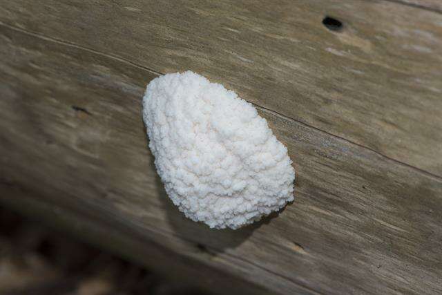
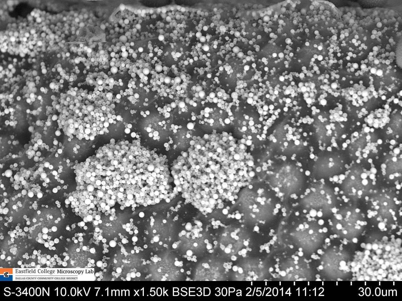
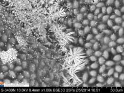
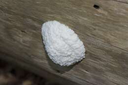
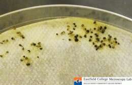
This is an image of multiple sporocarps from which most of the spores have been shed.
-
Eastfield College, Mesquite, TX
-
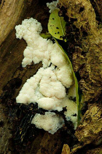
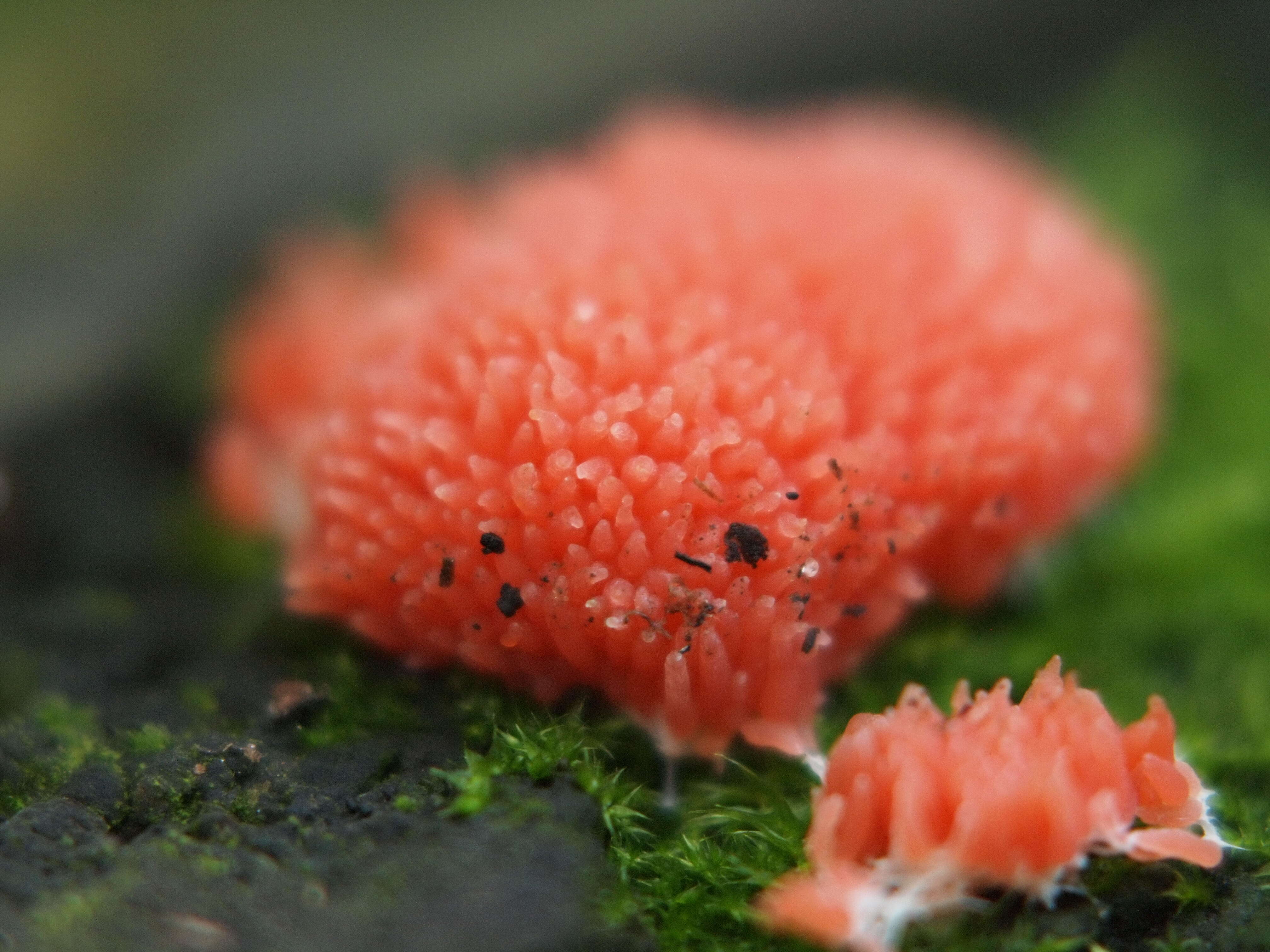
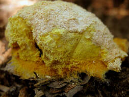
Tubifera ferruginosa
-
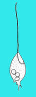
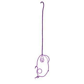
Mastigamoeba (ma-stig-a-me-ba) simplex Kent, 1880. Cells are about 8 microns long with very flexible amoeboid cell body. Pseudopodia are produced from the sides and posterior parts of the cell. One emergent flagellum is about 20 microns long and thickened. The flagellum is directed forwardly and beats stiffly. The nucleus is located in the anterior part of the cell. Rarely observed.
-
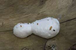
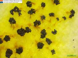
This is an image of multiple sporocarps.
-
Eastfield College, Mesquite, TX
-

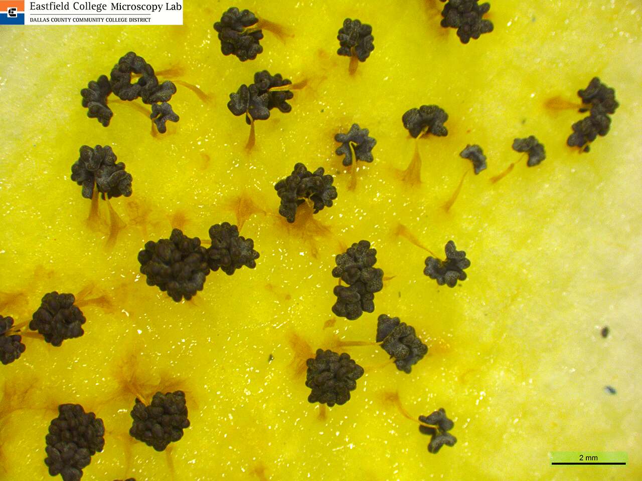
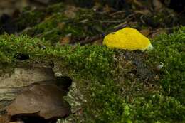
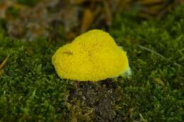
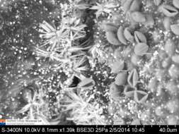
MUSHROOM INEDIBLE. Size was 2.5-20 cm, thickness 1-3cm. Mostly yellow, but can be of different colors. When mature gets darker. The touch is slimy and sticky. This fungus can be moved depending on the substrate on which grows, its PH value and position of the field. In that it takes its name, "creeping".Grows and develops waste material hidden in the trees, humus, etc.. Maturing exceeds the migration phase when the fruit appears (visible part of mushrooms). It is then possible to prorate plazmodium and the green parts of plants. At this stage, this fungus is not harmful to the green plant.For about a day growing body of fungus spores, dried and pectoral so slow exempted.If you touch something or the hook come out slow and then get the impression that this fungus looks like mold.Mainly located in the vicinity of houses and buildings, and can also be found in the forests. A copy of the image was found in an oak forest.Mainly to be found in the period from May to October.It is not edible.This fungus is actually mold. Before being included in the mushrooms, and now has established a special class to which it belongs.
-
Mastigamoeba simplex Kent, 1880. Flagellated and non-flagellated forms occur, rounded to elongate, 5 to 21 microns long, fagellated cells are usually ovoid to elongate, with the flagellum up to 40 microns long. The bulk of the nucleus is often located about one third of the cell away from the apex, although an extension of the nucleus connects to the base of the flagellum (this can be difficult to see). Inclusions may be present, although the anterior-most region of the cell is usually hyaline. Some eruptive formation of pseudopodia occurs in the region of the flagellar insertion. Fine, sometimes branching, pseudopodia may also form, especially from the posterior end, sometimes forming a pronounced tail by which the cell attaches to the substrate. When swimming, the flagellum is directed anteriorly. A contractile vacuole in freshwater isolates, usually located posteriorly, and filled by fusion of smaller vesicles.
-
Eastfield College, Mesquite, TX
-
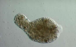
Mastigamoeba (massed-ig-a-me-ba) One of about 5 genera of pelobionts. These organisms are unusual in lacking mitochondria, dictyosomes and were for some time regarded as the most primitive of all eukaryotes. Most species which have been studied can adopt a variety of morphologies - including flagellates, amoebae, and cysts. The flagellates typically have one very long flagellum which beats in a fairly ineffectual fashion - and mostly progress by gliding. Body of cell most usually adopts an amoeboid form - such as that illustrated here. Typically found in habitats with little or no oxygen. Phase contrast.