-
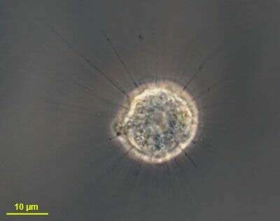
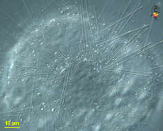
Phase contrast image of living cell, thin arms used for food capture.
-
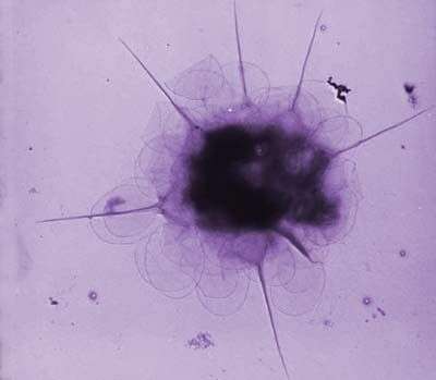
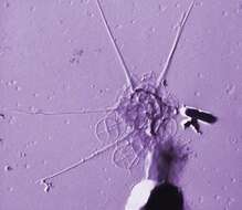
This image was made from samples taken during a scientific cruise in the Pacific. Water was filtered to concentrate the organisms that were present, then dried onto a thin sheet of plastic and then shadowed with a fine layer of metal to provide contrast. The preparation was then observed with an electron-microscope. This technique has been used to document the diversity of marine microbes, especially, protists in the oceans.
-
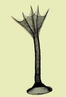
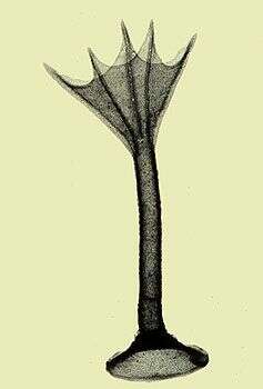
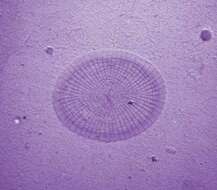
Transmission electron micrograph of a whole spine scale.
-
This image was made from samples taken during a scientific cruise in the Pacific. Water was filtered to concentrate the organisms that were present, then dried onto a thin sheet of plastic and then shadowed with a fine layer of metal to provide contrast. The preparation was then observed with an electron-microscope. This technique has been used to document the diversity of marine microbes, especially, protists in the oceans.
-
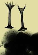
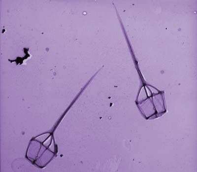
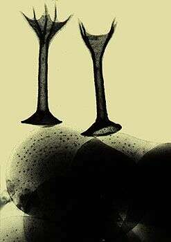
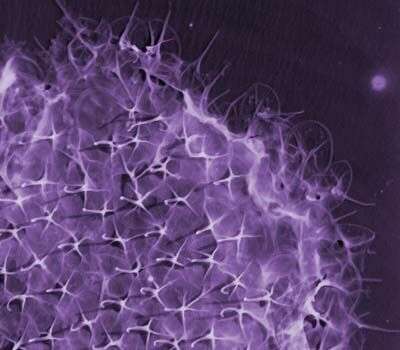
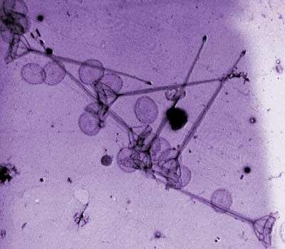
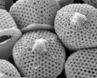
Transmission electron micrograph of whole spine scales and plate scales.
-
This image was made from samples taken during a scientific cruise in the Pacific. Water was filtered to concentrate the organisms that were present, then dried onto a thin sheet of plastic and then shadowed with a fine layer of metal to provide contrast. The preparation was then observed with an electron-microscope. This technique has been used to document the diversity of marine microbes, especially, protists in the oceans.
-
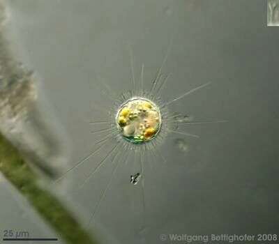
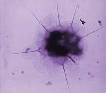
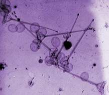
The picture shows the two types of actinomorphic siliceous spines (the shorter type is forked, see inset top right) and the tangential scales. Nucleus is clearly shown together with some food vacuoles. Sample from sphagnum pond Dosenmoor near Neumuenster (Schleswig-Holstein, Germany). Images were taken using Zeiss Universal with Olympus C7070 CCD camera.
-
This image was made from samples taken during a scientific cruise in the Pacific. Water was filtered to concentrate the organisms that were present, then dried onto a thin sheet of plastic and then shadowed with a fine layer of metal to provide contrast. The preparation was then observed with an electron-microscope. This technique has been used to document the diversity of marine microbes, especially, protists in the oceans.
-
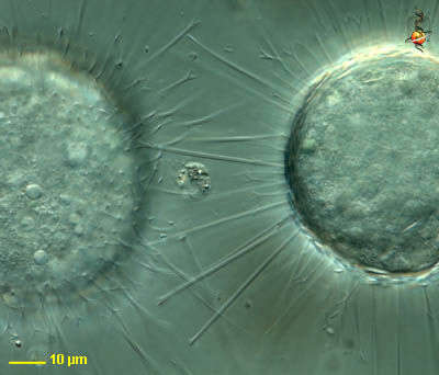
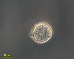
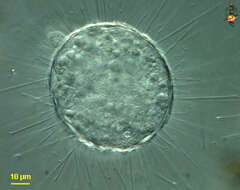
Acanthocystis turfacea (a-can-tho-sis-tis tur- fats-ee-a), a centrohelid heliozoon. Body coated with a layer of flattened siliceous scales and spine scales. Spine scales are of two lengths, and forked at their apices. This sample from moss. Nucleating site for axopodia just about evident in the cell to the right. Differential interference contrast.
-
This image was made from samples taken during a scientific cruise in the Pacific. Water was filtered to concentrate the organisms that were present, then dried onto a thin sheet of plastic and then shadowed with a fine layer of metal to provide contrast. The preparation was then observed with an electron-microscope. This technique has been used to document the diversity of marine microbes, especially, protists in the oceans.
-
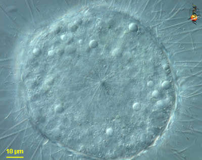
Acanthocystis turfacea (a-can-tho-sis-tis tur- fats-ee-a), a centrohelid heliozoon. Body coated with a layer of flattened siliceous scales and spine scales. Spine scales are of two lengths, and forked at their apex. This sample from moss. Differential interference contrast.
-
This image was made from samples taken during a scientific cruise in the Pacific. Water was filtered to concentrate the organisms that were present, then dried onto a thin sheet of plastic and then shadowed with a fine layer of metal to provide contrast. The preparation was then observed with an electron-microscope. This technique has been used to document the diversity of marine microbes, especially, protists in the oceans.
-
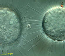
Acanthocystis turfacea (a-can-tho-sis-tis tur- fats-ee-a), a centrohelid heliozoon. Body coated with a layer of flattened siliceous scales and spine scales. Spine scales are of two lengths, and forked at their apices. Axonemes terminate on central granule or centroplast, shown here. This sample from moss. Differential interference contrast.
-
This image was made from samples taken during a scientific cruise in the Pacific. Water was filtered to concentrate the organisms that were present, then dried onto a thin sheet of plastic and then shadowed with a fine layer of metal to provide contrast. The preparation was then observed with an electron-microscope. This technique has been used to document the diversity of marine microbes, especially, protists in the oceans.
-
Acanthocystis turfacea (a-can-tho-sis-tis tur- fats-ee-a), a centrohelid heliozoon. Body coated with a layer of flattened siliceous scales and spine scales. Spine scales are of two lengths, and forked at their apices. This sample from moss. Differential interference contrast.
-
This image was made from samples taken during a scientific cruise in the Pacific. Water was filtered to concentrate the organisms that were present, then dried onto a thin sheet of plastic and then shadowed with a fine layer of metal to provide contrast. The preparation was then observed with an electron-microscope. This technique has been used to document the diversity of marine microbes, especially, protists in the oceans.
-
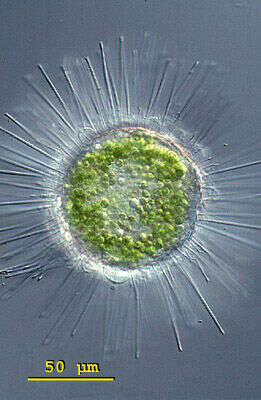
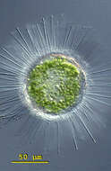
Acanthocystis turfacea (a-can-tho-sis-tis tur- fats-ee-a), a centrohelid heliozoon. Body coated with a layer of flattened siliceous scales and spine scales. Spine scales are of two lengths, and forked at their apex. The cytoplasm of this species contains often symbiotic algae. The body of this specimen measures 88 microns in diameter. This specimen was collected in a pond near Konstanz, Germany. Differential interference contrast.
-
This image was made from samples taken during a scientific cruise in the Pacific. Water was filtered to concentrate the organisms that were present, then dried onto a thin sheet of plastic and then shadowed with a fine layer of metal to provide contrast. The preparation was then observed with an electron-microscope. This technique has been used to document the diversity of marine microbes, especially, protists in the oceans.
-
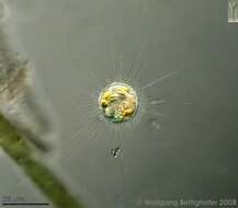
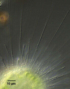
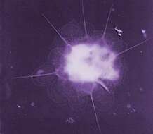
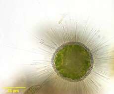
Portrait of Acanthocystis turfacea (Carter, 1863), a centroheliozoon with tangentially layered siliceous scales and two types of forked radial siliceous spines (one short, the other long) and radiating axopodia with extrusomes, all visible in this image. Chlorella endosymbionts are present. From standing fresh water near Boise, Idaho. Brightfield.
-
This image was made from samples taken during a scientific cruise in the Pacific. Water was filtered to concentrate the organisms that were present, then dried onto a thin sheet of plastic and then shadowed with a fine layer of metal to provide contrast. The preparation was then observed with an electron-microscope. This technique has been used to document the diversity of marine microbes, especially, protists in the oceans.
-
Portrait of Acanthocystis turfacea (Carter,1863), a centroheliozoon with tangentially layered siliceous scales and two types of forked radial siliceous spines (one short, the other long) and radiating axopodia with extrusomes, all visible in this image. Detail of radiating forked siliceous spines of Acanthocystis turfacea. . From standing fresh water near Boise, Idaho.DIC.
-
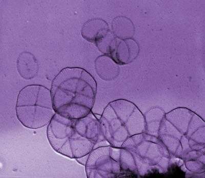
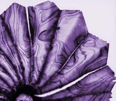
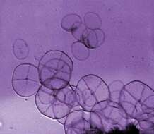
Detail of holococcoliths from the haploid life-cycle phase of S. pulchra
-
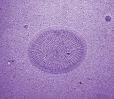
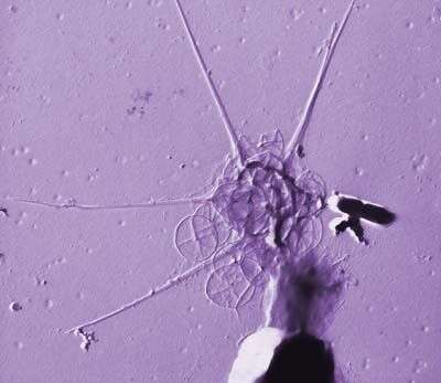
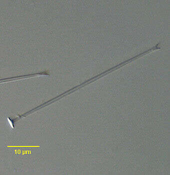
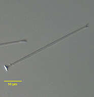
Detail of long forked radiating siliceous spine of Acanthocystis turfacea (Carter,1863), demonstrating the typical circular baseplate. From standing fresh water near Boise, Idaho.DIC.
-
This image was made from samples taken during a scientific cruise in the Pacific. Water was filtered to concentrate the organisms that were present, then dried onto a thin sheet of plastic and then shadowed with a fine layer of metal to provide contrast. The preparation was then observed with an electron-microscope. This technique has been used to document the diversity of marine microbes, especially, protists in the oceans.