-
Thomas Wesener, Daniel Minh-Tu Le, Stephanie F. Loria
Zookeys
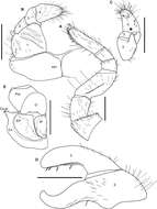
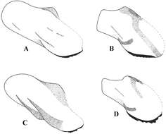
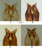
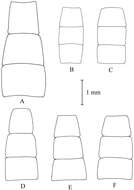
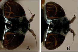
Figure 14.Sphaeromimus andohahela sp. n., A–D male holotype E female paratype. A left leg 9 B anterior telopod, anterior view C right anterior telopod, posterior view D left posterior telopod, posterior view E coxa and prefemur 2 with vulvae. Abbreviations: Cx = coxa; Cx-pr = coxal process; EP = external, lateral plate of vulva; IP = inner, mesal plate of vulva; O = operculum; Pre = prefemur; syn = syncoxite. Scale bars = 1 mm.
-
Antti Haarto, Gunilla Ståhls
Zookeys
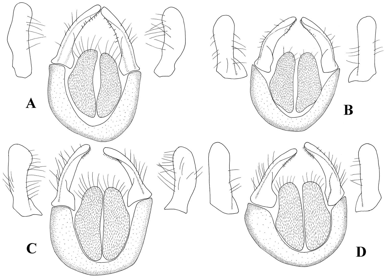
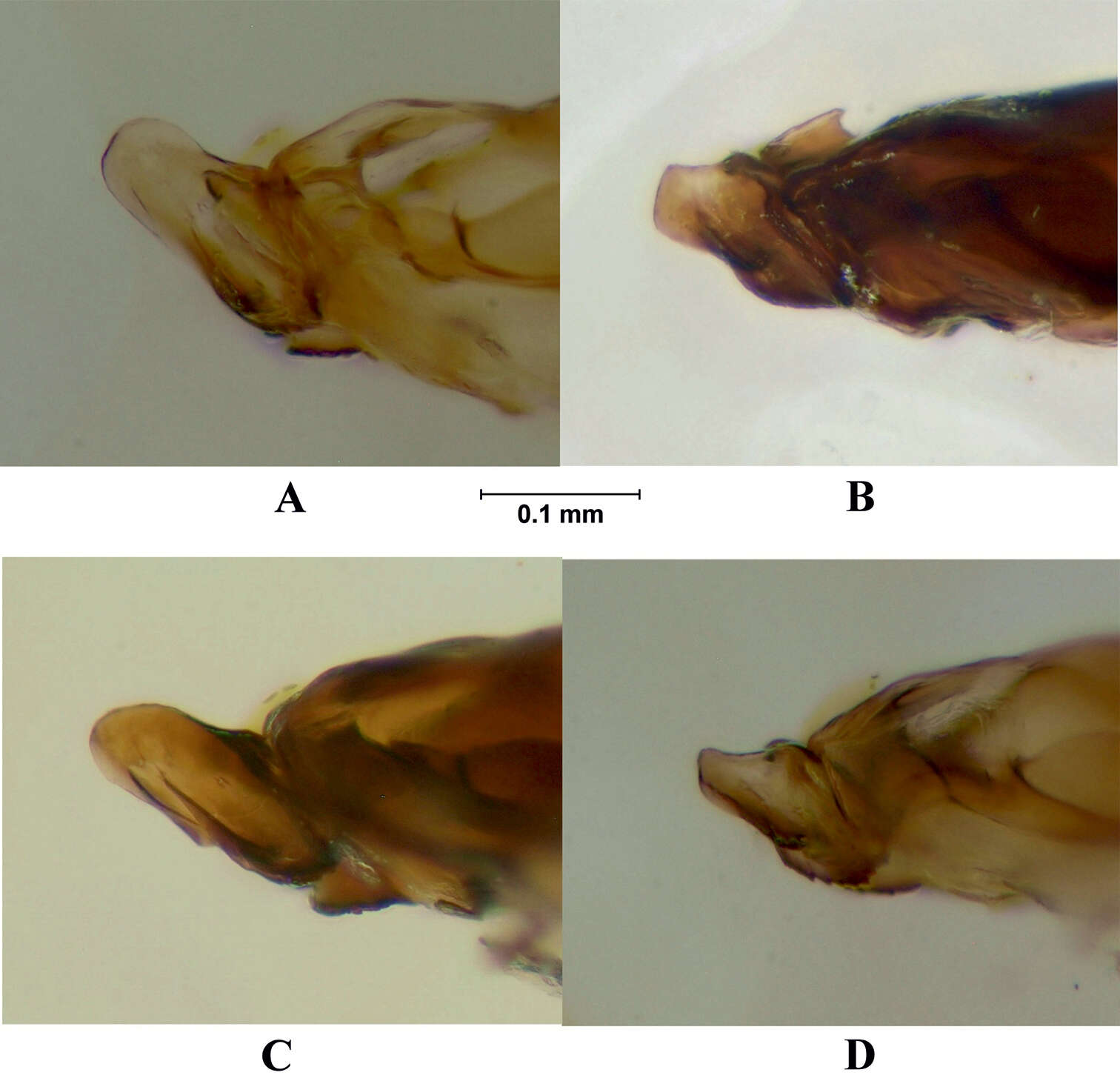
Figure 6.Cerci and surstyli, dorsal view and surstyli, lateral view. A Melanostoma scalare B Melanostoma certum C Melanostoma mellarium and D Melanostoma mellinum.
-
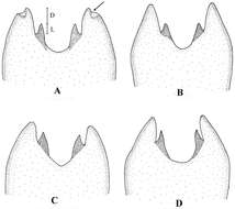
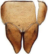
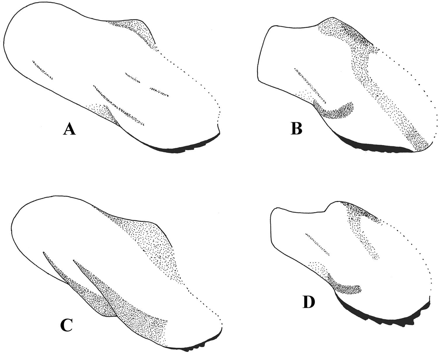
Ikelibeloha cristata, subgenital plates, ventrally (CAS) (Zahniser, J. N. & M. W. Nielson 2012, An extraordinary new genus and three new species of Acostemmini (Hemiptera: Cicadellidae: Deltocephalinae) from Madagascar with comments on the morphology and classification of the tribe Zootaxa. 3209: 28-52.)
-
Saša Širca, Gregor Urek, Stela Lazarova, Milka Elshishka, Vlada Peneva
Zookeys
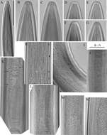
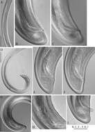
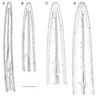
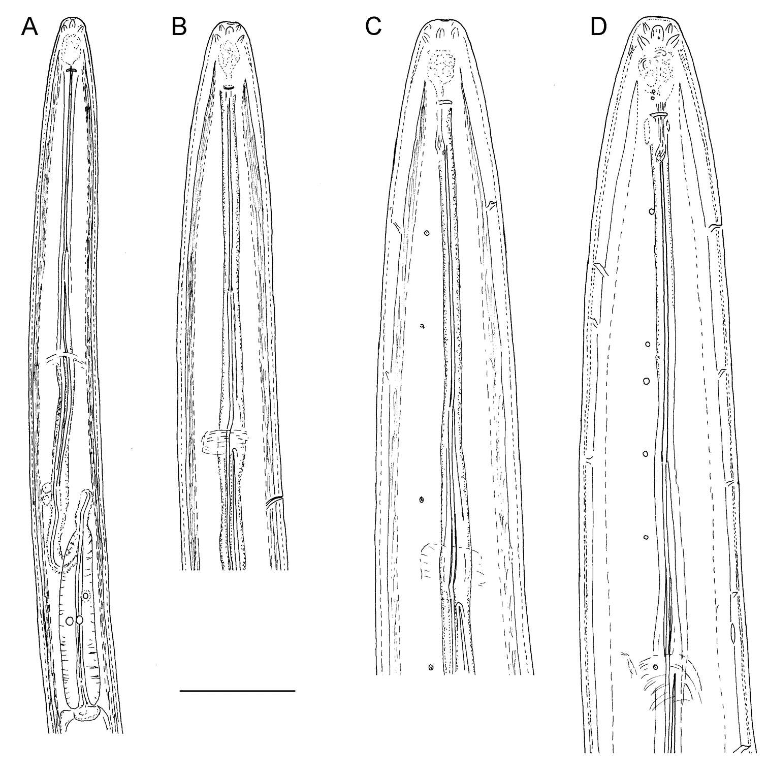
Figure 5.Longidorus carniolensis sp. n. Male: A, B Variation in tail shape. Scale bar: 50 μm.
-
Thomas Wesener, Daniel Minh-Tu Le, Stephanie F. Loria
Zookeys
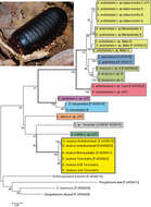
Figure 20.Maximum likelihood tree obtained from the COI dataset after 1000 bootstrap replicates under the GTR+I+G model. Habitus photograph shows Sphaeromimus andohahela from Manantantely. Colours used to separate species. Green colours = mid-elevation rainforest; Blue & Red colours, littoral and lowland rainforests; Yellow colours = southern spiny forest. See table 1 for more details about sequenced specimens.
-
Antti Haarto, Gunilla Ståhls
Zookeys
Figure 7.Postgonite and anterior part of hypandrium, lateral view. A Melanostoma scalare B Melanostoma certum C Melanostoma mellarium and D Melanostoma mellinum.
-
Saša Širca, Gregor Urek, Stela Lazarova, Milka Elshishka, Vlada Peneva
Zookeys
Figure 6.Longidorus carniolensis sp. n. Male: A Anterior region B, C Head region D–G Amphidial fovea H Vestigium (white arrow), excretory pore (thick arrow) and ventral pores (slender arrows) I Ejaculatory glands (marked by arrows) J Lateral field K, L Pharyngeal bulb with glandular bodies (marked by arrows) M, N Sperm cells at different stage of development. Scale bars: A 200 μm; B–N 50 μm.
-
Thomas Wesener, Daniel Minh-Tu Le, Stephanie F. Loria
Zookeys
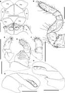
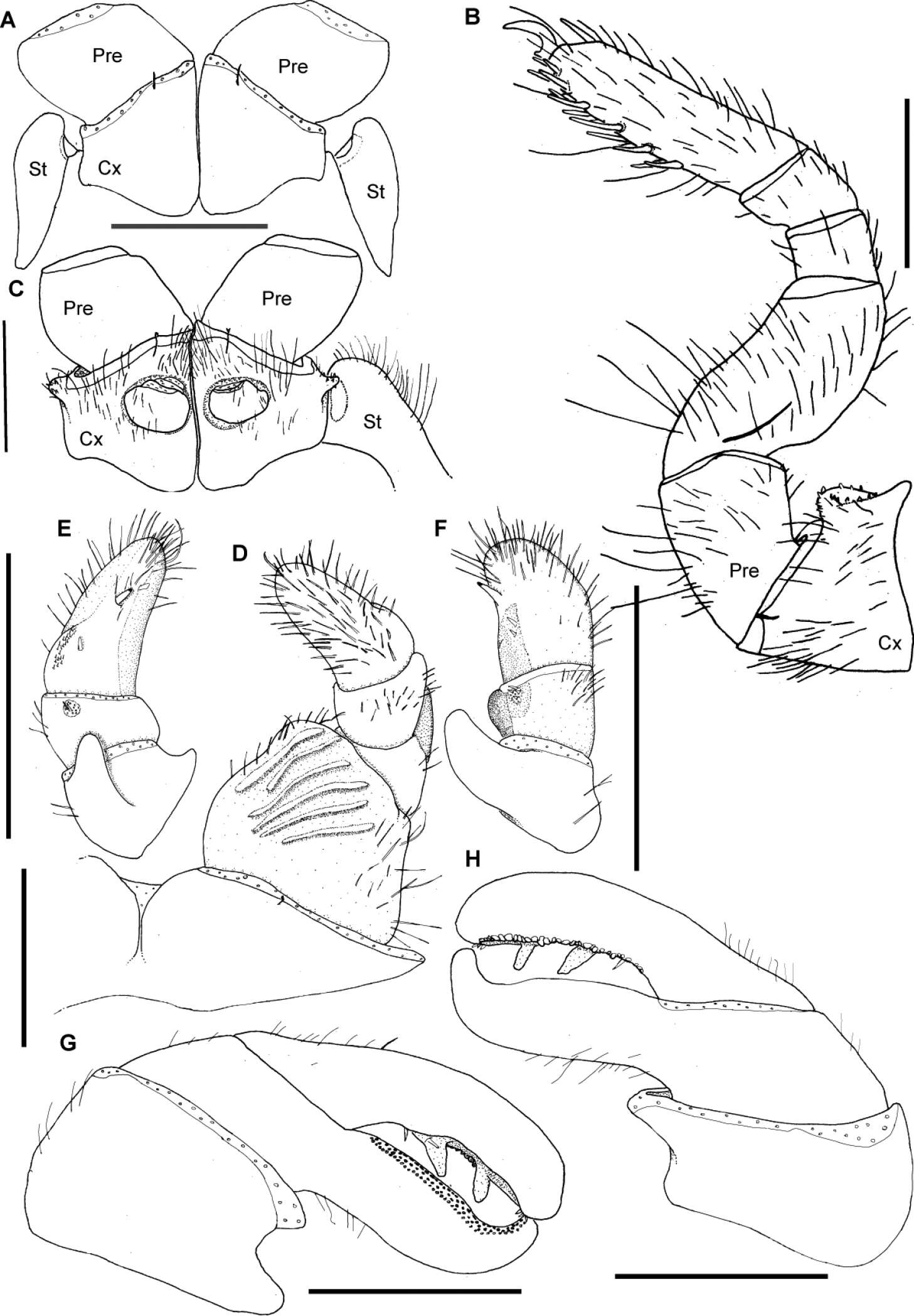
Figure 15.Sphaeromimus ivohibe sp. n., holotype. A coxae and prefemora 1 with stigmatic plates B left leg 9 C coxae and prefemora 2 with gonopore and stigmatic plate D right anterior telopod, anterior view E right anterior telopod, posterior view ♀ right anterior telopod, lateral view G left posterior telopod, anterior view H left posterior telopod, posterior view. Abbreviations: Cx = coxa; Pre = prefemur; St = stigmatic plate. Scale bars = 1 mm.
-
Antti Haarto, Gunilla Ståhls
Zookeys
Figure 8.Postgonite, lateral view. A Melanostoma scalare B Melanostoma certum C Melanostoma mellarium and D Melanostoma mellinum.
-
Saša Širca, Gregor Urek, Stela Lazarova, Milka Elshishka, Vlada Peneva
Zookeys
Figure 7.Longidorus carniolensis sp. n. Male: A Posterior genital branch B, C, E, F Tail and copulatory apparatus – different optical sections D, G Posterior end H Rectum (marked by arrow), spicules and lateral piece I Partly protracted spicules. Scale bars: A, D, G – 200 μm; B, C, E–F, H, I – 50 μm.
-
Thomas Wesener, Daniel Minh-Tu Le, Stephanie F. Loria
Zookeys
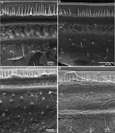
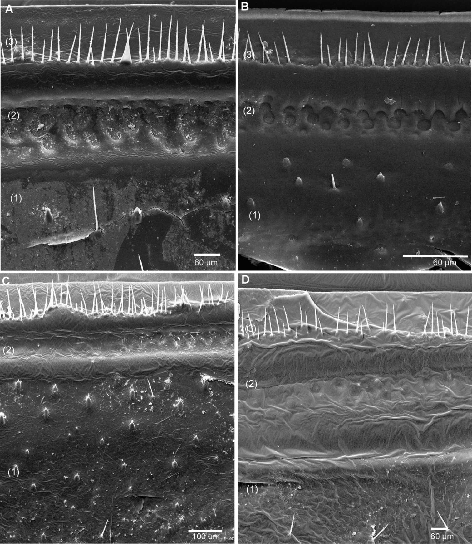
Figure 16.SEM, Endoterga of mid-body tergite. A Sphaeromimus ivohibe sp. n., paratype B Sphaeromimus saintelucei sp. n., holotype from Isaka-Ivondro C Sphaeromimus andrahomana sp. n., holotype D Sphaeromimus andrahomana cave specimen. Abbreviations: (1) = inner area with large spines and long setae; (2) = area with cuticular patterns; (3) = outer area with row(s) of marginal bristles and tergite margin.
-
Antti Haarto, Gunilla Ståhls
Zookeys
Figure 9.Postgonite, ventral view. A Melanostoma scalare B Melanostoma certum C Melanostoma mellarium and D Melanostoma mellinum.
-
Saša Širca, Gregor Urek, Stela Lazarova, Milka Elshishka, Vlada Peneva
Zookeys
Figure 8.Longidorus carniolensis sp. n. Juveniles: A Neck region of first stage B–D Spear region of second, third and fourth stage. Scale bar: 50 μm.
-
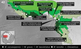
Figure 42.Distribution of the species of Monodius convexipennis, Monodius gravis, Monodius laevistriatus, Monodius lamottei and Eleoselinus gen. n. The division of Afrotropical Realm into ecoregions was adopted after Olson et al. 2001. Different colors were used to distinguish the adjacent ecoregions.
-
Antti Haarto, Gunilla Ståhls
Zookeys
Figure 10.Hypandrium, lateral view. A Melanostoma scalare B Melanostoma certum C Melanostoma mellarium and D Melanostoma mellinum.
-
Saša Širca, Gregor Urek, Stela Lazarova, Milka Elshishka, Vlada Peneva
Zookeys
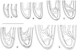
Figure 9.Longidorus carniolensis sp. n. Evolution of the tail. A–D Tail of first–fourth juvenile stage E Tail of female. Scale bar: 100 μm.
-
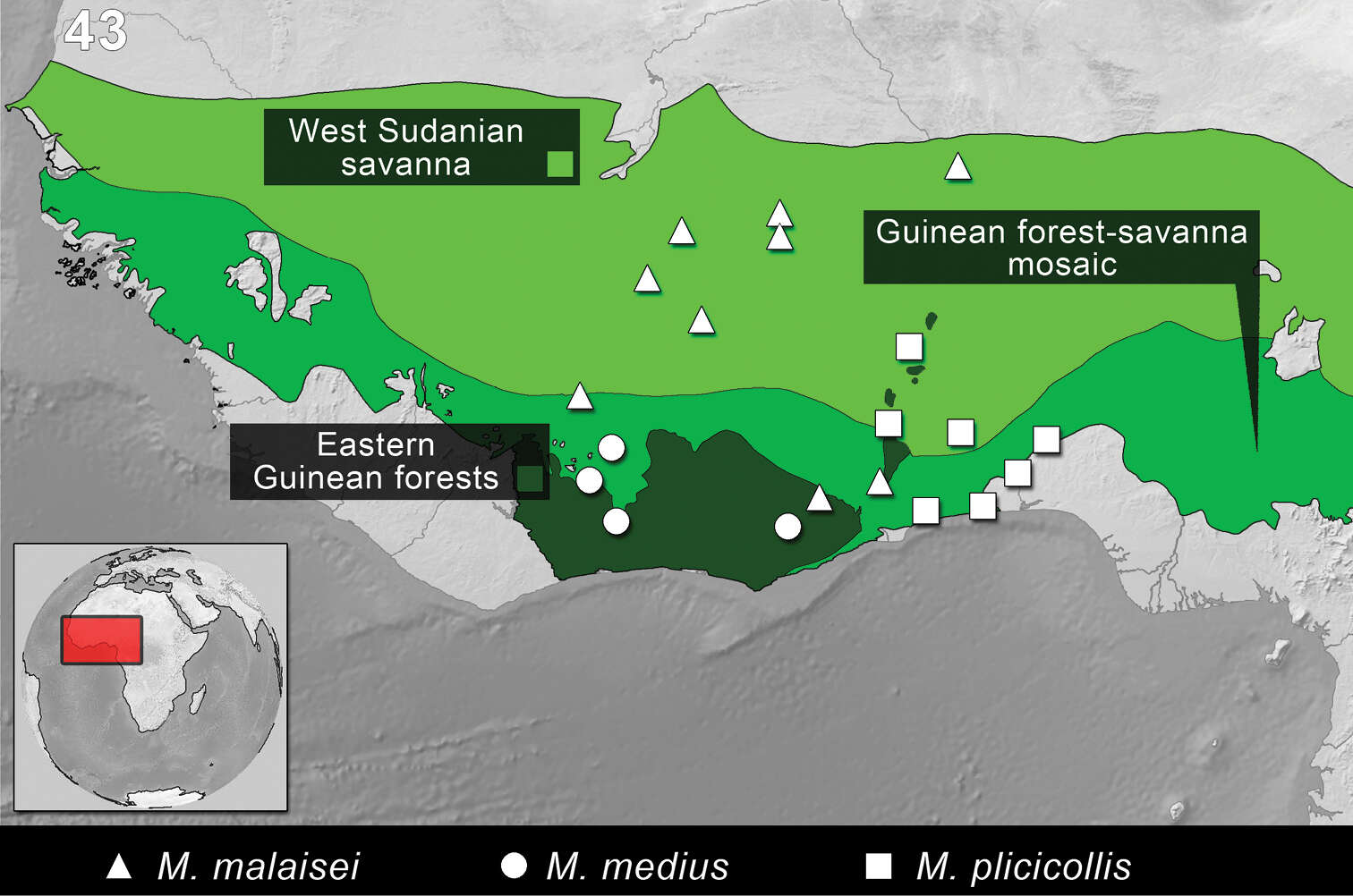
Figure 43.Distribution of the species of Monodius malaisei, Monodius medius and Monodius plicicollis. The division of Afrotropical Realm into ecoregions was adopted after Olson et al. 2001. Different colors were used to distinguish the adjacent ecoregions.
-
Antti Haarto, Gunilla Ståhls
Zookeys
Figure 11.Hypandrium, ventral view, shape of postgonites A Melanostoma scalare B Melanostoma certum C Melanostoma mellarium and D Melanostoma mellinum.
-
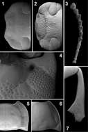
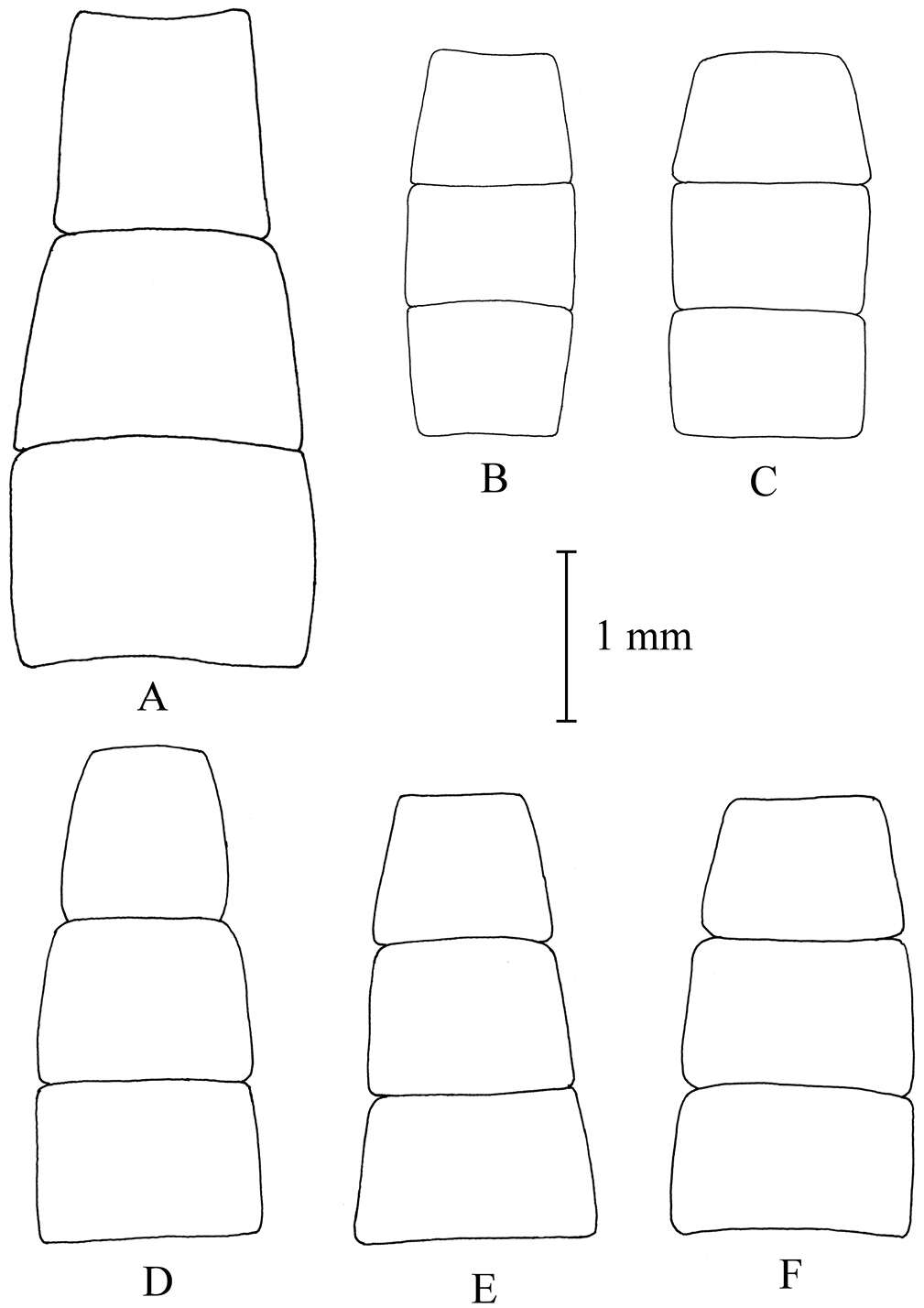
Figures 1–7.Head, dorsal view (1, 2), ventral view (4); antenna (3); pronotal disc (5, 6); mesotibia (7). Ectateus calcaripes (3), Ectateus crenatus (2), Ectateus ghesquierei (5), Monodius medius (7), Monodius plicicollis (4), Selinus planus (1, 6).
-
Antti Haarto, Gunilla Ståhls
Zookeys
Figure 12.Shape of female sterna 2–4. A Melanostoma scalare, B and C Melanostoma certum D Melanostoma mellarium and E and F Melanostoma mellinum.
-
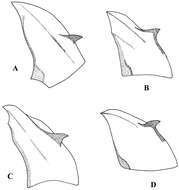
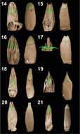
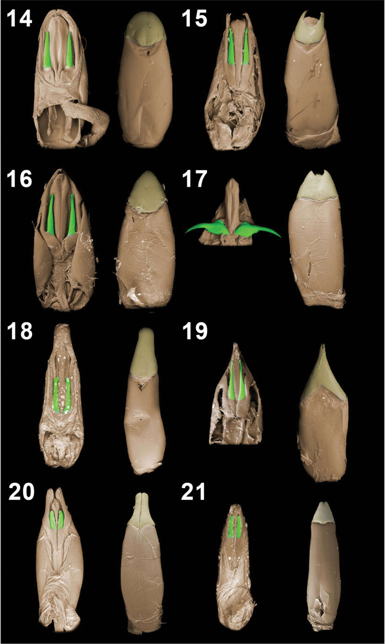
Figures 14–21.Aedeagal tegmina (dorsal and vental views). Monodius gravis (14), Monodius plicicollis (15), Monodius convexipennis (16), Monodius malaisei (17), Monodius lamottei (18), Monodius laevistriatus (19), Ectateus calcaripes (20), Selinus striatus (21).
-
Antti Haarto, Gunilla Ståhls
Zookeys
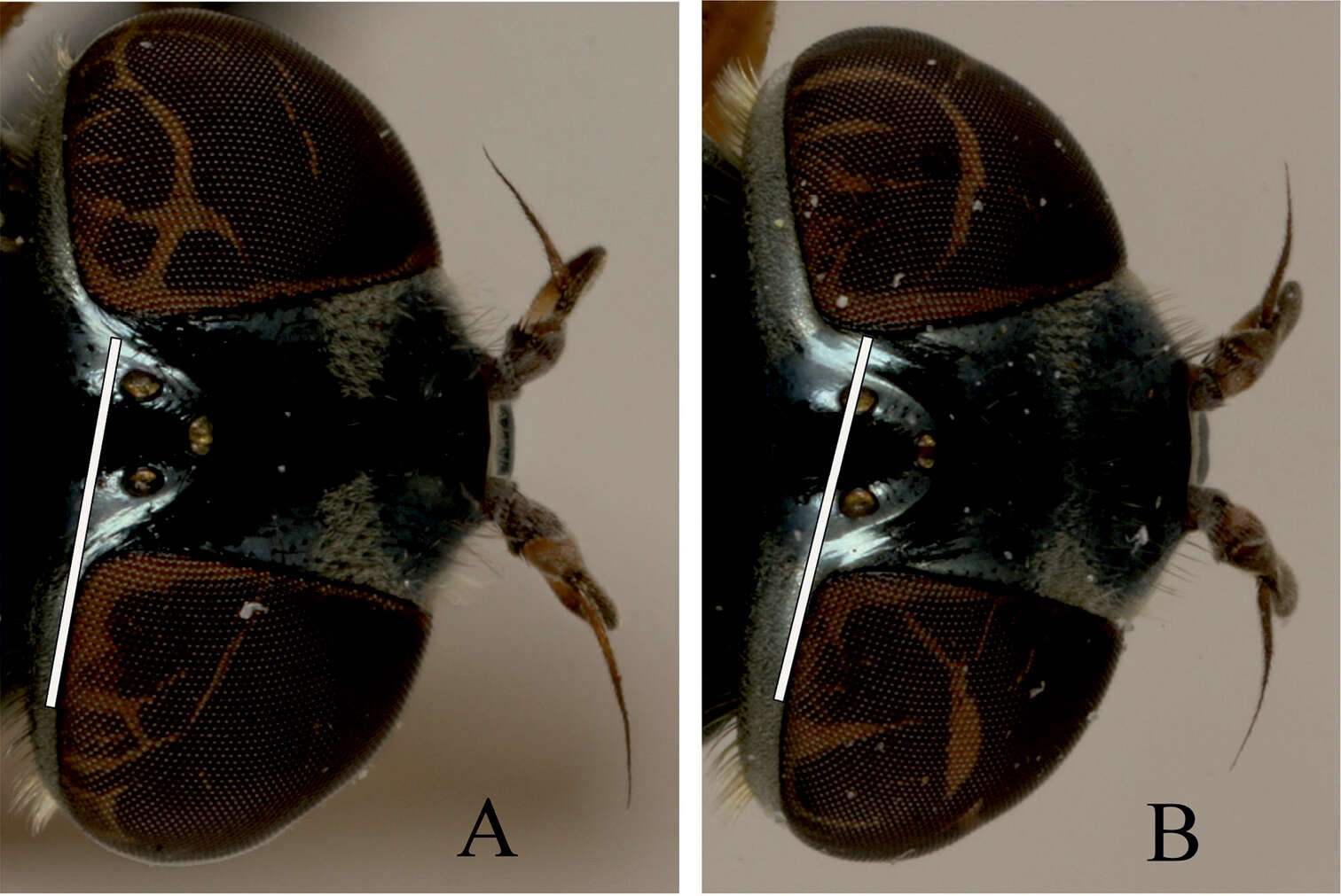
Figure 13.Dorsal view of female head. Position of posterior ocellus as compared to the hind eye line of female. A Melanostoma mellarium and B Melanostoma mellinum.
-
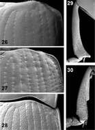
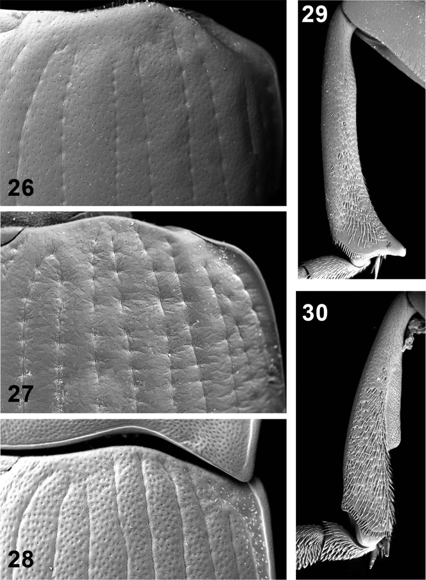
Figures 26–30.Elytral disc (26, 27, 28); male mesotibia (29); male protibia (30). Monodius gravis (26, 29, 30), Ectateus calcaripes (27), Ectateus lamottei (28).
-
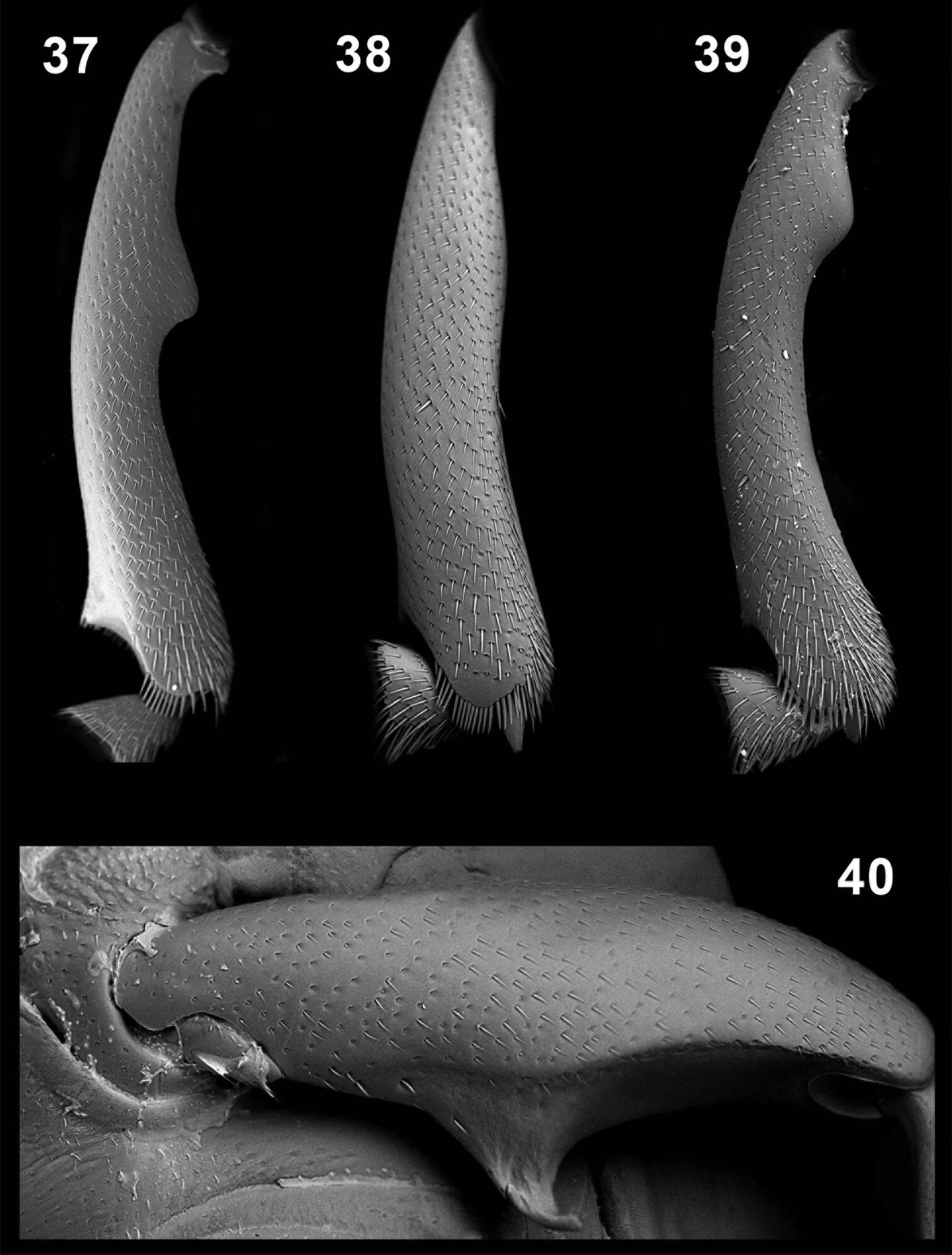
Figures 37–40.Male protibiae (37–39); male mesofemora (40). Monodius convexipennis (37), Monodius malaisei (38), Selinus planus (39), Ectateus laevistriatus (40).