-
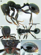
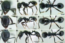
Figure 119.Stenamma megamanni holotype worker (CASENT0622851) A Profile B Face C Dorsum D Anterior clypeal margin in anterodorsal view E Gaster.
-
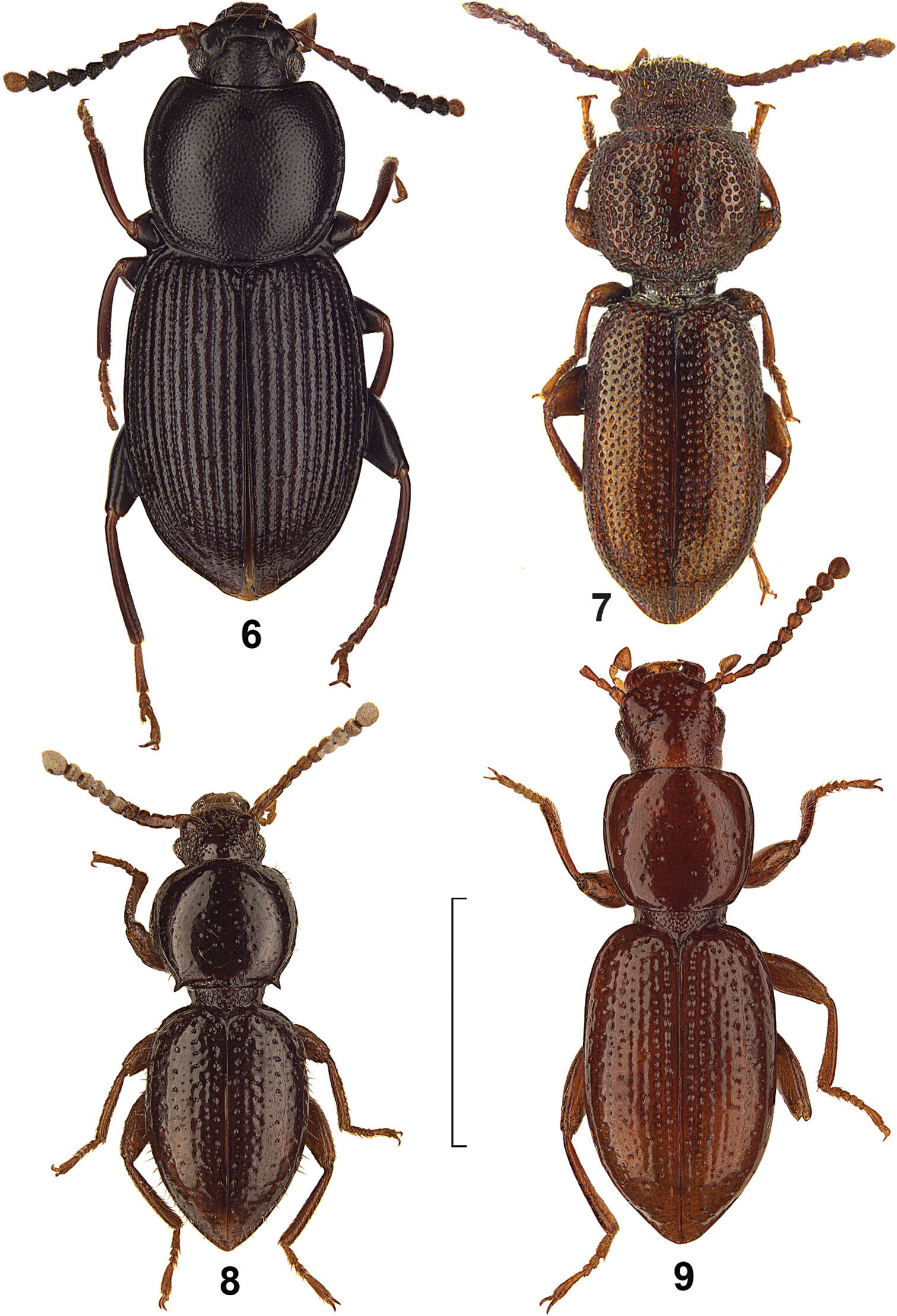
Figures 6–9.Dorsal view of Hovadelium and Mimolaena species. 6 Hovadelium elongatum, non-type SMNS 7 Mimolaena clarissae, holotype MZUF 8 Mimolaena janaki sp. n., holotype SMNS. 9 Mimolaena pauliani, holotype MNHN. – Scale line 2 mm.
-
Donald M. Windsor, Guillaume J. Dury, Fernando A. Frieiro-Costa, Susanne Lanckowsky, Jacques M. Pasteels
Zookeys
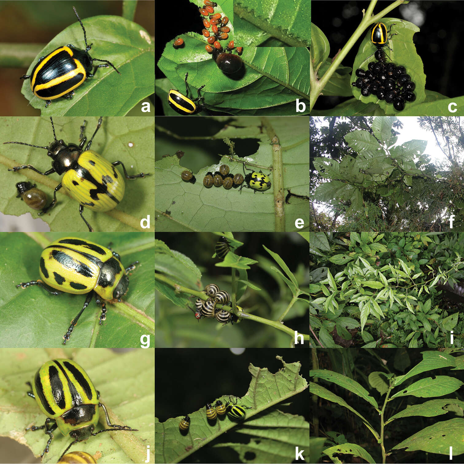
Figure 3.Maternal care providing Proseicela species, a Proseicela vittata adult (Photo by D.W.) b Proseicela vittata female and larvae from two cohorts. Insert shows detail of vein pinching along approximately 1cm of the primary vein (Photo by D.W.) c Proseicela vittata female with late stage larvae (Photo by D.W.) d Proseicela bicruciata adult female, (photo by G.D.) e Proseicela bicruciata female tending larvae (photo by G.D.) f Proseicela bicruciata food plant, Solanum abitaguense (photo by G.D.) g Proseicela spectabilis adult (photo by G.D.) h Proseicela spectabilis with nearly full-grown larval brood and tachinid parasitoid (photo by G.D.) i. Proseicela spectabilis host plant, Solanum sp. (photo by G.D.) j Proseicela sp. n. adult female (photo by G.D.) k the same female tending three feeding larvae feeding on Cuatresia sp. (Solanaceae) (photo by G.D.) l wider view of the host plant (photo by G.D.).
-
Lizhi Huo, Xingmin Wang, Xiaosheng Chen, Shunxiang Ren
Zookeys
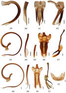
Figures 39–52.39–42 Aspidimerus nigritus (Pang & Mao), male genitalia: 39 penis 40 apex of penis 41 tegmen, ventral view 42 tegmen, lateral view 43–47 Aspidimerus esakii Sasaji, male genitalia: 43 penis 44 apex of penis 45 apex of penis, ventral view 46 tegmen, ventral view 47 tegmen, lateral view 48–52 Aspidimerus menglensis Huo & Ren, sp. n. 48–51 male genitalia: 48 penis 49 apex of penis 50 tegmen, ventral view 51 tegmen, lateral view 52 female genitalia: ovipositor. Scale bars: 0.1mm.
-
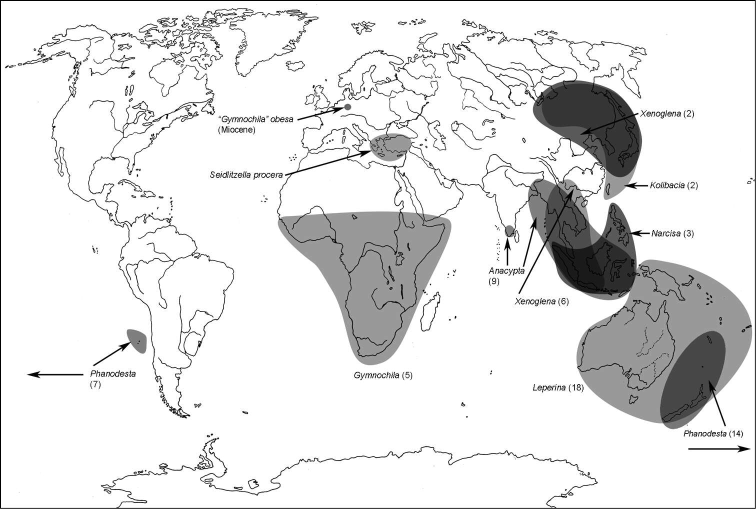
Map 4.A distribution of the tribe Gymnochilini.
-
Francisco Hita Garcia, Brian L. Fisher
Zookeys
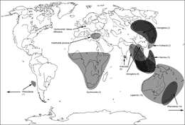
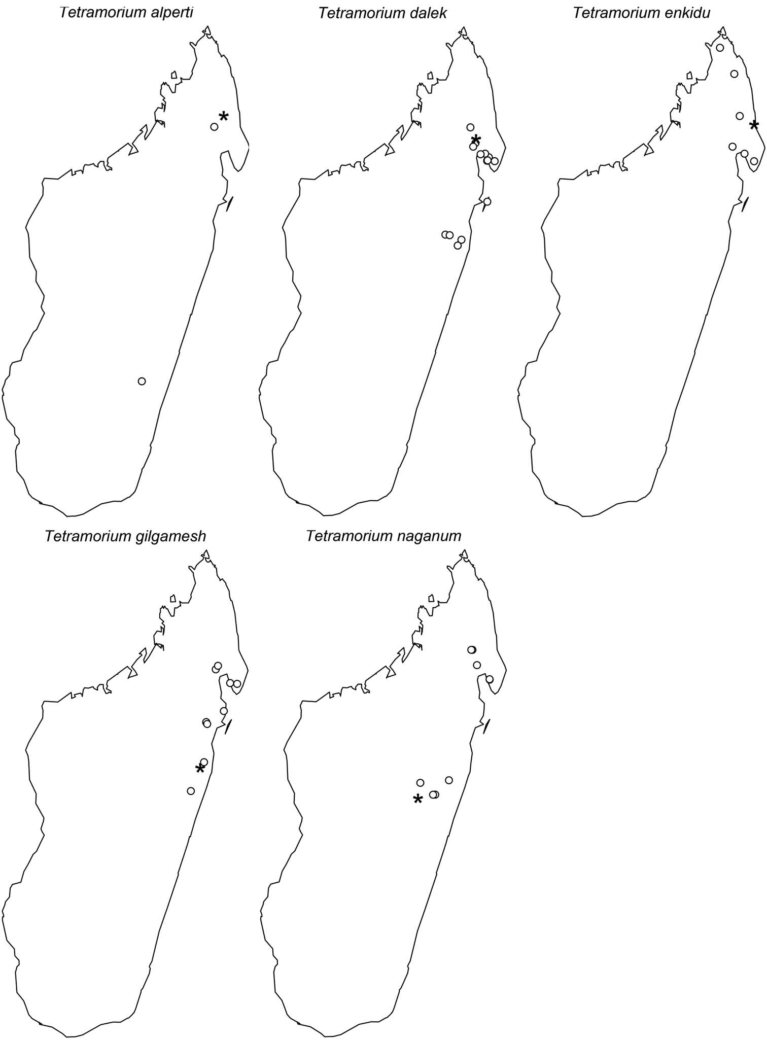
Figure 3.Map of sub-Saharan Africa showing the known distribution ranges of the five members of the Tetramorium decem species group: Tetramorium decem (filled circle), Tetramorium raptor (empty circle), Tetramorium uelense (filled square), Tetramorium ultor (empty square), and Tetramorium venator (star).
-
Francisco Hita Garcia, Brian L. Fisher
Zookeys
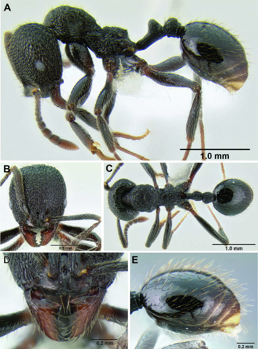
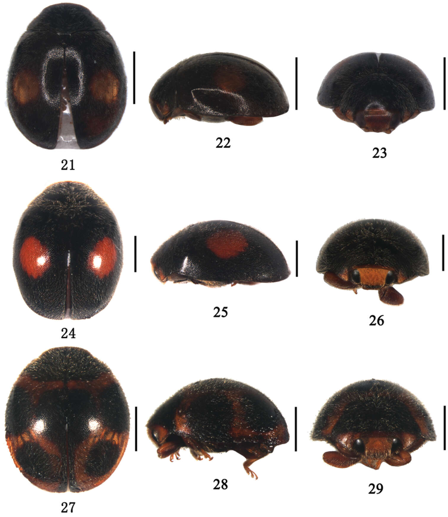
Figure 5.Tetramorium alperti holotype worker (CASENT0042547). A Body in profile B Body in dorsal view C Head in full-face view.
-
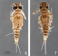
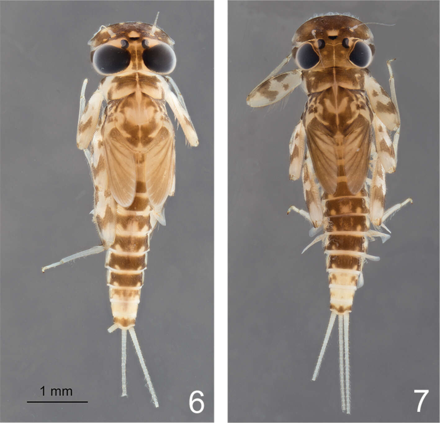
Figures 6–7.Rhithrogeniella ornata Ulmer, 1939. 6 Male nymph 7 Female nymph with slight color variations.
-
Alicia E. Timm, John W. Brown
Zookeys
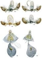
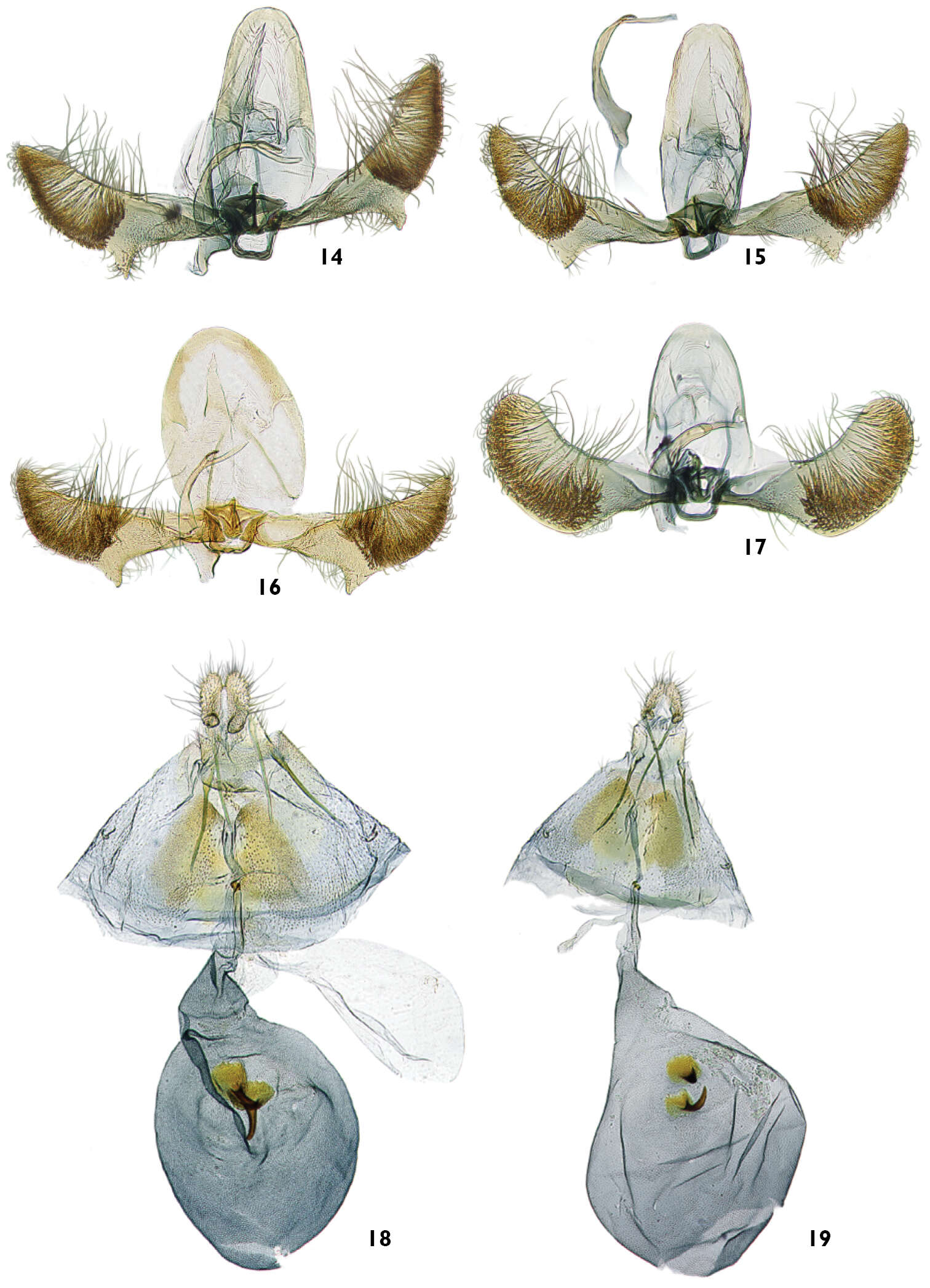
Figures 14–19.Male and female genitalia of Thaumatovalva. 14 Male of Thaumatovalva deprinsorum (USNM slide 144,491) 15 Male of Thaumatovalva albolineana (USNM slide 144,488) 16 Male of Thaumatotibia spinai (slide 2656) 17 Male of Thaumatovalva limbata (USNM slide 144,490) 18 Female of Thaumatovalva deprinsorum (USNM slide 144,492) 19 Female of Thaumatovalva albolineana (USNM slide 144,489).
-
Donald R. Davis, David L. Wagner
Zookeys
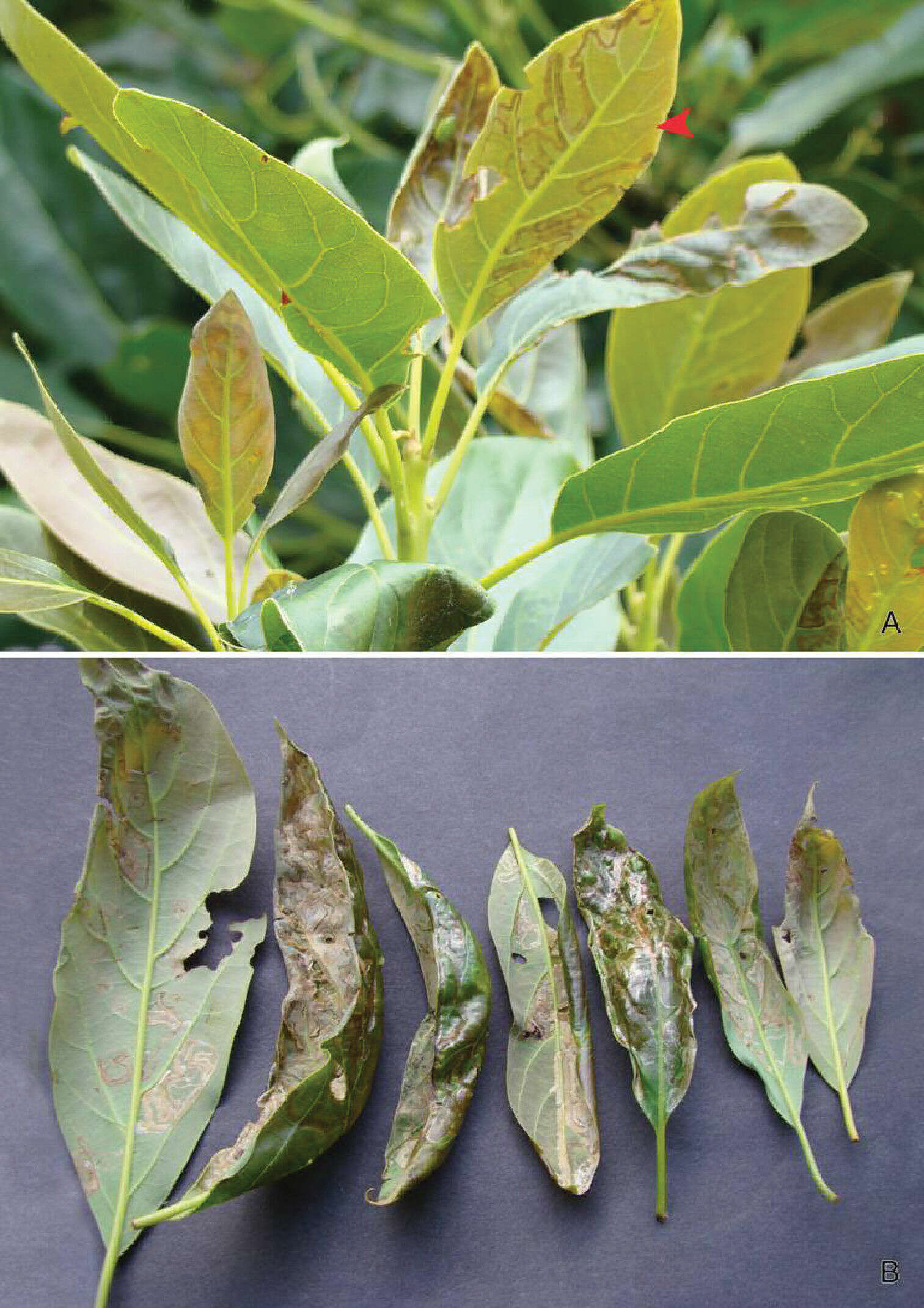
Figure 4.Leafmines of Phyllocnistis perseafolia sp. n. on Persea americana. A General habitus, note lower side mine (arrow) B Leaf damage caused by upper and lower side larval mining.
-
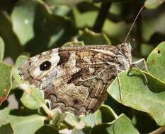
Montejaque, Malaga Province, Andalucia. Spain
-
Photo by Luxmmi Varathan & Craig Perl.
-
Protochiasmus mysticus, profemur (MZSP)
-
Michel P. Valim, Jason D. Weckstein
Zookeys
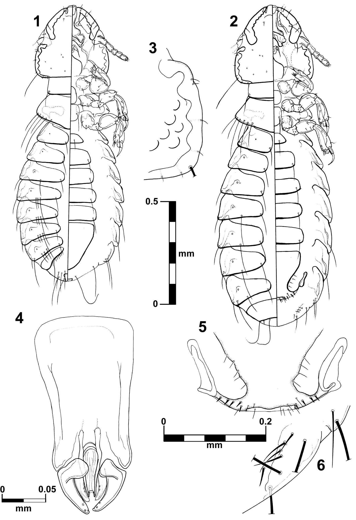
Figures 1–6.Brueelia sueta sp. n.: male, dorso-ventral views (1); female, dorso-ventral views (2); temporal carina (3); male genitalia (4); female vulvar margin (5); female gonapophysis (6).
-
Juli Pujade-Villar, Paul Hanson, Claudia A. Medina, Miguel Torres, George Melika
Zookeys
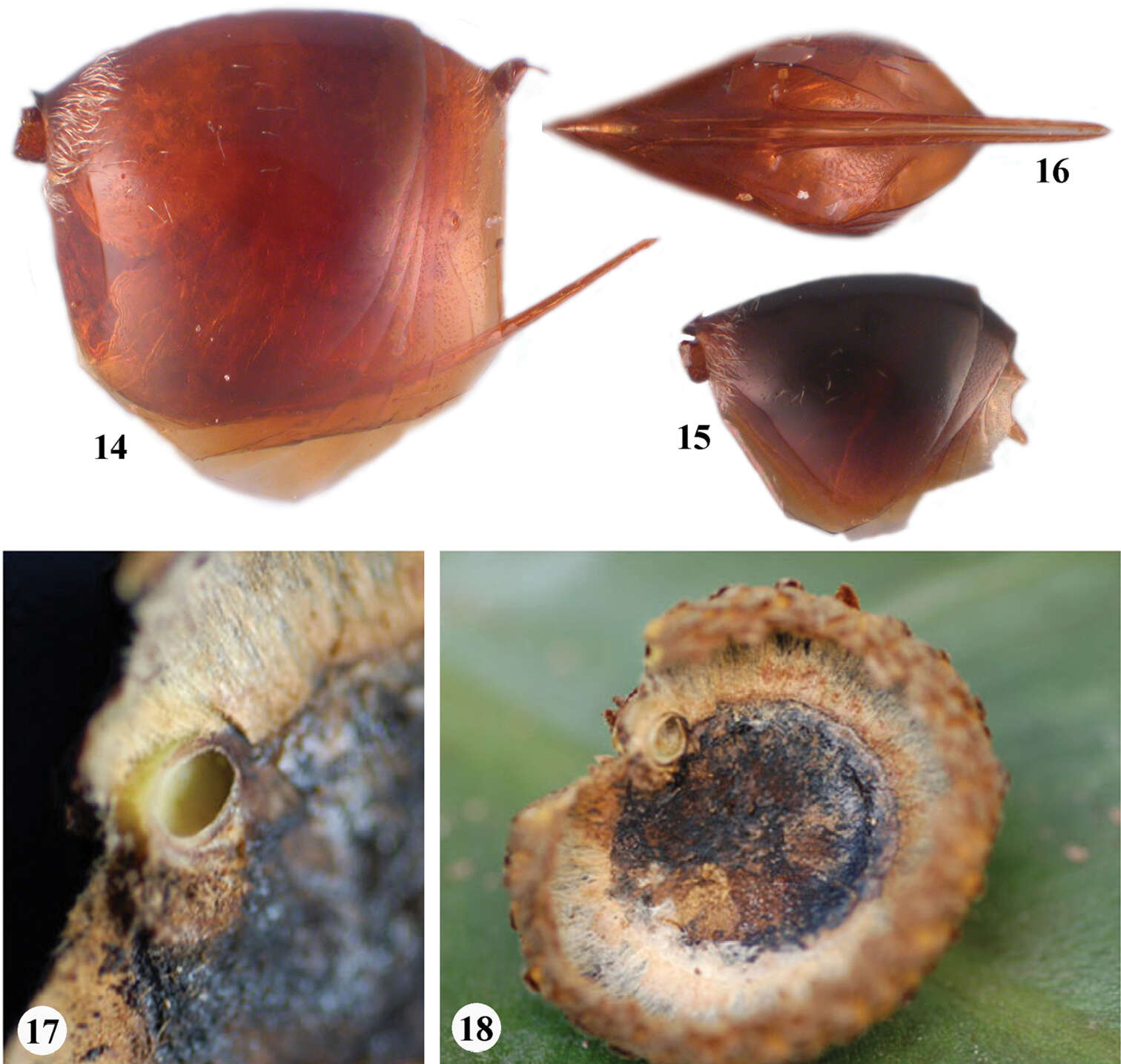
Figures 14–18.Zapatella grahami 14 metasoma, female (lateral view) 15 metasoma, male (lateral view) 16 ventral spine of hypopygium (ventral view) 17–18 gall.
-
Pierfilippo Cerretti, D. Monty Wood, James E. O’Hara
Zookeys
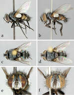
Figure 1.Neoethilla gen. n.ignobilis (New Mexico) a–d habitus a male in lateral view b female in lateral view c male in dorsal view d female in dorsal view d–e head in frontal view d male e female.
-
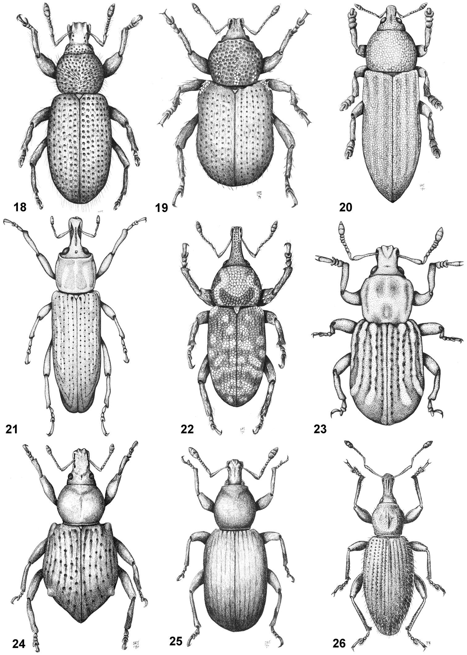
Figures 18–26.Habitus of representative Listroderini. 18 Adioristidius hirsutus 19 Puranius nigrinus 20 Haversiella albolimbata 21 Listronotus bosqi 22 Neopachytychius squamosus 23 Falklandiellus suffodens 24 Falklandiopsis magellanica 25 Falklandius antarcticus 26 Gromilus veneris.
-
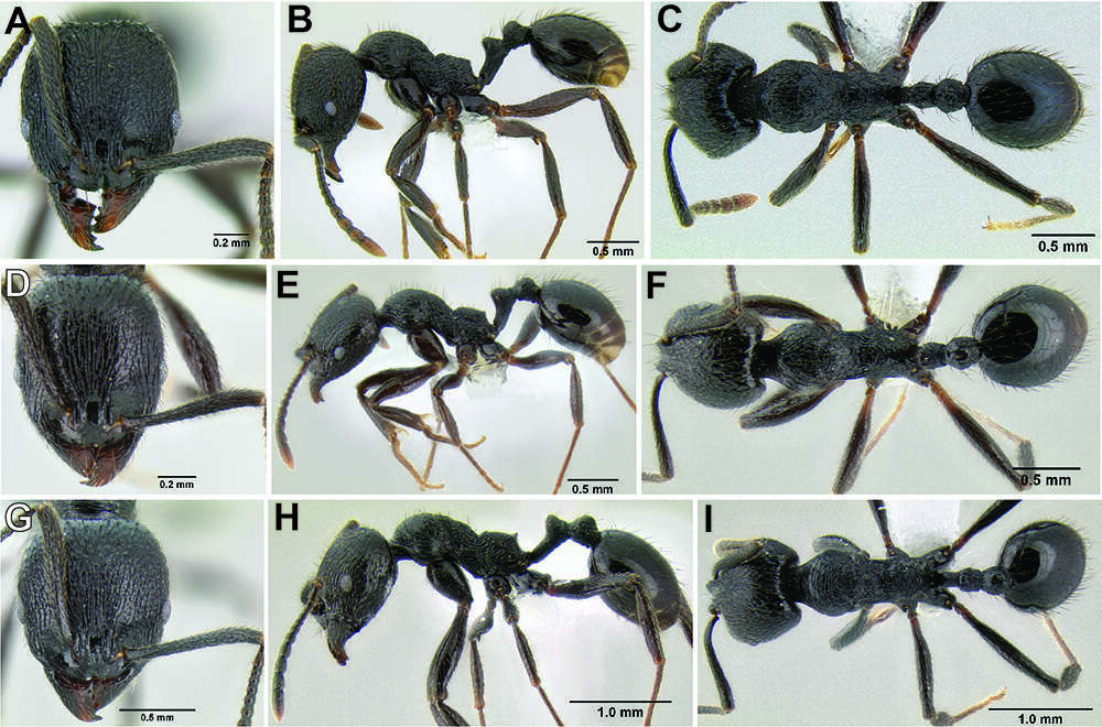
Figure 120.Stenamma megamanni worker variants. Face, profile, dorsal views A–C Variant 1 (CASENT0621836) D–F Variant 2 (CASENT0606734) G–I Variant 3 (CASENT0603924).
-
Figures 1–10.1–2. Symploce torchaceus Feng & Woo, male: 1 holotype, dorsal view 2 same, ventral view 3–4 Symploce bispot Feng and Woo, male: 3 holotype, dorsal view 4 same, ventral view 5–6 Symploce sphaerica sp. n., male: 5 holotype, dorsal view 6 same, ventral view 7–8 Symploce paramarginata sp. n., male: 7 holotype, dorsal view 8 same, ventral view 9–10 Symploce evidens sp. n., male: 9 holotype, dorsal view 10 same, ventral view. Scale bars = 1.0 cm.
-
Lizhi Huo, Xingmin Wang, Xiaosheng Chen, Shunxiang Ren
Zookeys
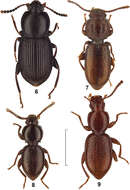
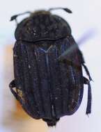
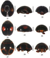
Figures 21–29.21–23 Aspidimerus guangxiensis Yu, 21 dorsal view 22 lateral view 23 frontal view 24–26 Aspidimerus matsumurai Sasaji, 24 dorsal view 25 lateral view 26 frontal view 27–29 Aspidimerus kabakovi Hoàng 27 dorsal view 28 lateral view 29 frontal view. Scale bars: 1.0mm.
-
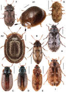
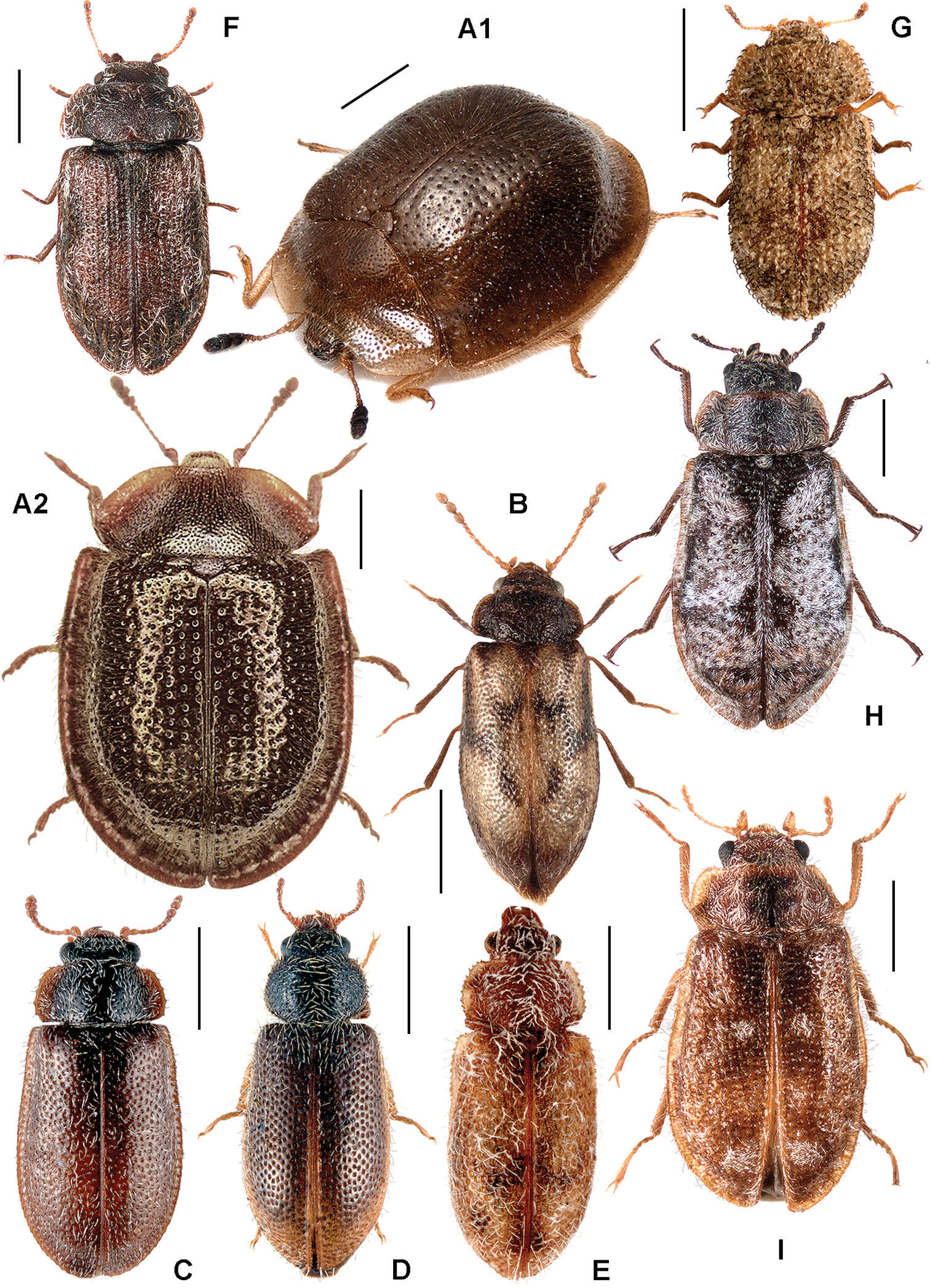
Figure 10.A Thymalus limbatus B Phloiophilus edwardsi C Eronyxa marginicollis D Decamerus haemorhoidalis E Diontolobus punctatipennis F Afrocyrona ciskeiensis G Afrocyrona dwesae H Grynoma sp., New Zealand I Grynoma diluta.
-
Francisco Hita Garcia, Brian L. Fisher
Zookeys
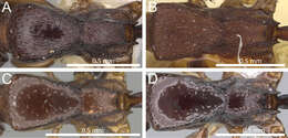
Figure 4.Mesosoma in dorsal view. A Tetramorium raptor (CASENT0195628) B Tetramorium uelense (CASENT0914084) C Tetramorium ultor (CASENT0235465) D Tetramorium venator (CASENT0401714).
-
Francisco Hita Garcia, Brian L. Fisher
Zookeys
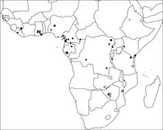
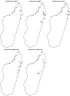
Figure 61.Geographic distribution maps for the species of the Tetramorium naganum species group. Star symbols represent type localities while circles represent non-type localities.
-
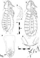
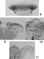
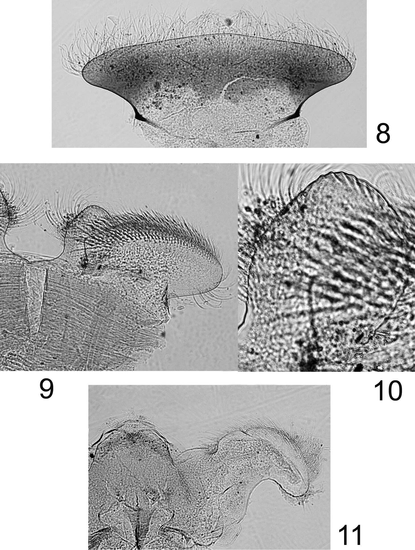
Figures 8–11.Rhithrogeniella ornata Ulmer, 1939, nymphal mouthparts. 8 Labrum in dorsal view 9 Left glossae and paraglossae of the labium 10 Detail of the glossae from 9 11 Hypopharynx, ventral view lingua and left superlingua.