-
Donald R. Davis, David L. Wagner
Zookeys
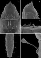
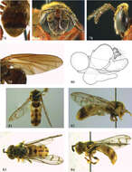
Figure 14.Phyllocnistis perseafolia sp. n. pupa. A Head, ventral view (176 µm) B Head, lateral view (200 µm) C Dorsal spines of abdominal tergum 5 (76 µm) D Lateral view of C (60 µm) E Abdominal terga 4-10 (100 µm) F Lateral seta of abdominal segment 6 (10 µm). (Length of bar scales shown in parentheses.)
-
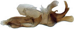
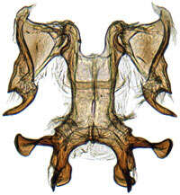
Mukaria maculata, aedeagus, connective, and style, laterally (INHS)
-
Michel P. Valim, Jason D. Weckstein
Zookeys
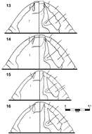
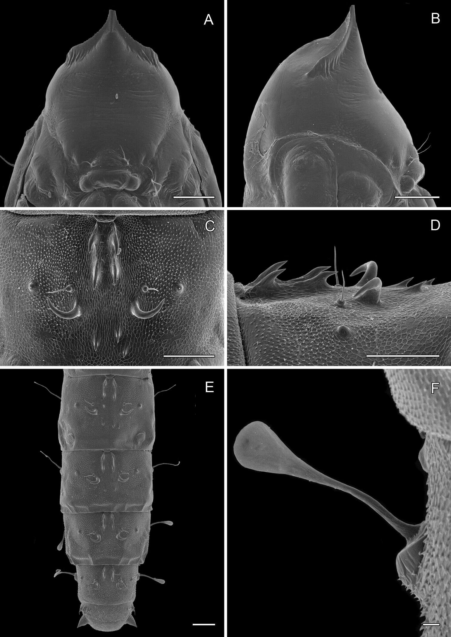
Figures 13–16.Brueelia sueta sp. n.: male preantenal region, dorso-ventral views (13); female preantenal region, dorso-ventral views (14); Brueelia cicchinoi sp. n.: male preantenal region, dorso-ventral views (15); female preantenal region, dorso-ventral views (16).
-
Juli Pujade-Villar, Paul Hanson, Claudia A. Medina, Miguel Torres, George Melika
Zookeys
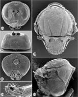
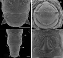
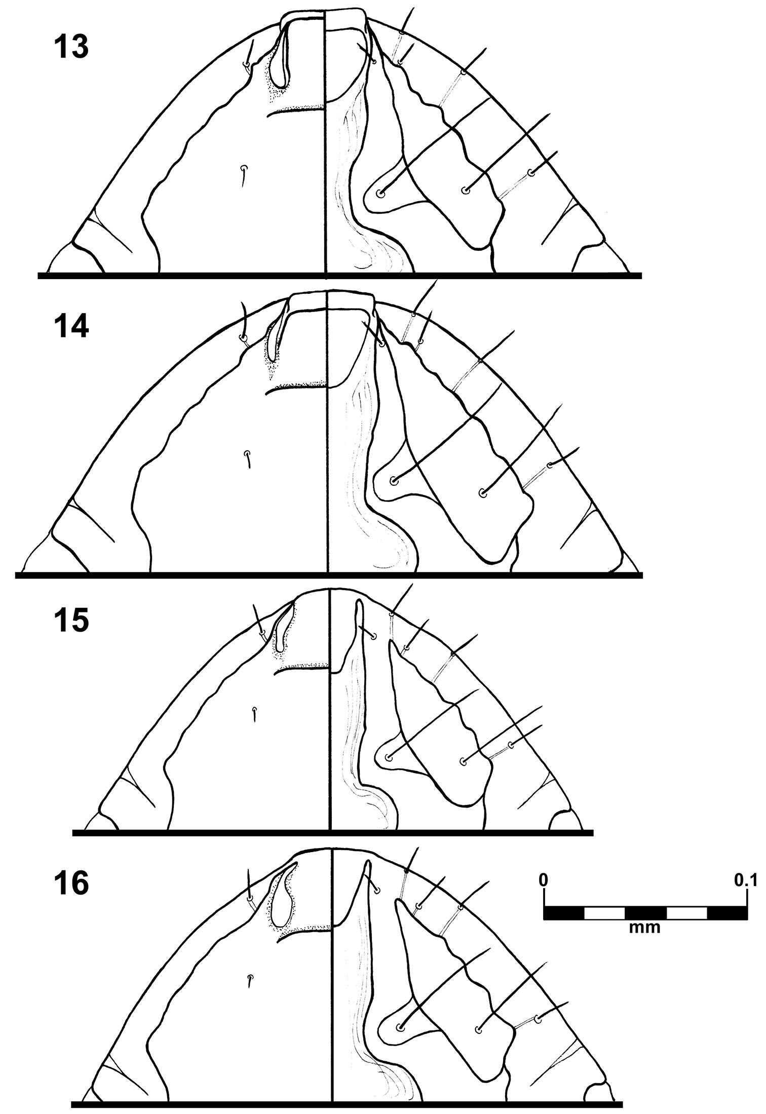
Figures 19–24.Zapatella nievesaldreyi, female 19 head (anterior view) 20 head (dorsal view) 21 head (posterior view) 22 mesosoma (dorsal view) 23 mesosoma (lateral view) 24 tarsal claw.
-
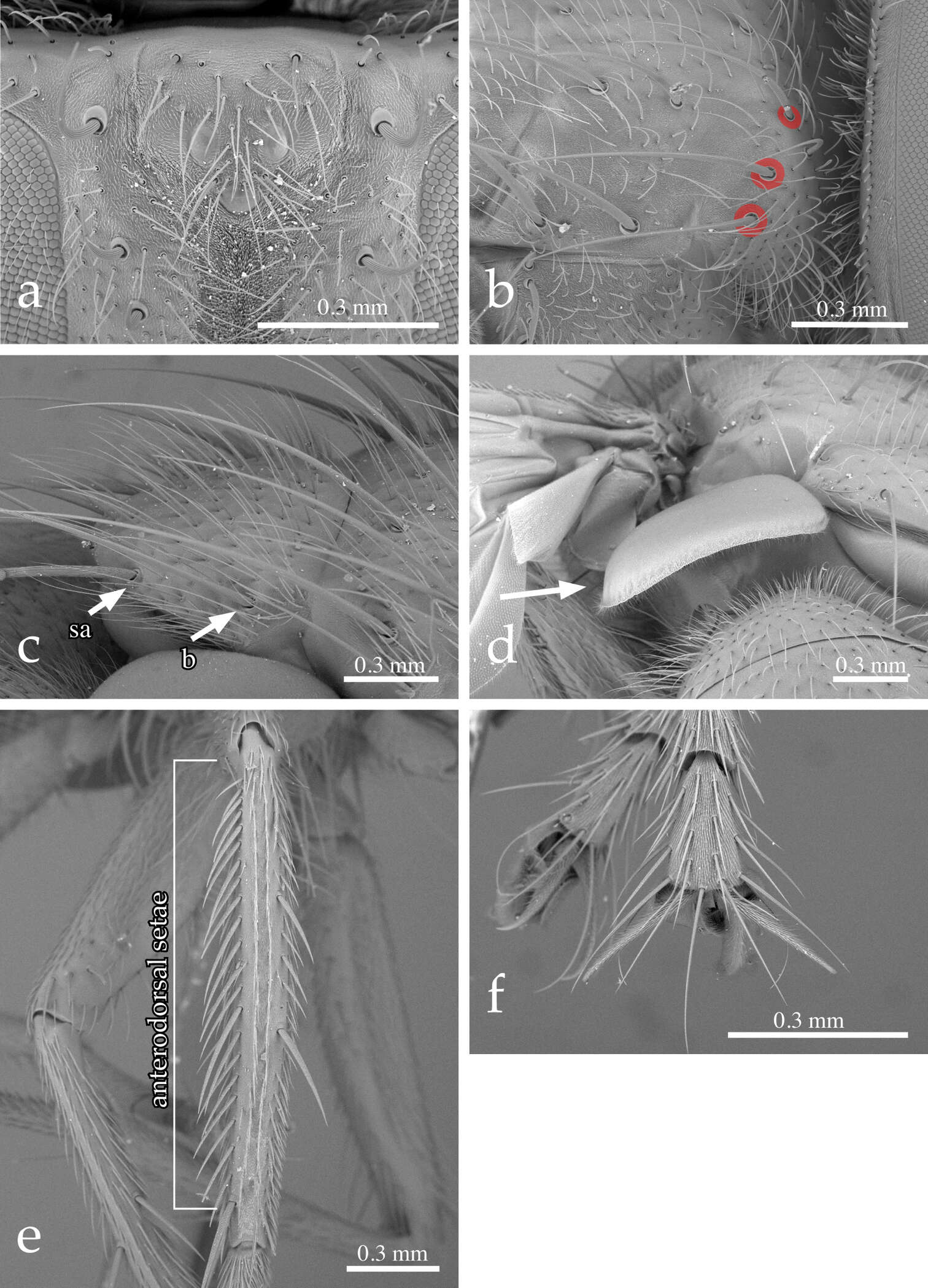
Pierfilippo Cerretti, D. Monty Wood, James E. O’Hara
Zookeys
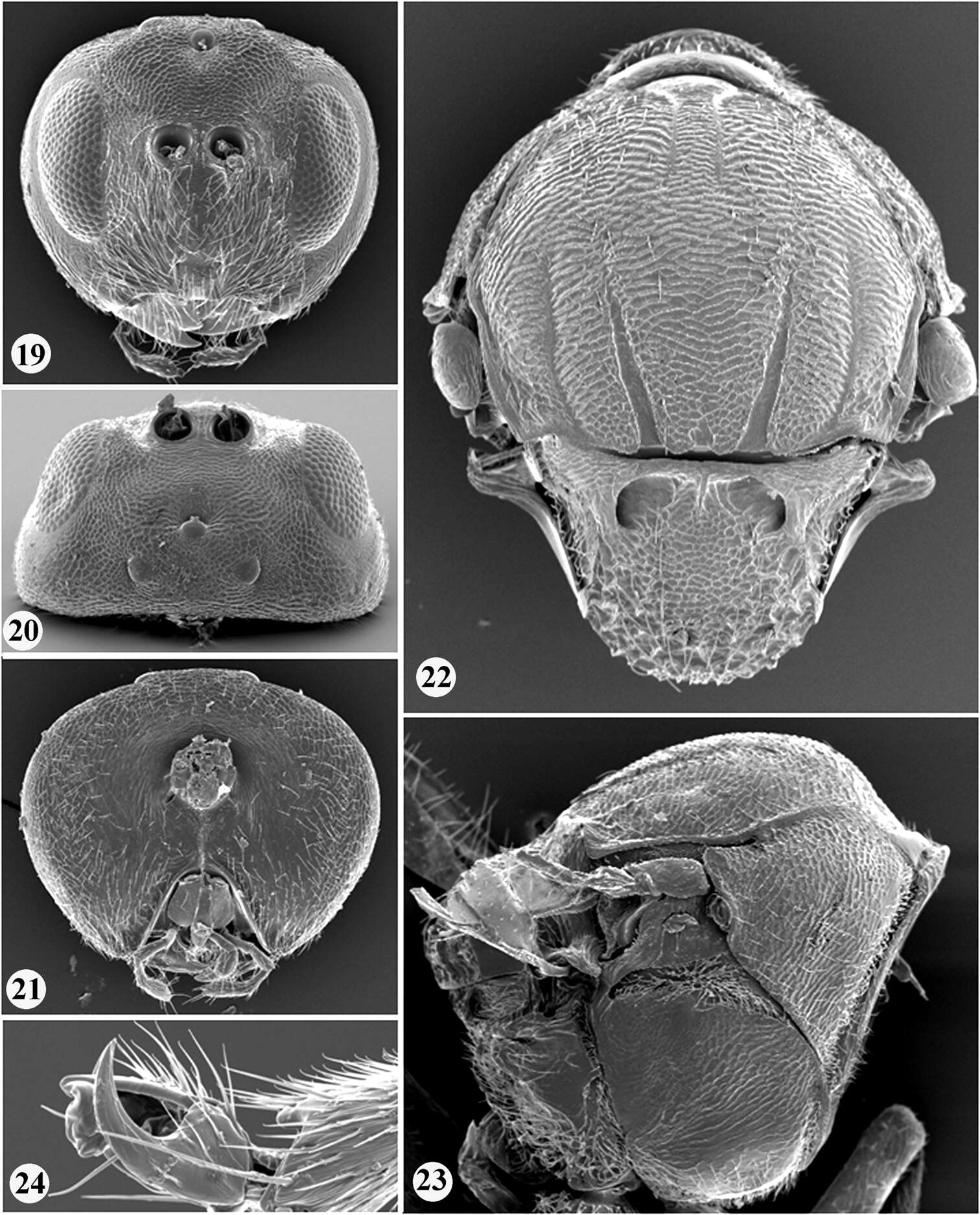
Figure 2.Neoethilla gen. n.ignobilis (male, New Mexico) a vertex in anterodorsal view b right postpronotum and part of presutural portion of scutum in laterodorsal view [circles indicate basal postpronotal saetae] c scutellum in laterodorsal view [b = basal scutellar seta; sa = subapical scutellar seta] d lower calypter in posterior view e left hind tibia in dorsal view f fore claws.
-
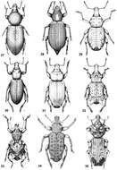
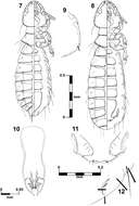
Figures 27–35.Habitus of representative Listroderini. 27 Lanteriella microphtalma 28 Telurus caudiculatus 29 Acrorius papallacta 30 Acrostomus bruchi 31 Antarctobius lacunosus 32 Germainiellus dentipennis 33 Lamiarhinus aelficus 34 Listroderes annulipes 35 Philippius superbus.
-
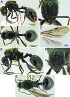
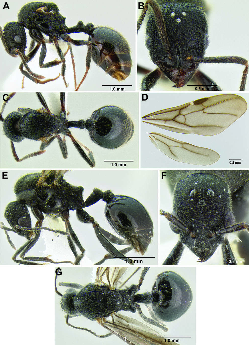
Figure 121.Stenamma megamanni A Paratype queen (CASENT0604840), profile B Same, face C Same, dorsum D Same, wings E Male (CASENT000007293), profile F Same, face G Same, dorsum.
-
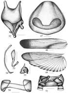
Figures 47–55.Symploce evidens sp. n. 47 pronotum 48 tegmen 49 hind wing 50 abdominal tergum 9 and lateral plates, ventral view 51 supra-anal plate and paraprocts, ventral view 52 subgenital plate, dorsal view 53 hook-like phallomere 54 median phallomere 55 right phallomere. Scale bars = 1.0 mm (Fig. 47), 2.0 mm (Figs 48–49), 0.5 mm (Figs 50–55).
-
Lizhi Huo, Xingmin Wang, Xiaosheng Chen, Shunxiang Ren
Zookeys
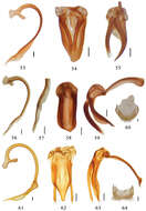
Figures 53–64.53–55 Aspidimerus guangxiensis Yu, male genitalia: 53 penis 54 tegmen, ventral view 55 tegmen, lateral view 56–60 Aspidimerus matsumurai Sasaji 56–59 male genitalia: 56 penis 57 apex of penis 58 tegmen, ventral view 59 tegmen, lateral view 60 female genitalia: ovipositor 61–64 Aspidimerus kabakovi Hoàng 61–63 male genitalia 61 penis 62 tegmen, ventral view 63 tegmen, lateral view 64 female genitalia: ovipositor. Scale bars: 0.1mm.
-
Map 12.A distribution of the tribe Ancyronini.
-
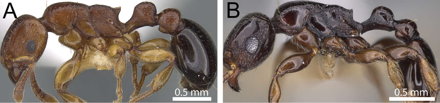
Francisco Hita Garcia, Brian L. Fisher
Zookeys
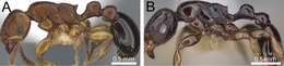
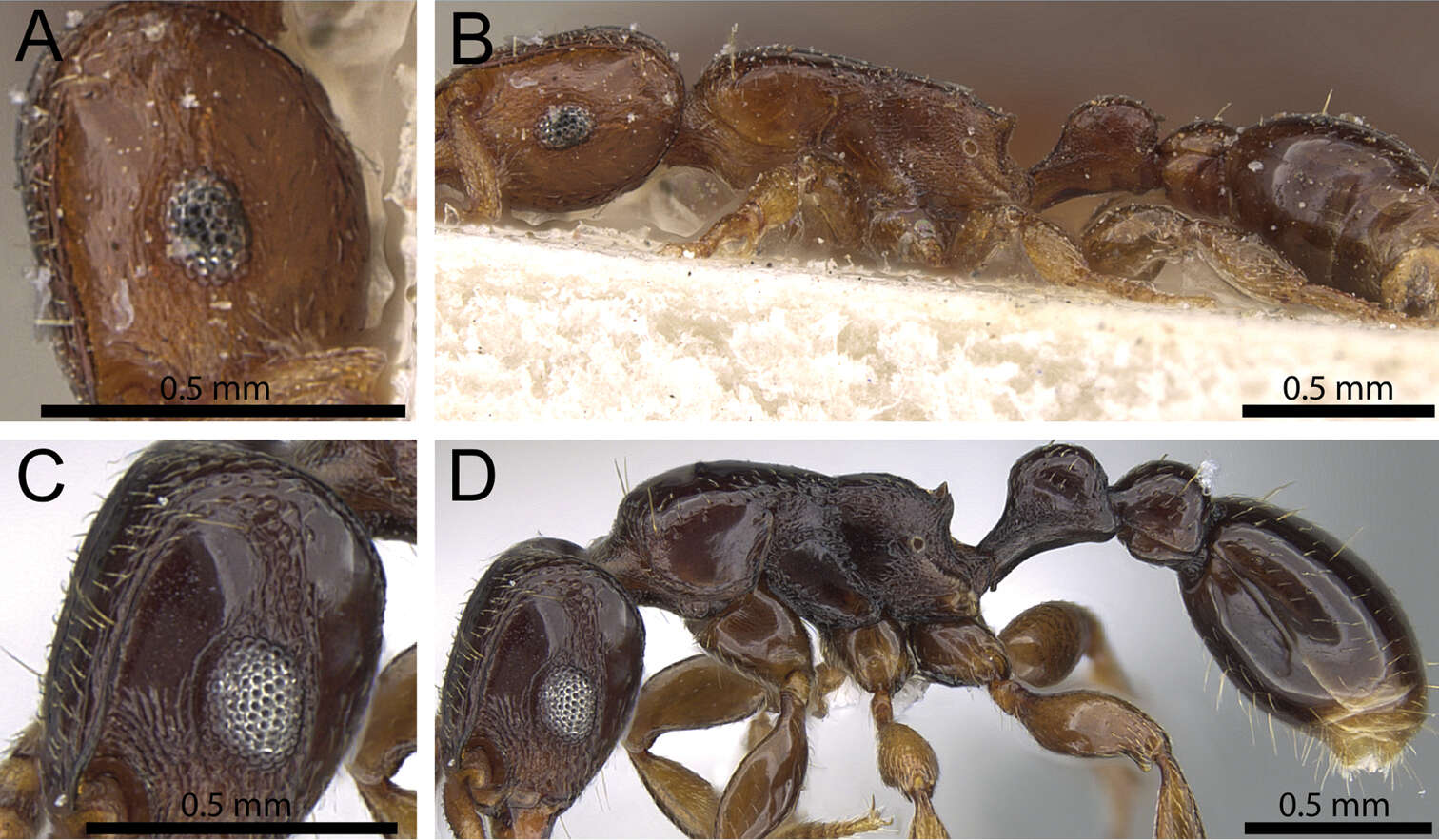
Figure 6.Body in profile. A Tetramorium decem (CASENT0914088) B Tetramorium venator (CASENT0195574).
-
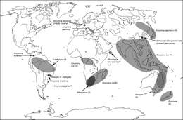
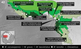
Figure 42.Distribution of the species of Monodius convexipennis, Monodius gravis, Monodius laevistriatus, Monodius lamottei and Eleoselinus gen. n. The division of Afrotropical Realm into ecoregions was adopted after Olson et al. 2001. Different colors were used to distinguish the adjacent ecoregions.
-
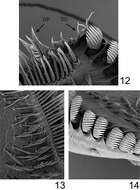
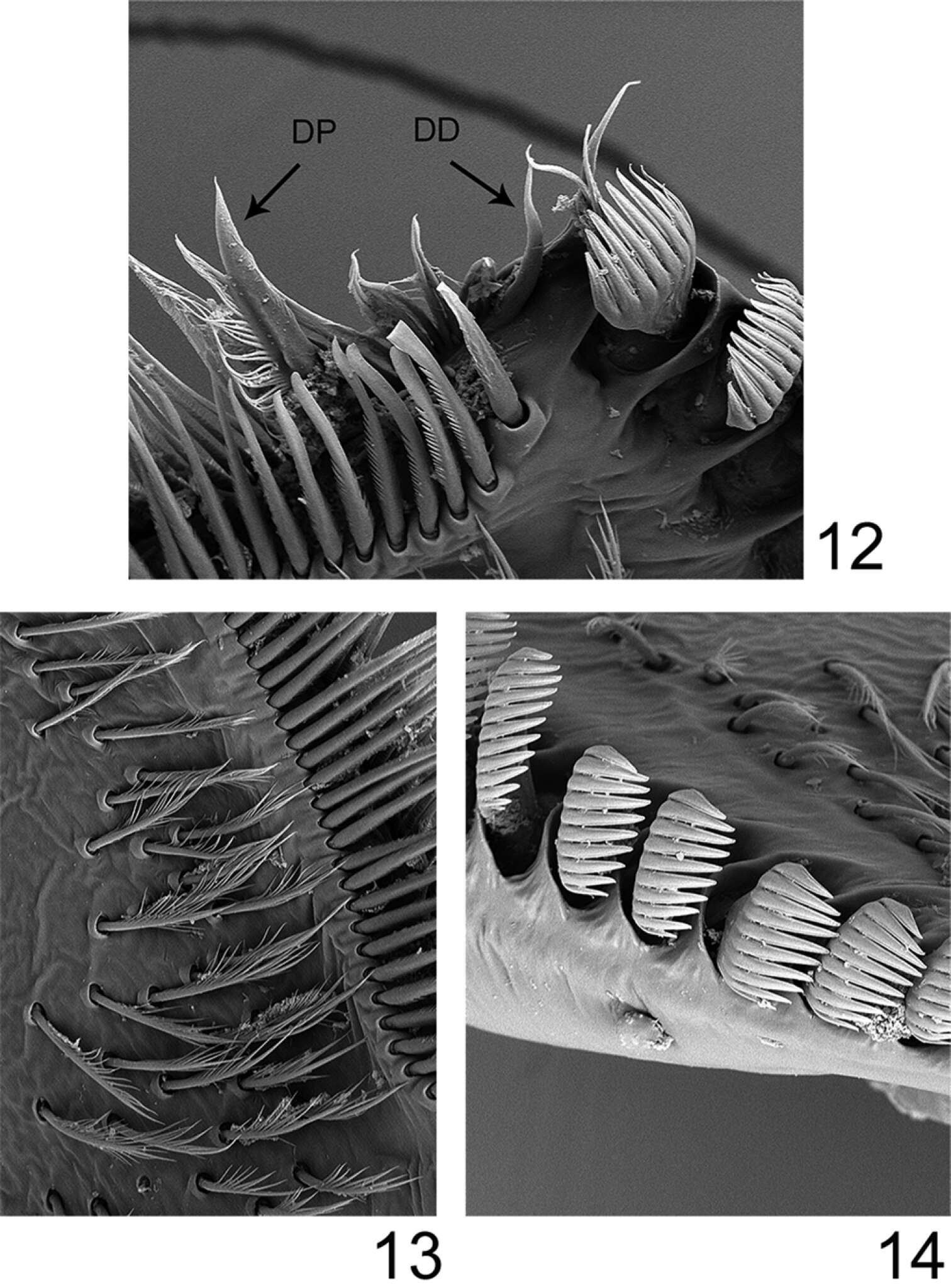
Figures 12–14.Rhithrogeniella ornata Ulmer, 1939, SEM pictures of the maxilla. 12 Dentisetae (DP: proximal dentiseta, DD: distal dentiseta) 13 Fimbriate setae on the ventral surface 14 Comb-shape setae on the crown of the galea-lacinia.
-
Donald R. Davis, David L. Wagner
Zookeys
Figure 15.Phyllocnistis perseafolia sp. n. pupa. A Abdominal terga 7-10 (100 µm) B Caudal end of abdomen (100 µm) C Abdominal sterna 6-10 (100 µm) D Spinules of sternum 6 in longitudinal rows (100 µm). (Length of bar scales shown in parentheses.)
-
Mukaria maculata, connective, style, and aedeagus, ventrally (INHS)
-
Michel P. Valim, Jason D. Weckstein
Zookeys
Figures 7–12.Brueelia cicchinoi sp. n.: male, dorso-ventral views (7); female, dorso-ventral views (8); temporal carina (9); male genitalia (10); female vulvar margin (11); female gonapophysis (12).
-
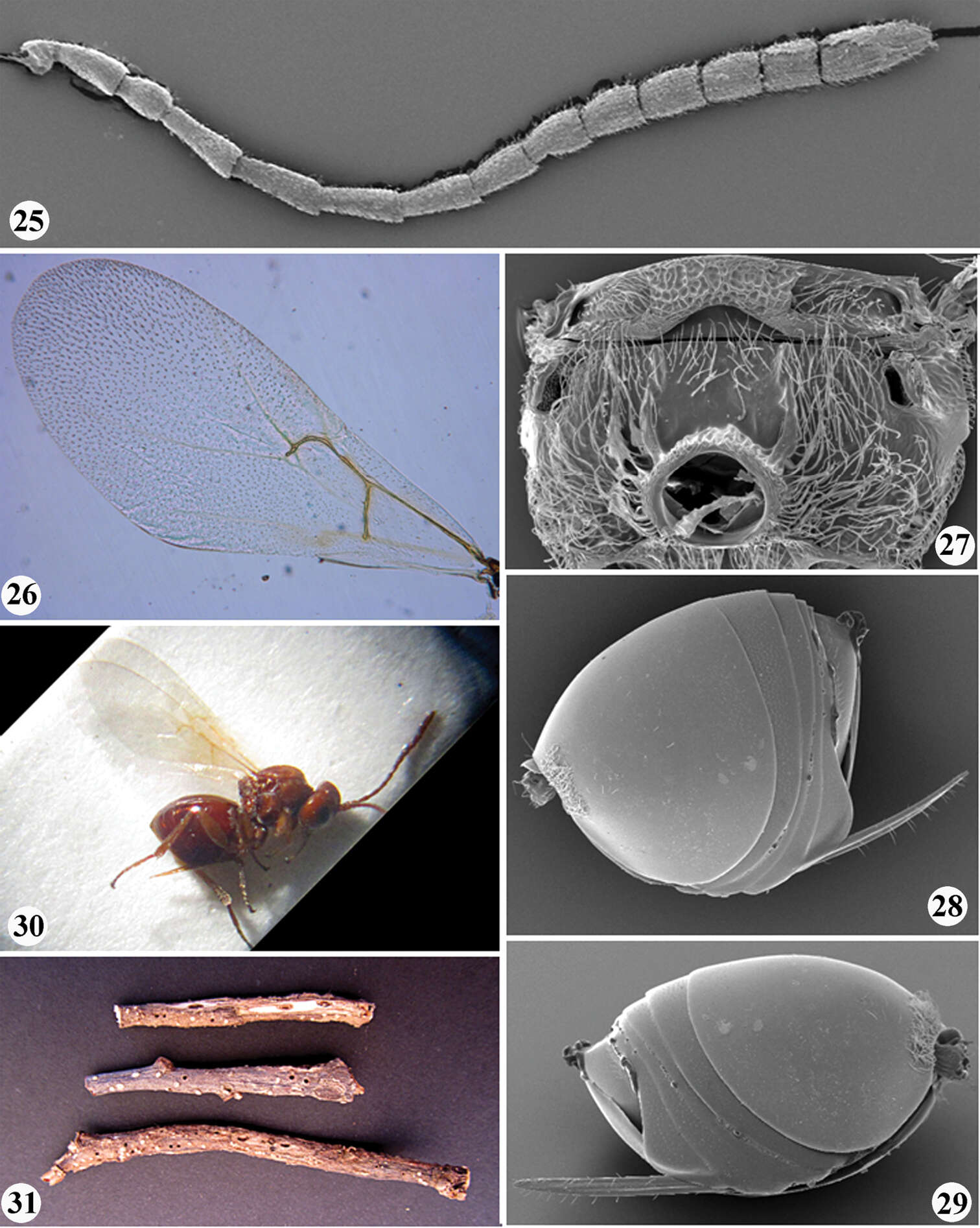
Juli Pujade-Villar, Paul Hanson, Claudia A. Medina, Miguel Torres, George Melika
Zookeys
Figures 25–31.Zapatella nievesaldreyi,female: 25 antenna 26 forewing 27 metascutellum and propodeum (posterodorsal view) 28 metasoma (lateral view) 29 metasoma with ventral spine of hypopygium (lateral view) 30 female habitus (lateral view) 31 twigs with galls.
-
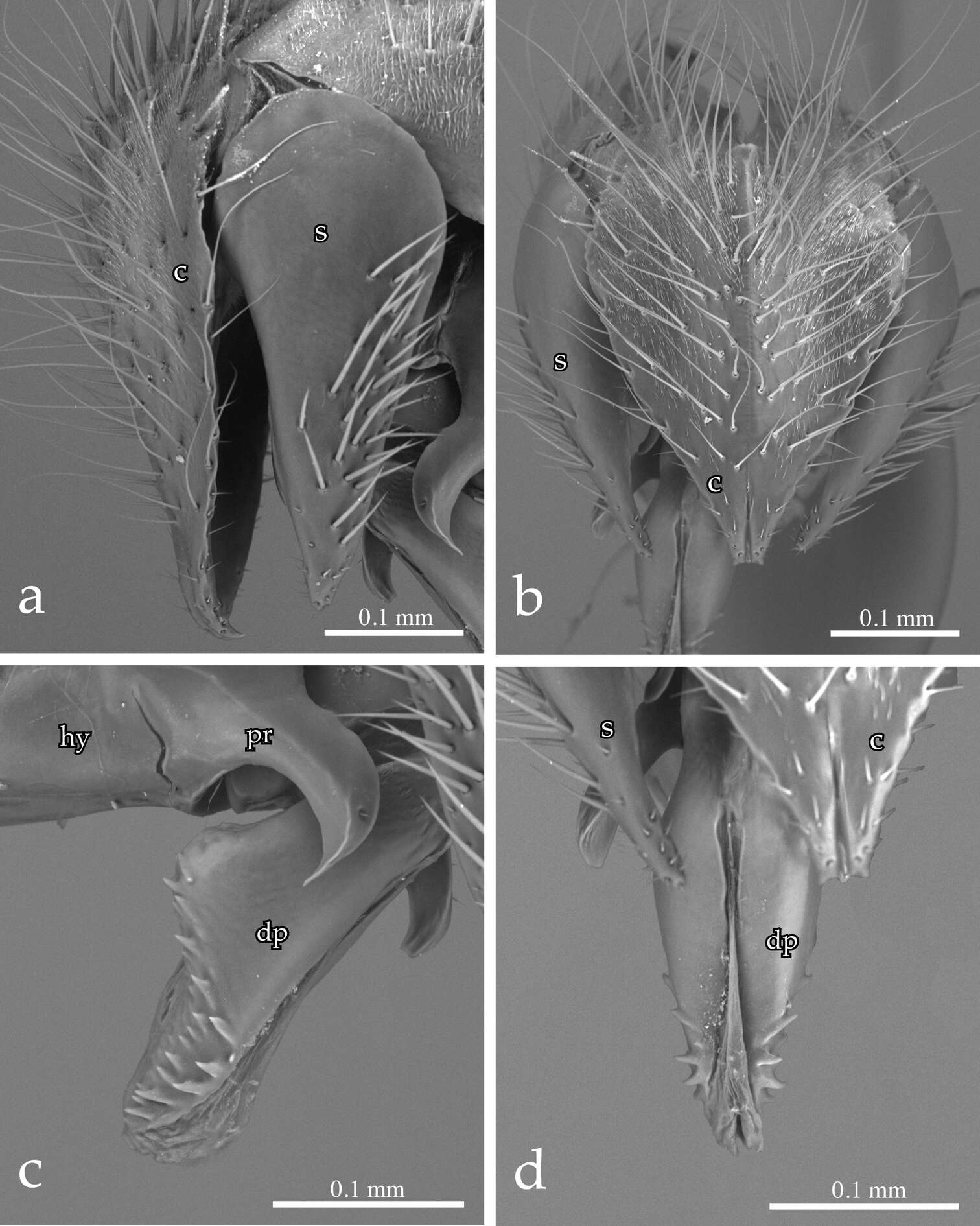
Pierfilippo Cerretti, D. Monty Wood, James E. O’Hara
Zookeys
Figure 4.Neoethilla gen. n.ignobilis (male, New Mexico) [c = cerci; hy = hypandrium; dp = distiphallus; pr = pregonite; s = surstylus] a cerci and right surstylus in lateral view b epandrial complex in posterior view c distiphallus and pregonite in left lateral view d distiphallus in dorsal view.
-
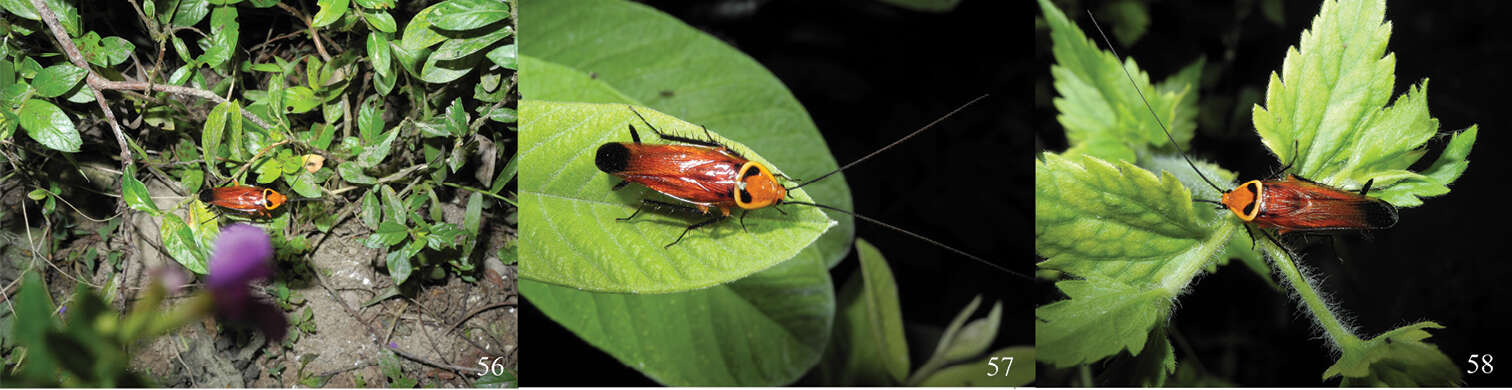
Menno Reemer, Gunilla Ståhls
Zookeys
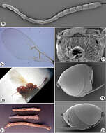
Figures 94–100.94–96 Heliodon, male genitalia: 94 Heliodon doris (holotype) 95 Heliodon tiber (holotype) 96 Heliodon gloriosus (holotype of Microdon aurivesta Hull, jun. syn.) 97–98 Hypselosyrphus amazonicus female (holotype Microdon scutellaris Shannon): 97 habitus dorsal 98 habitus lateral 99–100 Hypselosyrphus trigonus male (holotype): 99 head frontal 100 head lateral.
-
Figure 121.Stenamma megamanni A Paratype queen (CASENT0604840), profile B Same, face C Same, dorsum D Same, wings E Male (CASENT000007293), profile F Same, face G Same, dorsum.
-
Figures 56–58.Symploce evidens sp. n. in Mountain Qixianling, Baoting County, Hainan Province, 2 May 2013 (photographs by Keliang Wu).
-
Lizhi Huo, Xingmin Wang, Xiaosheng Chen, Shunxiang Ren
Zookeys
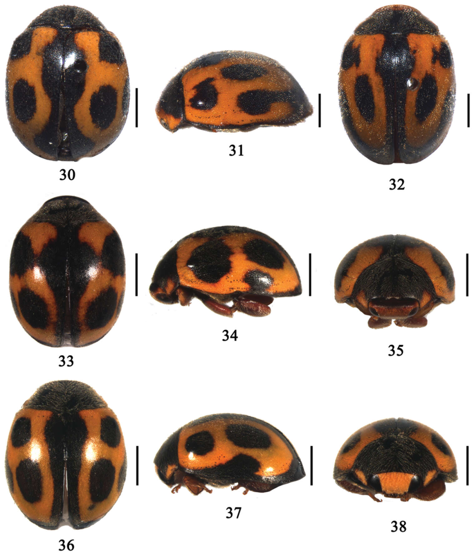
Figures 30–38.30 Aspidimerus decemmaculatus Pang & Mao, dorsal view; 31–32 Aspidimerus mouhoti Crotch, 31 lateral view 32 dorsal view 33–35 Aspidimerus zhenkangicus Huo & Ren, sp. n. 33 dorsal view 34 lateral view 35 frontal view 36–38 Aspidimerus ruficrus Gorham, 36 dorsal view 37 lateral view 38 frontal view. Scale bars: 1.0mm.
-
Bernhard Seifert, Isabelle Kleeberg, Barbara Feldmeyer, Tobias Pamminger, Evelien Jongepier, Susanne Foitzik
Zookeys
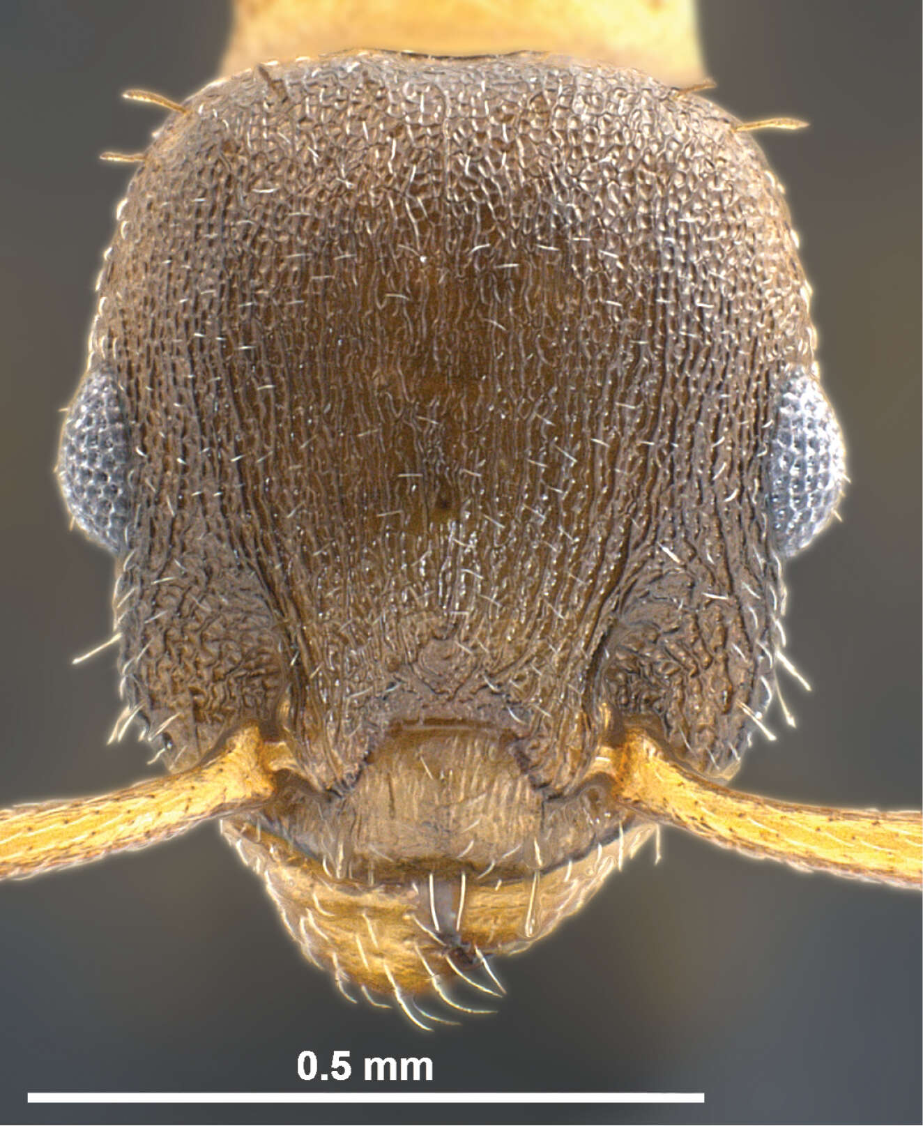
Figure 1.Temnothorax pilagens sp. n., worker, head of holotype in dorsal view.
-
Francisco Hita Garcia, Brian L. Fisher
Zookeys
Figure 7.Head and body in profile. A, B Tetramorium ultor (CASENT0235465) C, D Tetramorium venator (CASENT0401714).