-
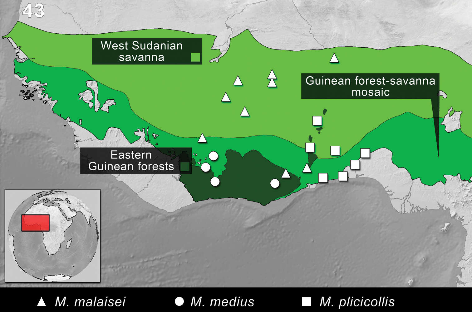
Figure 43.Distribution of the species of Monodius malaisei, Monodius medius and Monodius plicicollis. The division of Afrotropical Realm into ecoregions was adopted after Olson et al. 2001. Different colors were used to distinguish the adjacent ecoregions.
-
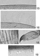
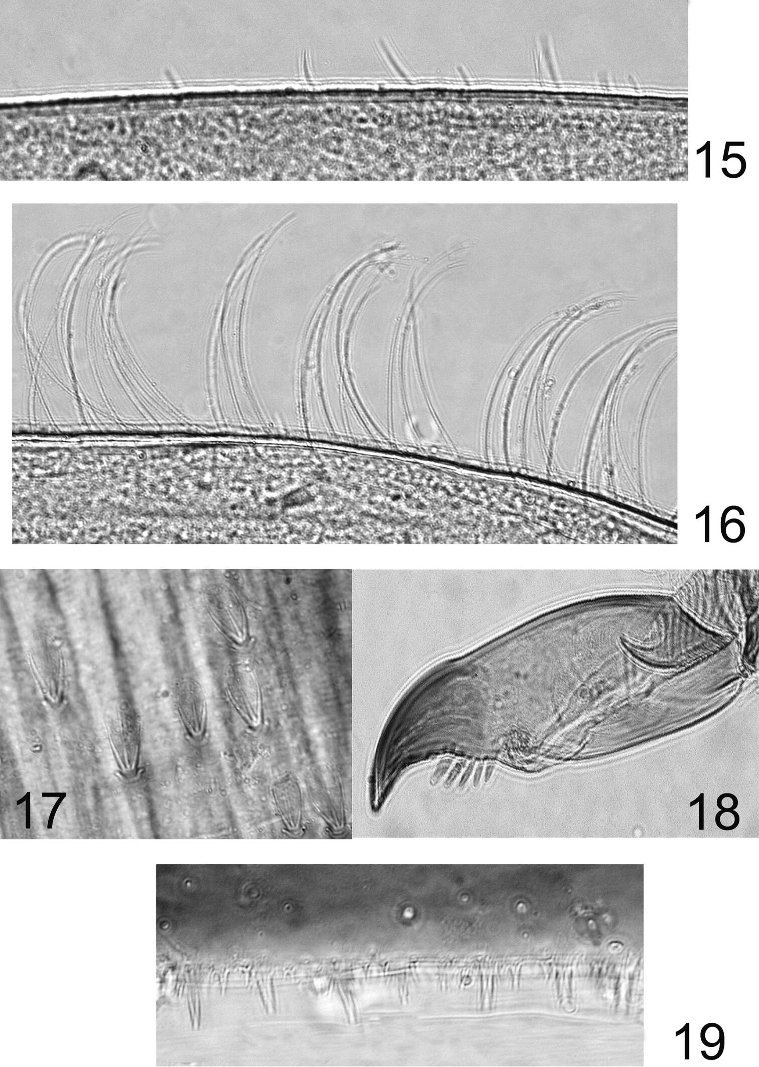
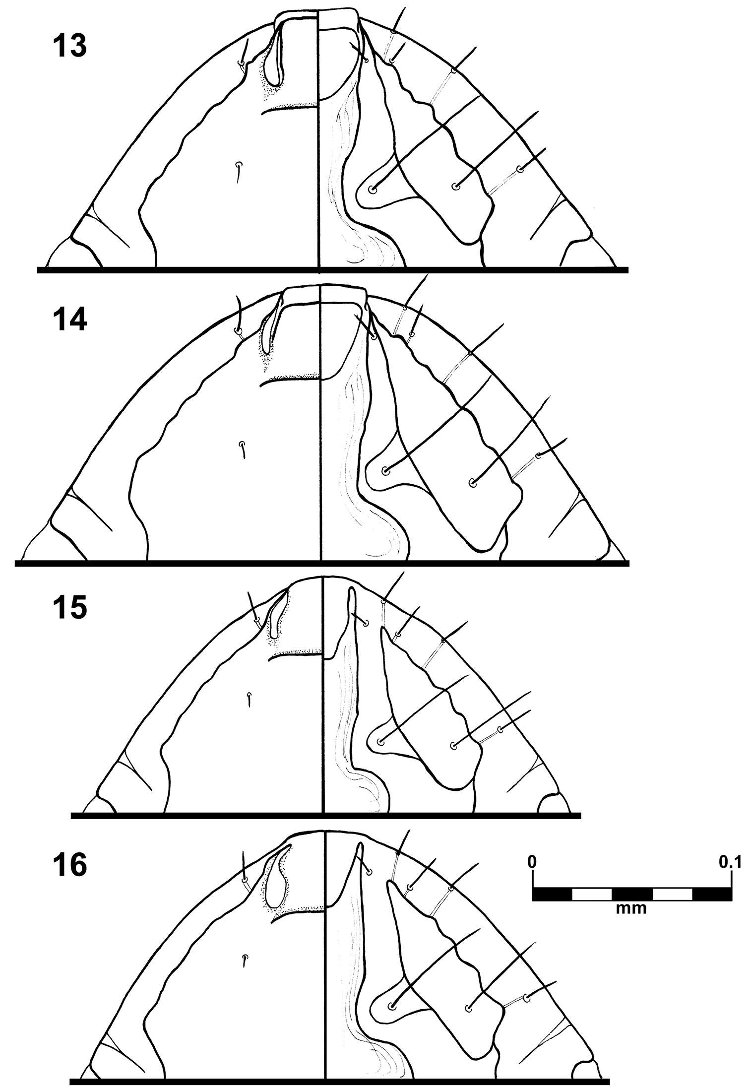
Figures 15–19.Rhithrogeniella ornata Ulmer, 1939. 15 Outer margin of the fore tibia 16 Outer margin of the hind tibia 17 Bristles on the dorsal surface of hind femur 18 Tarsal claw 19 Posterior margin of tergite V.
-
Donald R. Davis, David L. Wagner
Zookeys
Figure 19. Phyllocnistis perseafolia sp. n. genitalia. A Male, ventral view B Mesal view of valva C Aedeagus D Female, lateral view E Ventral view of D segments 7-10.
-
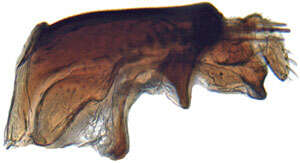
Mukaria maculata, subgenital plates and valve, ventrally (INHS)
-
Michel P. Valim, Jason D. Weckstein
Zookeys
Figures 13–16.Brueelia sueta sp. n.: male preantenal region, dorso-ventral views (13); female preantenal region, dorso-ventral views (14); Brueelia cicchinoi sp. n.: male preantenal region, dorso-ventral views (15); female preantenal region, dorso-ventral views (16).
-
Juli Pujade-Villar, Paul Hanson, Claudia A. Medina, Miguel Torres, George Melika
Zookeys
Figures 32–38.Zapatella cryptica, female 32 head (anterior view) 33 head (dorsal view) 34 head (posterior view) 35 antenna 36 hind coxa 37 mesosoma (lateral view) 38 mesosoma (dorsal view).
-
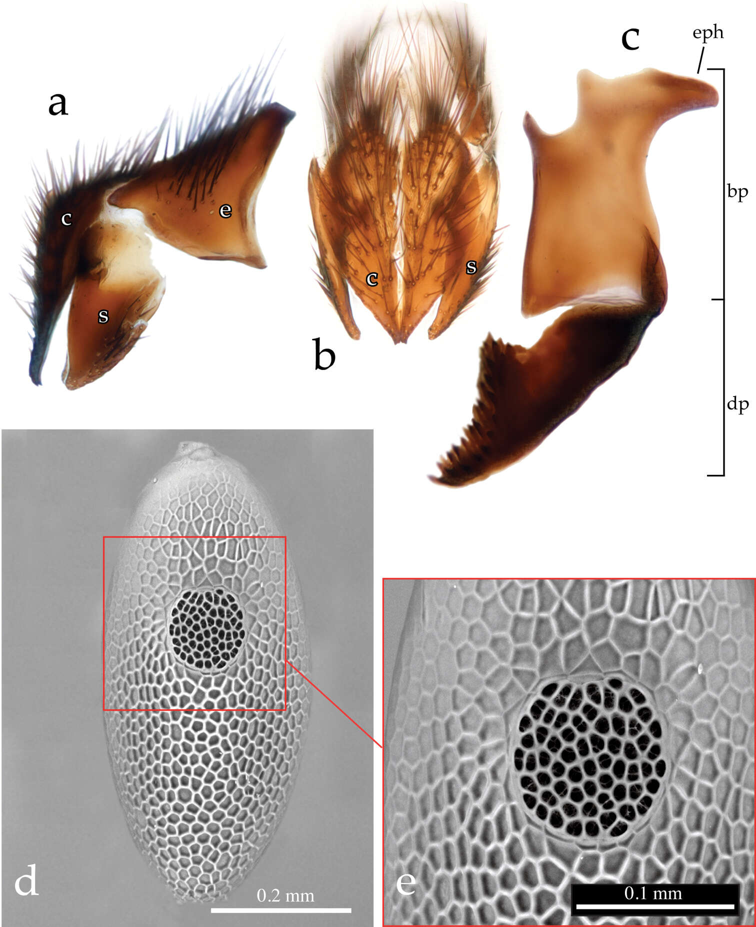
Pierfilippo Cerretti, D. Monty Wood, James E. O’Hara
Zookeys
Figure 5.Neoethilla gen. n. ignobilis a–c terminalia (male, Idaho) [c = cerci; bp = basiphallus; dp = distiphallus; e = epnadrium; eph = epiphallus] a epandrial complex in lateral view b epandrial complex in posterior view c phallus in lateral view d–e egg in dorsal view (Massachusetts) d habitus e detail of operculum.
-
Menno Reemer, Gunilla Ståhls
Zookeys
Figures 101–108.101–102 Hypselosyrphus, male genitalia: 101 Heliodon amazonicus (Peru, coll. RMNH) (cercus missing) 102 Heliodon analis (holotype) 103–106 Indascia gracilis male (holotype): 103 habitus dorsal 104 habitus lateral 105 wing 106 genitalia 107–108 Indascia gigantica male (holotype): 107 habitus dorsal 108 habitus lateral.
-
Figure 122.Distribution map of Stenamma megamanni.
-
Lizhi Huo, Xingmin Wang, Xiaosheng Chen, Shunxiang Ren
Zookeys
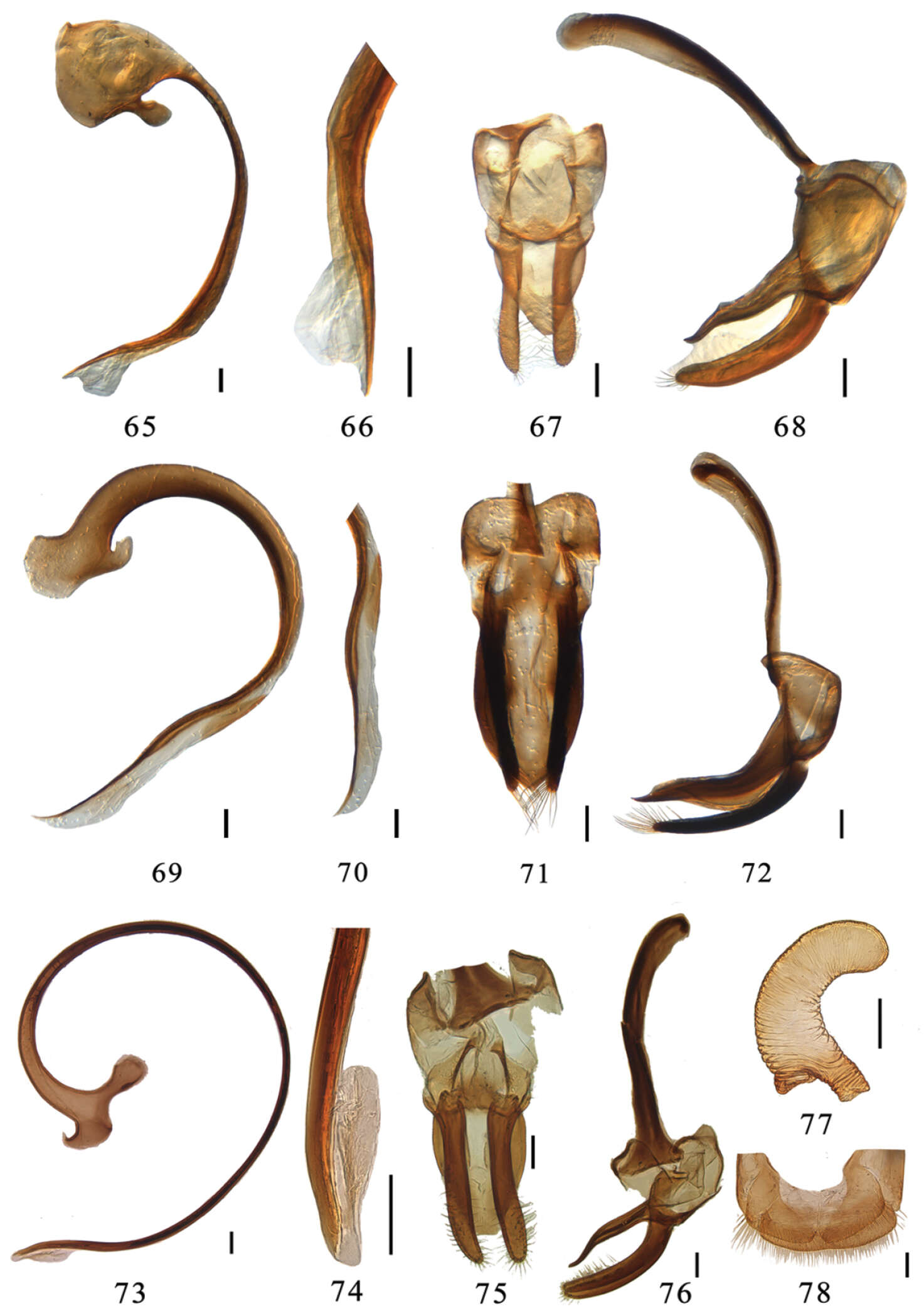
Figures 65–78.65–68 Aspidimerus decemmaculatus Pang & Mao, male genitalia: 65 penis 66 apex of penis 67 tegmen, ventral view 68 tegmen, lateral view 69–72 Aspidimerus mouhoti Crotch, male genitalia: 69 penis 70 apex of penis 71 tegmen, ventral view 72 tegmen, lateral view 73–78 Aspidimerus zhenkangicus Huo & Ren, sp. n.73–76 male genitalia: 73 penis 74 apex of penis 75 tegmen, ventral view 76 tegmen, lateral view 77–78 female genitalia: 77 spermatheca 78 ovipositor. Scale bars: 0.1mm.
-
Bernhard Seifert, Isabelle Kleeberg, Barbara Feldmeyer, Tobias Pamminger, Evelien Jongepier, Susanne Foitzik
Zookeys
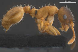
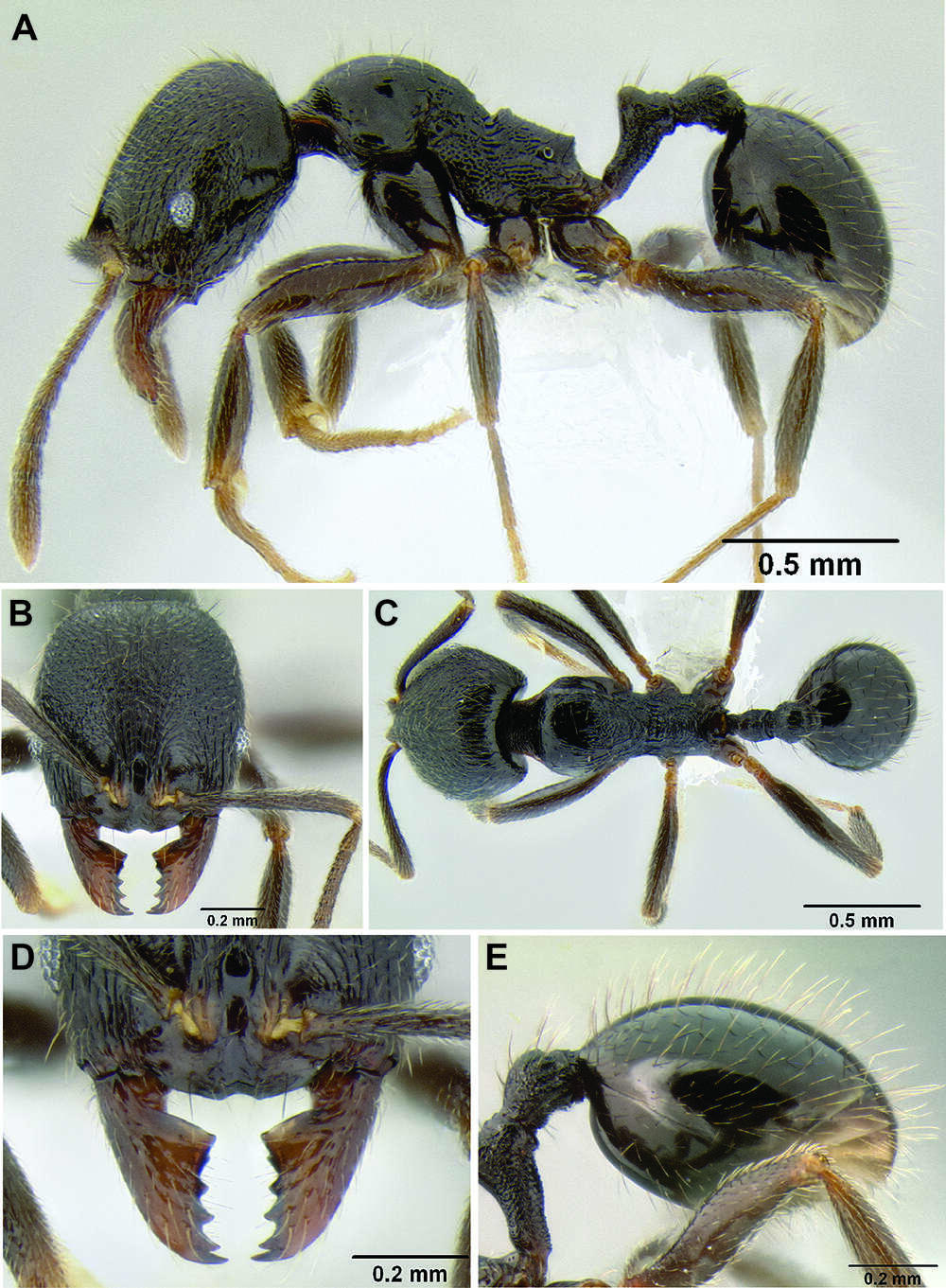
Figure 3.Temnothorax pilagens sp. n., worker, holotype in lateral view.
-
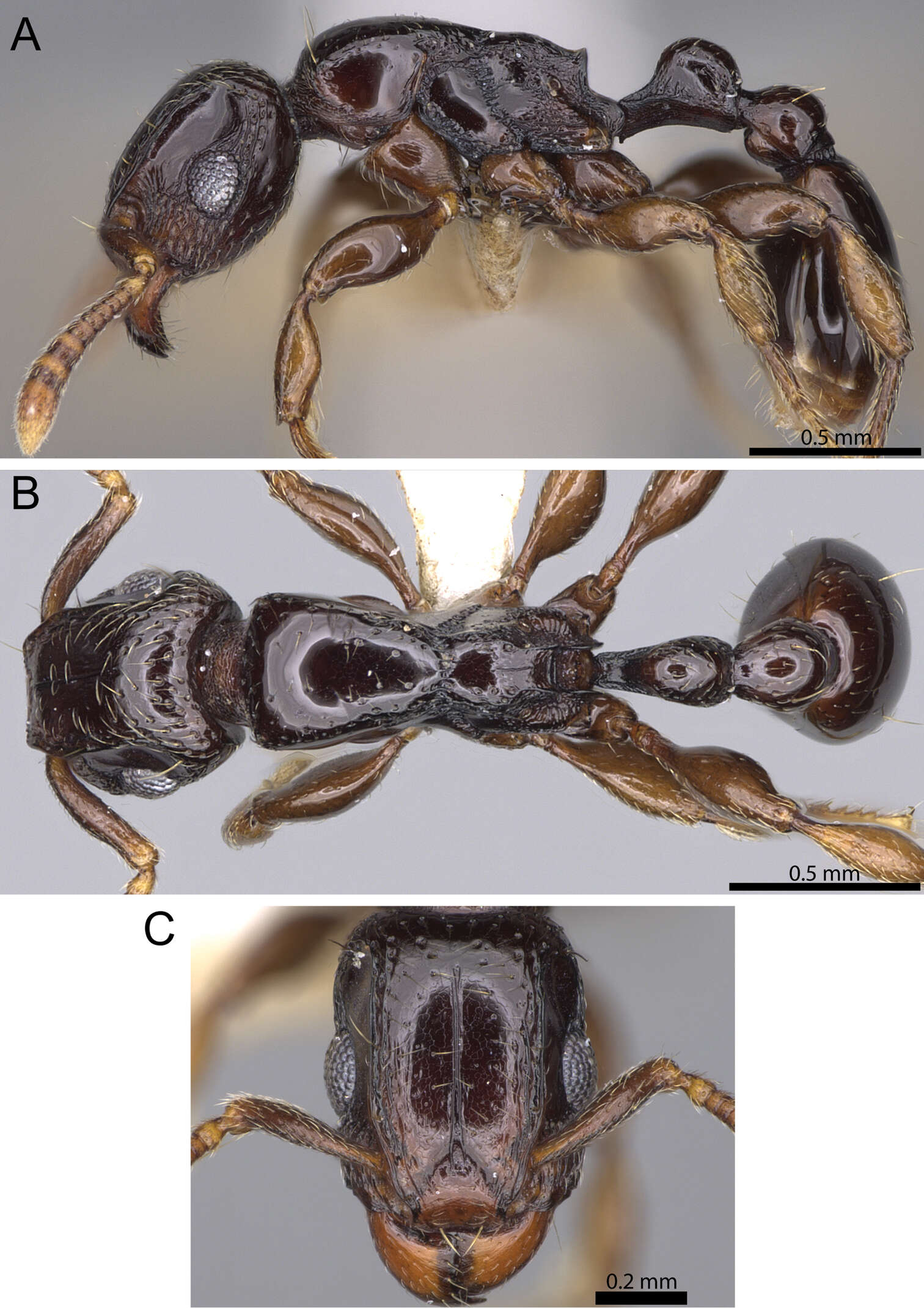
Francisco Hita Garcia, Brian L. Fisher
Zookeys
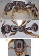
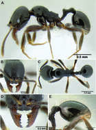
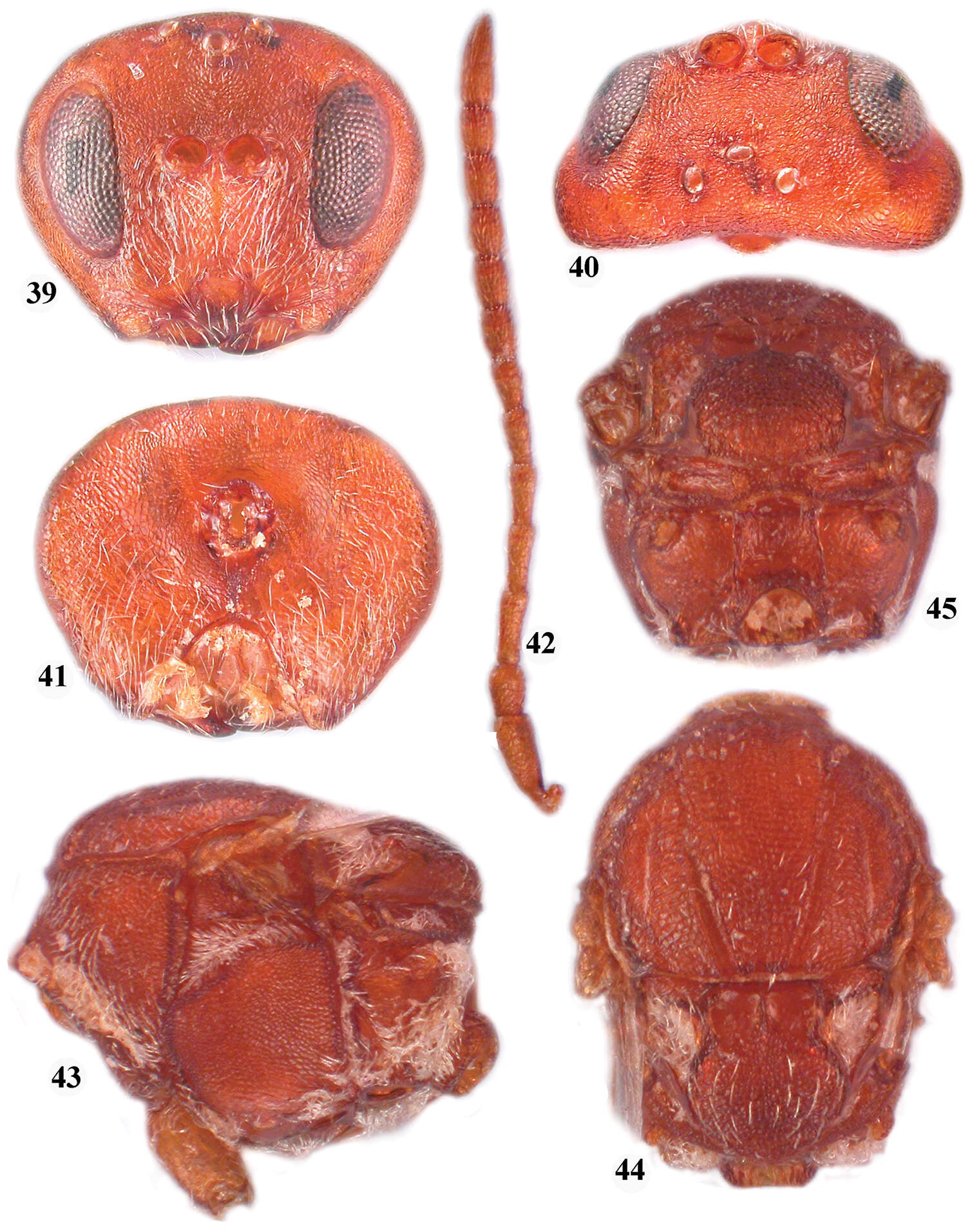
Figure 12.Tetramorium venator holotype worker (CASENT0195574). A Body in profile B Body in dorsal view C Head in full-face view.
-
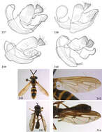
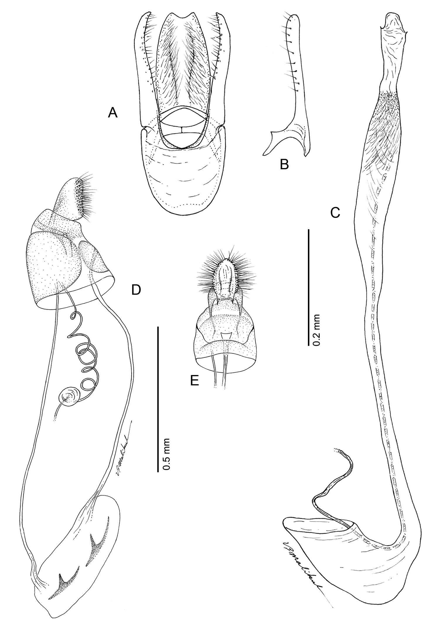
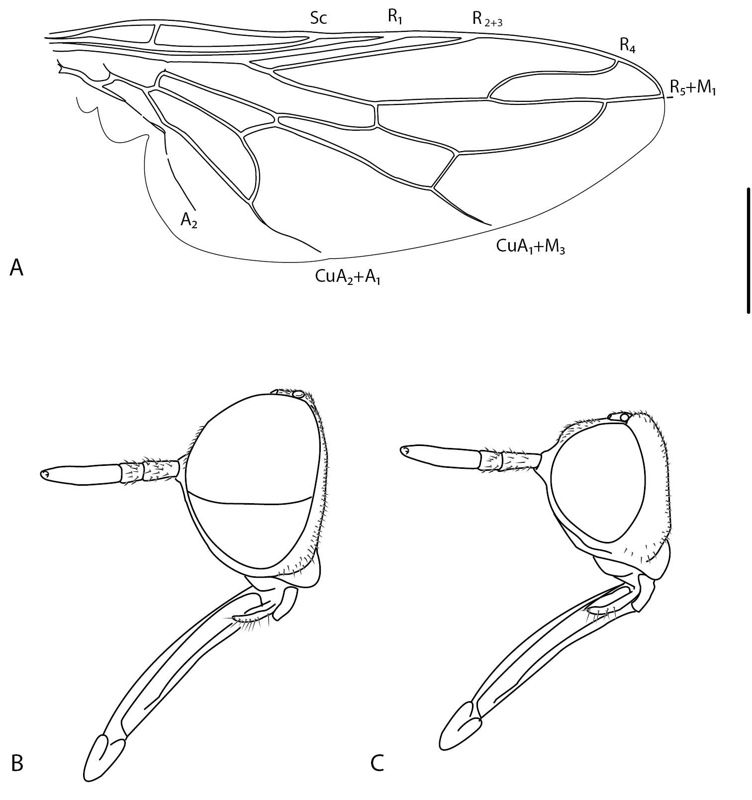
Figures 1–7.Head, dorsal view (1, 2), ventral view (4); antenna (3); pronotal disc (5, 6); mesotibia (7). Ectateus calcaripes (3), Ectateus crenatus (2), Ectateus ghesquierei (5), Monodius medius (7), Monodius plicicollis (4), Selinus planus (1, 6).
-
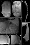
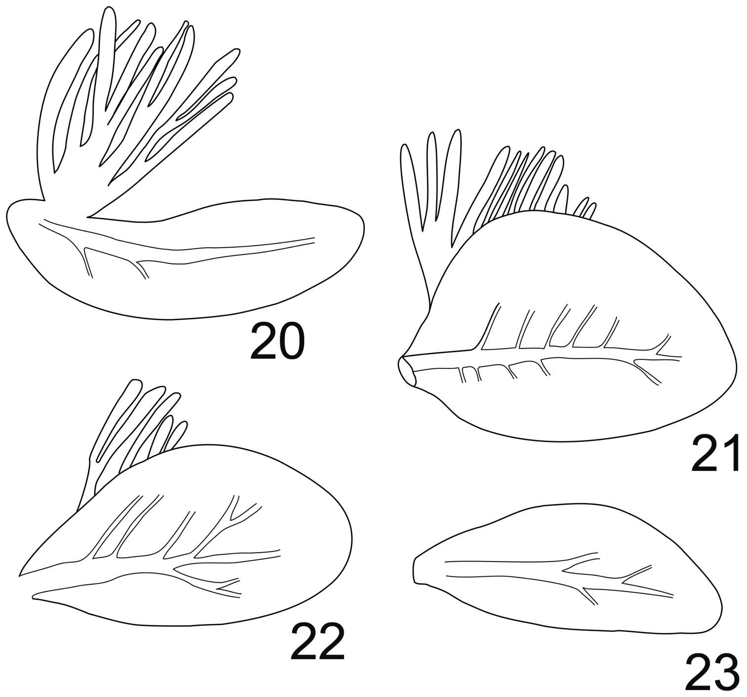
Figures 20–23.Rhithrogeniella ornata Ulmer, 1939. 20 Gill I 21 Gill IV 22 Gill VI 23 Gill VII.
-
Donald R. Davis, Jurate De Prins
Zookeys
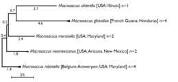
Figure 1.Compressed subtree sequenced data for cytochrome c oxidase I (COI) of Macrosaccus, derived from 13 samples among 5 species based upon neighbor-joining analysis with Kimura 2-parameter model. Numbers above branches indicate branch length. Sequence lengths obtained for all samples were 658bp each.
-
Mukaria maculata, pygofer, laterally (INHS)
-
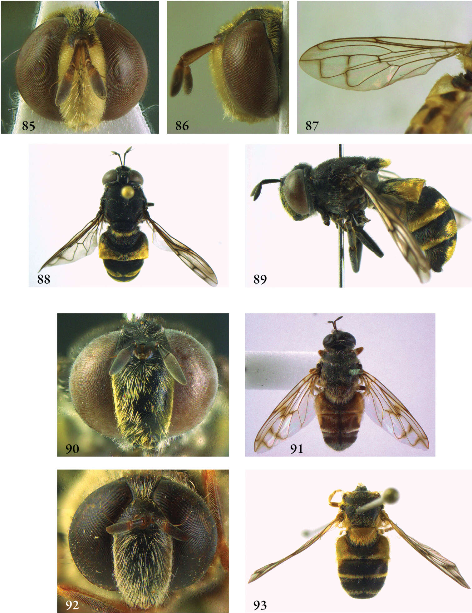
Shaun L. Winterton, Babak Gharali
Zookeys
Figure 1.Iranotrichia insolita sp. n.: A. wing; B. male head, lateral; C, female head, lateral. Scale line = 0.2 mm.
-
Juli Pujade-Villar, Paul Hanson, Claudia A. Medina, Miguel Torres, George Melika
Zookeys
Figures 39–45.Zapatella herberti, female 39, head (anterior view) 40 head (dorsal view) 41 head (posterior view) 42 antenna 43 mesosoma (lateral view) 44 mesosoma (dorsal view) 45 metascutellum and propodeum (posterodorsal view).
-
Maurizio Biondi, Paola D’Alessandro
Zookeys
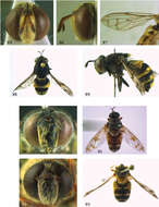
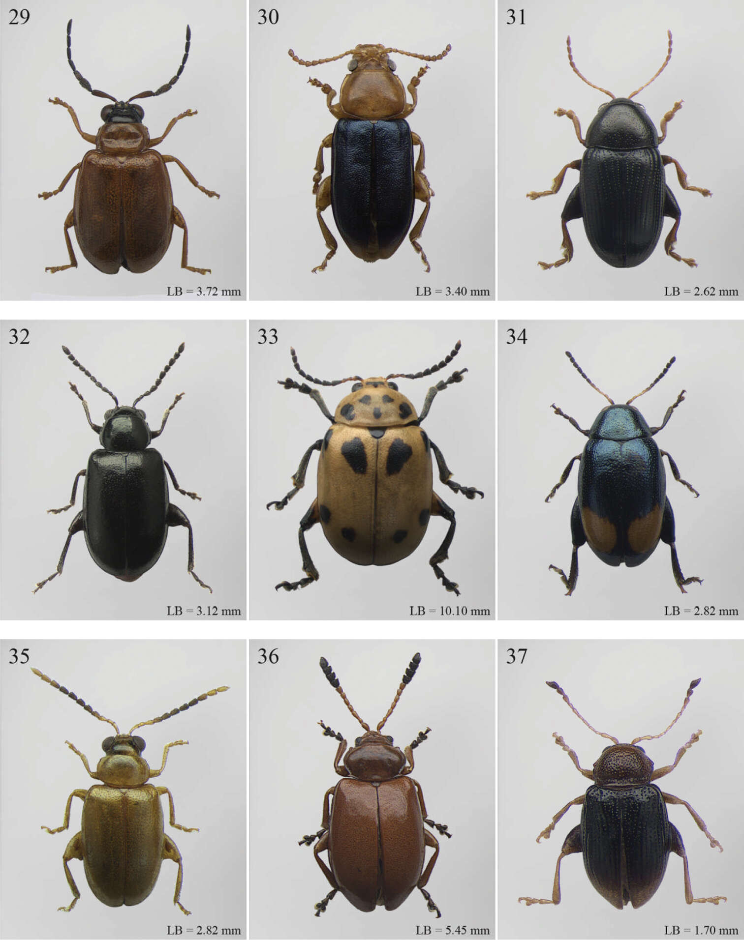
Figures 29–37.Habitus. 29 Chaillucola formicicornis Bechyné 30 Chirodica chalcoptera Germar 31 Collartaltica cryptostoma Bechyné 32 Decaria abdominalis Jacoby 33 Diamphidia nigroornata Stål 34 Dibolia bimaculata Jacoby 35 Dimonikaea descarpentriesi Bechyné 36 Diphaulacosoma laevipenne Jacoby 37 Djallonia maindra Bechyné.
-
Menno Reemer, Gunilla Ståhls
Zookeys
Figures 251–259.251–252 Oligeriops iridomyrmex female (syntype): 251 head frontal 252 head lateral 253 Oligeriops dimorphon male (Australia, coll. USNM), genitalia lateral 254–255 Omegasyrphus pallipennis male (USA, California, coll. RMNH) 254 habitus dorsal 255 habitus lateral 256 Omegasyrphus coarctatus male (USA, Virginia, coll. RMNH), genitalia lateral. 257–258 Paragodon paragoides female (Costa Rica, coll. RMNH): 257 habitus lateral 258 wing.
-
Figure 125.Stenamma muralla holotype worker (CASENT0621311) A Profile B Face C Dorsum D Anterior clypeal margin in anterodorsal view E Gaster.
-
Lizhi Huo, Xingmin Wang, Xiaosheng Chen, Shunxiang Ren
Zookeys
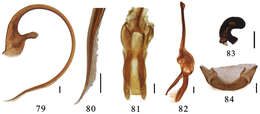
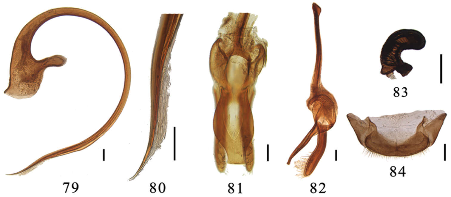
Figures 79–84.Aspidimerus ruficrus Gorham, 79–82 male genitalia: 79 penis 80 apex of penis 81 tegmen, ventral view 82 tegmen, lateral view 83–84 female genitalia: 83 spermatheca 84 ovipositor. Scale bars: 0.1mm.
-
Bernhard Seifert, Isabelle Kleeberg, Barbara Feldmeyer, Tobias Pamminger, Evelien Jongepier, Susanne Foitzik
Zookeys
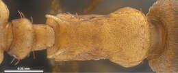
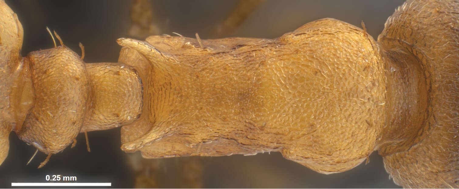
Figure 5.Temnothorax pilagens sp. n., worker, mesosoma of holotype in dorsal view.
-
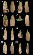
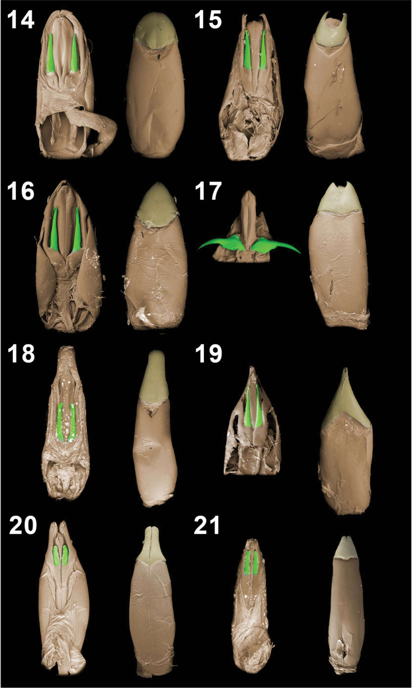
Figures 14–21.Aedeagal tegmina (dorsal and vental views). Monodius gravis (14), Monodius plicicollis (15), Monodius convexipennis (16), Monodius malaisei (17), Monodius lamottei (18), Monodius laevistriatus (19), Ectateus calcaripes (20), Selinus striatus (21).