-
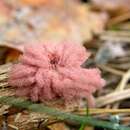
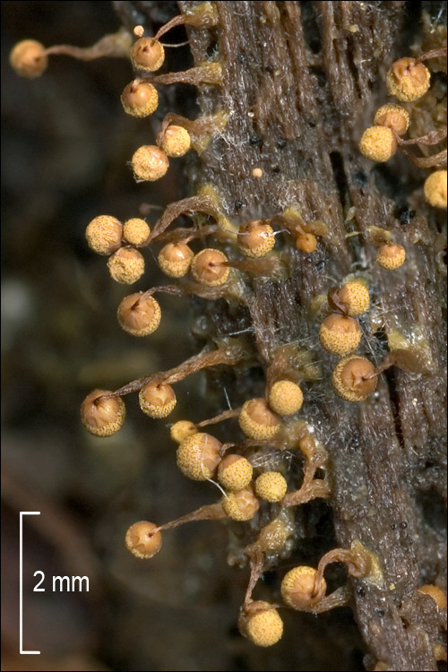
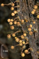
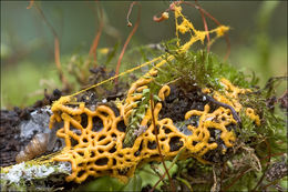
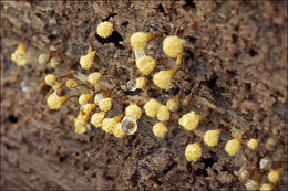
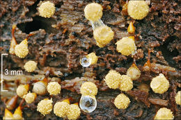
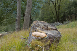
Habitat: mixed wood in a ravine, cretaceous clastic rock (flysh), rain protected by trees canopies and tall herb, in shade, very humid, precipitations ~3.000 mm/year, average temperature 8-10 deg C, altitude 450 m (1.500 feet), alpine phytogeographical region. - Substratum: fallen, debarked and completely rotten deciduous tree trunk. - Comment: Eventually Cribraria tenella but stalks seem too robust. C. atrofusca seems to be another option. Determination uncertain. No microscopic verification. - Ref: http://www.myxomycetes.it/foto_big.php?foto=Cribraria/cribraria-tenella.jpg
-
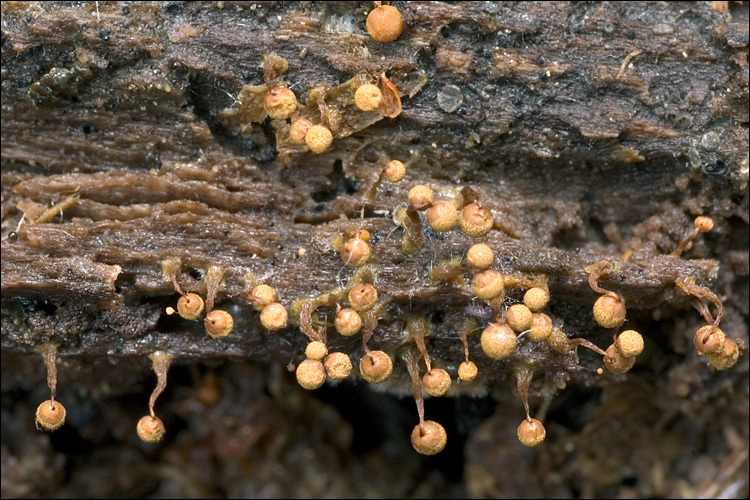
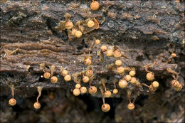
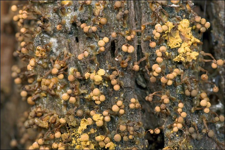
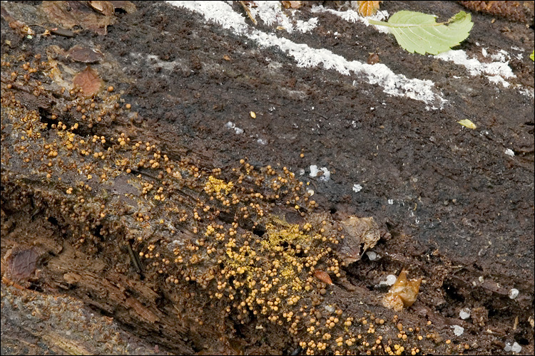
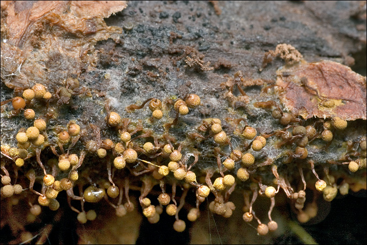
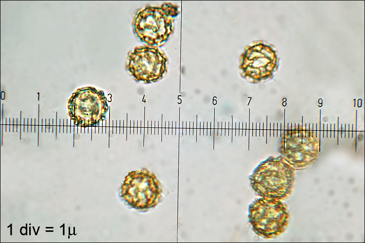
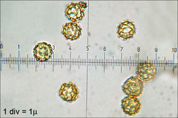
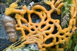
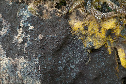
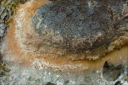
Habitat: mixed woodland, nearly flat ground, cretaceous clastic rock (flysh), rain protected by trees canopies, in full shade, average precipitations ~3.000 mm/year, average temperature 8-10 deg C, altitude 445 m (1.450 feet), alpine phytogeographical region. - Substratum: fallen old deciduous tree, probably Acer sp., partly debarked, covered with mosses. - Magnification 1.000x Oil, Motic B1-211, Nikon D70, Nikkor 50mm/f1.8, in water.
-
Habitat: mixed woodland, nearly flat ground, cretaceous clastic rock (flysh), rain protected by trees canopies, in full shade, average precipitations ~3.000 mm/year, average temperature 8-10 deg C, altitude 445 m (1.450 feet), alpine phytogeographical region. - Substratum: fallen old deciduous tree, probably Acer sp., partly debarked, covered with mosses.
-
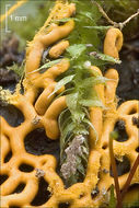
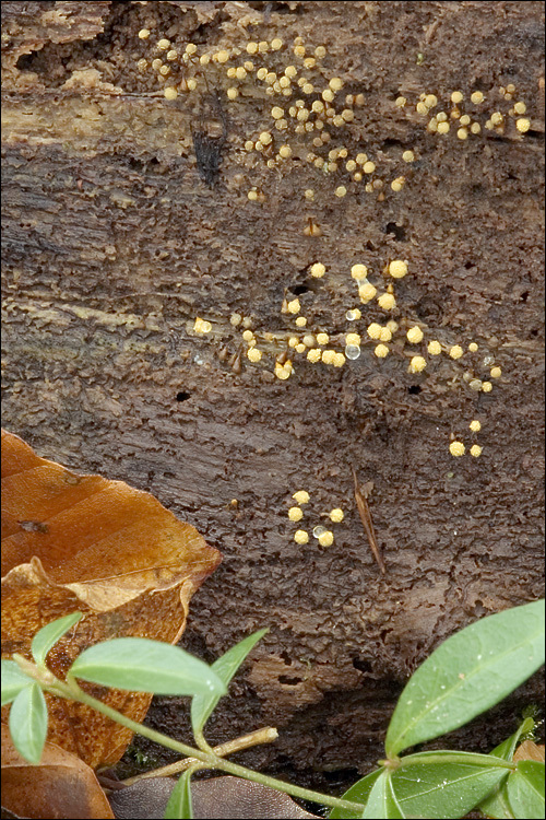
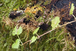
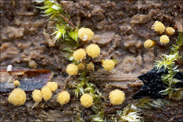
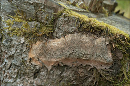
Slo.: ? Determination uncertain. Recognized by sight, no microscopic verification. Habitat: mixed forest, shady, humid and cool place, N exposed, on horizontal to vertical surface, precipitations >3.000 mm/year, partly protected from direct rain, average temperature 6-8 deg C. Substrates: heavily rotten, debarked log of a deciduous tree
-

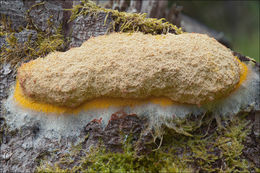
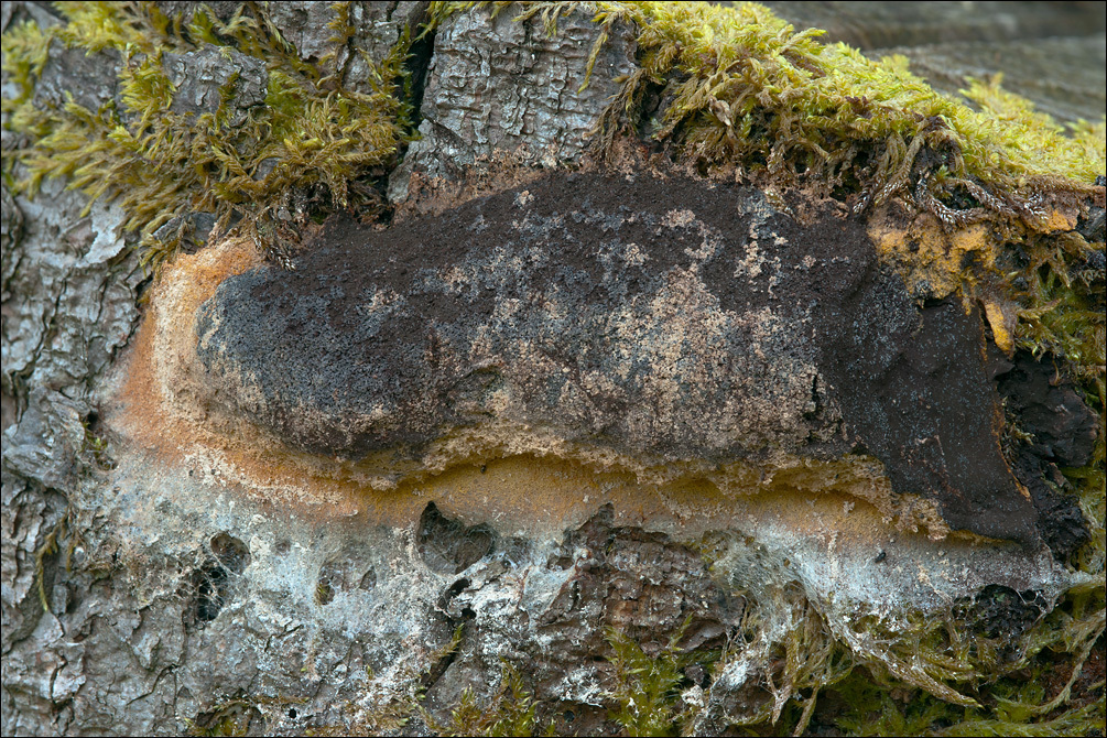
Slo.: reslov cvet - syn. Mucorsepticus L., Reticularia septica (L.) With., Aethalium septicum (L.) Fr., Fuligo varians Sommerf. - The aethalium picture taken on July 21. 2014.(Figs.13 - 16) - Habitat: old partly tree overgrown pasture, near mixed wood edge; moderately southeast inclined foot of an old overgrown scree slope; open, dry, sunny place; shallow, skeletal, calcareous ground, exposed to direct rain, average precipitations ~ 3.000 mm/year, average temperature 7-9 deg C, elevation 630 m (2.070 feet), alpine phytogeographical region. - Substratum: a stump of Picea abies cut down three years ago. - Comment: Myxomycetes are poorly known yet very interesting creatures. For decades they have been shuffled back and forth between the animal and plants kingdoms until recognized as separate creatures. They are not animals because they proliferate by spores. They are also not plants since they crumble around (an animal like ability) and fix themselves firmly to substrate only at the end of their life cycle. They don't produce their own food like plants but feed by 'hunting' (actually engulfing) bacteria and tiny bits of other organic matter, which is another animal like feature. The first stage in their development cycle, which is observable in the field, is called plasmodium. Earlier stages (from myxoflagellates, myxoamoebae, to zygote) are microscopic and can be observed only in labs. Plasmodium is a single giant living cell, a clump of protoplasm filled with thousands of cell nuclei, crawling around, eating bacteria and growing. Some of such plasmodia are the largest cells of living creatures known. In some species they can measure several meters across or weight up to 20 kg. That it much larger than ostrich's eggs, which are popularly considered as 'largest living cells'! Plasmodia could be found in the field, some are even brightly colored and easy to spot; however, it is almost impossible to determine to which species they belong. Plasmodium of Fuligo septica is commonly described like disgusting mucus, spilled scrambled eggs, dogs vomiting and other 'benevolent' portrayals. - When the time is right or delicate environmental conditions required for growth worsen plasmodia eventually evolves (usually almost completely) into sporocarps of different forms. These are bodies producing spores and then vanishing. In genus Fuligo sporocarp is a cushion like aethalium sitting on a thin whitish, 'fibrous' layer called hypothallus (Fig.14.). These'cushions' are what one usually finds in the field. But, other Myxomycetes develop also many other forms of sporocarps full of beauty, delicacy and imagination. Aethalia of Fuligo septica are usually covered with a kind of crust called cortex, which is brittle and soon crumbles away. In humid conditions it may not fully develop (Ref.1). Inside a mature aethalium there is a mesh of thin tubes or fibers called capillitium and zillions of dark brown spores. Fuligo septica has characteristic nodes on capillitial tubes, which are clearly seen on Fig. 4M. In due course the aethalium decomposes almost entirely into spore mass (Fig.17., 18.), which are sooner or later blown or washed away (Fig.20. taken about three weeks after the first photo). Size and shape of spores and structure of their surface are important traits for species determination. - Cushion-shaped aethalium measured approximately 14 x 5 cm and was about 3 cm thick. Spores are minutely warty and globose to subglobose. Dimensions: 8 [8,4 ; 8,7] 9,1 x 7,4 [8 ; 8,2] 8,7 microns; Q = [1 ; 1,07] 1,1; N = 25; C = 95%; Me = 8,5 x 8,1 microns; Qe = 1,05. Olympus CH20, NEA 100x/1.25, magnification 1.000 x, oil (spores), NEA 40x/0.65, magnification 400x (capillitium), NEA 10x/0.25, magnification 100x (hypothallus); in water, living material. AmScope MA500 digital camera. Spore sample taken on July 23. 2014. - Herbarium: Mycotheca and lichen herbarium (LJU-Li) of Slovenian Forestry Institute, Vena pot 2, Ljubljana, Index Herbariorum LJF - Ref.: (1) B. Ing, The Myxomycetes of Britain and Ireland, The Richmond Publ. Co.Ltd, (1999), p 246 (2) S.L.Stephenson and H.Stempen, Myxomycetes, Timber Press Inc.(2000), p123. (3) http://www.hiddenforest.co.nz/slime/family/physaraceae/physa02.htm (4) M. Poulain, M. Meyer, J. Bozzonet, Les Myxomycetes, FMBDS (2011), Vol.1., p128; Vol.2., p168.
-
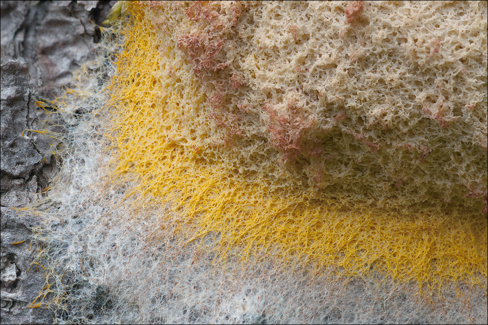
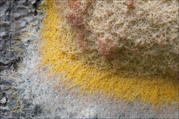
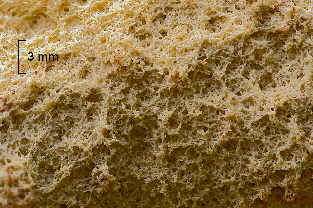
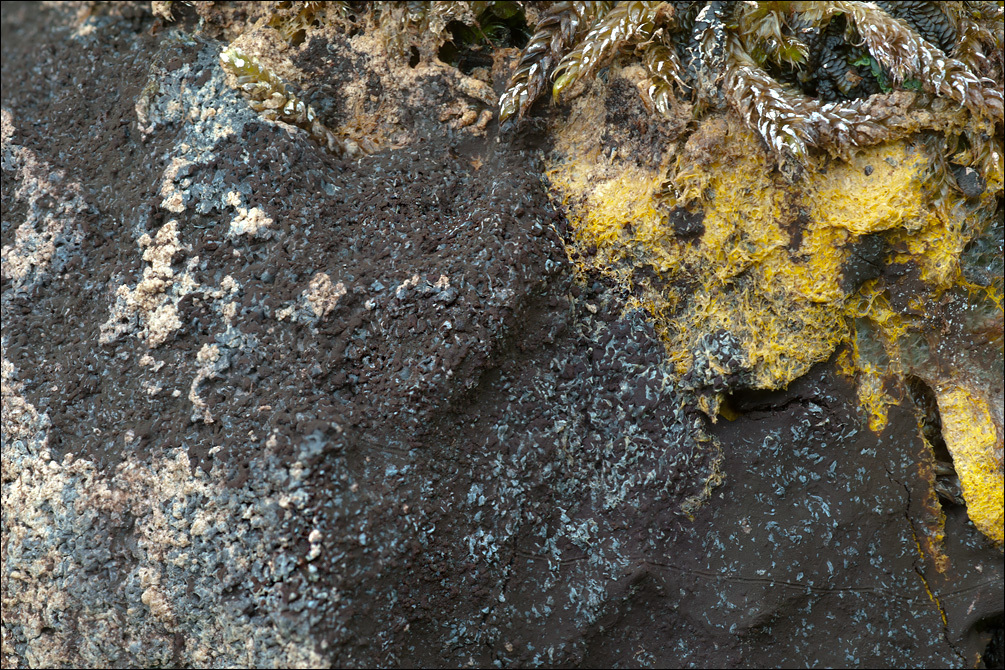
Slo.: reslov cvet - syn. Mucorsepticus L., Reticularia septica (L.) With., Aethalium septicum (L.) Fr., Fuligo varians Sommerf. - The aethalium after two days. (Figs. 17 - 19) - Habitat: old partly tree overgrown pasture, near mixed wood edge; moderately southeast inclined foot of an old overgrown scree slope; open, dry, sunny place; shallow, skeletal, calcareous ground, exposed to direct rain, average precipitations ~ 3.000 mm/year, average temperature 7-9 deg C, elevation 630 m (2.070 feet), alpine phytogeographical region. - Substratum: a stump of Picea abies cut down three years ago. - Comment: Myxomycetes are poorly known yet very interesting creatures. For decades they have been shuffled back and forth between the animal and plants kingdoms until recognized as separate creatures. They are not animals because they proliferate by spores. They are also not plants since they crumble around (an animal like ability) and fix themselves firmly to substrate only at the end of their life cycle. They don't produce their own food like plants but feed by 'hunting' (actually engulfing) bacteria and tiny bits of other organic matter, which is another animal like feature. The first stage in their development cycle, which is observable in the field, is called plasmodium. Earlier stages (from myxoflagellates, myxoamoebae, to zygote) are microscopic and can be observed only in labs. Plasmodium is a single giant living cell, a clump of protoplasm filled with thousands of cell nuclei, crawling around, eating bacteria and growing. Some of such plasmodia are the largest cells of living creatures known. In some species they can measure several meters across or weight up to 20 kg. That it much larger than ostrich's eggs, which are popularly considered as 'largest living cells'! Plasmodia could be found in the field, some are even brightly colored and easy to spot; however, it is almost impossible to determine to which species they belong. Plasmodium of Fuligo septica is commonly described like disgusting mucus, spilled scrambled eggs, dogs vomiting and other 'benevolent' portrayals. - When the time is right or delicate environmental conditions required for growth worsen plasmodia eventually evolves (usually almost completely) into sporocarps of different forms. These are bodies producing spores and then vanishing. In genus Fuligo sporocarp is a cushion like aethalium sitting on a thin whitish, 'fibrous' layer called hypothallus (Fig.14.). These'cushions' are what one usually finds in the field. But, other Myxomycetes develop also many other forms of sporocarps full of beauty, delicacy and imagination. Aethalia of Fuligo septica are usually covered with a kind of crust called cortex, which is brittle and soon crumbles away. In humid conditions it may not fully develop (Ref.1). Inside a mature aethalium there is a mesh of thin tubes or fibers called capillitium and zillions of dark brown spores. Fuligo septica has characteristic nodes on capillitial tubes, which are clearly seen on Fig. 4M. In due course the aethalium decomposes almost entirely into spore mass (Fig.17., 18.), which are sooner or later blown or washed away (Fig.20. taken about three weeks after the first photo). Size and shape of spores and structure of their surface are important traits for species determination. - Cushion-shaped aethalium measured approximately 14 x 5 cm and was about 3 cm thick. Spores are minutely warty and globose to subglobose. Dimensions: 8 [8,4 ; 8,7] 9,1 x 7,4 [8 ; 8,2] 8,7 microns; Q = [1 ; 1,07] 1,1; N = 25; C = 95%; Me = 8,5 x 8,1 microns; Qe = 1,05. Olympus CH20, NEA 100x/1.25, magnification 1.000 x, oil (spores), NEA 40x/0.65, magnification 400x (capillitium), NEA 10x/0.25, magnification 100x (hypothallus); in water, living material. AmScope MA500 digital camera. Spore sample taken on July 23. 2014. - Herbarium: Mycotheca and lichen herbarium (LJU-Li) of Slovenian Forestry Institute, Vena pot 2, Ljubljana, Index Herbariorum LJF - Ref.: (1) B. Ing, The Myxomycetes of Britain and Ireland, The Richmond Publ. Co.Ltd, (1999), p 246 (2) S.L.Stephenson and H.Stempen, Myxomycetes, Timber Press Inc.(2000), p123. (3) http://www.hiddenforest.co.nz/slime/family/physaraceae/physa02.htm (4) M. Poulain, M. Meyer, J. Bozzonet, Les Myxomycetes, FMBDS (2011), Vol.1., p128; Vol.2., p168.
-

Slo.: reslov cvet - syn. Mucorsepticus L., Reticularia septica (L.) With., Aethalium septicum (L.) Fr., Fuligo varians Sommerf. - The aethalium after 21 days after first photographing. (Fig. 20) - Habitat: old partly tree overgrown pasture, near mixed wood edge; moderately southeast inclined foot of an old overgrown scree slope; open, dry, sunny place; shallow, skeletal, calcareous ground, exposed to direct rain, average precipitations ~ 3.000 mm/year, average temperature 7-9 deg C, elevation 630 m (2.070 feet), alpine phytogeographical region. - Substratum: a stump of Picea abies cut down three years ago. - Comment: Myxomycetes are poorly known yet very interesting creatures. For decades they have been shuffled back and forth between the animal and plants kingdoms until recognized as separate creatures. They are not animals because they proliferate by spores. They are also not plants since they crumble around (an animal like ability) and fix themselves firmly to substrate only at the end of their life cycle. They don't produce their own food like plants but feed by 'hunting' (actually engulfing) bacteria and tiny bits of other organic matter, which is another animal like feature. The first stage in their development cycle, which is observable in the field, is called plasmodium. Earlier stages (from myxoflagellates, myxoamoebae, to zygote) are microscopic and can be observed only in labs. Plasmodium is a single giant living cell, a clump of protoplasm filled with thousands of cell nuclei, crawling around, eating bacteria and growing. Some of such plasmodia are the largest cells of living creatures known. In some species they can measure several meters across or weight up to 20 kg. That it much larger than ostrich's eggs, which are popularly considered as 'largest living cells'! Plasmodia could be found in the field, some are even brightly colored and easy to spot; however, it is almost impossible to determine to which species they belong. Plasmodium of Fuligo septica is commonly described like disgusting mucus, spilled scrambled eggs, dogs vomiting and other 'benevolent' portrayals. - When the time is right or delicate environmental conditions required for growth worsen plasmodia eventually evolves (usually almost completely) into sporocarps of different forms. These are bodies producing spores and then vanishing. In genus Fuligo sporocarp is a cushion like aethalium sitting on a thin whitish, 'fibrous' layer called hypothallus (Fig.14.). These'cushions' are what one usually finds in the field. But, other Myxomycetes develop also many other forms of sporocarps full of beauty, delicacy and imagination. Aethalia of Fuligo septica are usually covered with a kind of crust called cortex, which is brittle and soon crumbles away. In humid conditions it may not fully develop (Ref.1). Inside a mature aethalium there is a mesh of thin tubes or fibers called capillitium and zillions of dark brown spores. Fuligo septica has characteristic nodes on capillitial tubes, which are clearly seen on Fig. 4M. In due course the aethalium decomposes almost entirely into spore mass (Fig.17., 18.), which are sooner or later blown or washed away (Fig.20. taken about three weeks after the first photo). Size and shape of spores and structure of their surface are important traits for species determination. - Cushion-shaped aethalium measured approximately 14 x 5 cm and was about 3 cm thick. Spores are minutely warty and globose to subglobose. Dimensions: 8 [8,4 ; 8,7] 9,1 x 7,4 [8 ; 8,2] 8,7 microns; Q = [1 ; 1,07] 1,1; N = 25; C = 95%; Me = 8,5 x 8,1 microns; Qe = 1,05. Olympus CH20, NEA 100x/1.25, magnification 1.000 x, oil (spores), NEA 40x/0.65, magnification 400x (capillitium), NEA 10x/0.25, magnification 100x (hypothallus); in water, living material. AmScope MA500 digital camera. Spore sample taken on July 23. 2014. - Herbarium: Mycotheca and lichen herbarium (LJU-Li) of Slovenian Forestry Institute, Vena pot 2, Ljubljana, Index Herbariorum LJF - Ref.: (1) B. Ing, The Myxomycetes of Britain and Ireland, The Richmond Publ. Co.Ltd, (1999), p 246 (2) S.L.Stephenson and H.Stempen, Myxomycetes, Timber Press Inc.(2000), p123. (3) http://www.hiddenforest.co.nz/slime/family/physaraceae/physa02.htm (4) M. Poulain, M. Meyer, J. Bozzonet, Les Myxomycetes, FMBDS (2011), Vol.1., p128; Vol.2., p168.
-
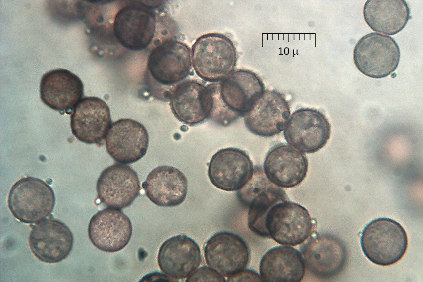
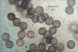
Slo.: reslov cvet - syn. Mucorsepticus L., Reticularia septica (L.) With., Aethalium septicum (L.) Fr., Fuligo varians Sommerf. - Spores at magnification 1000x. Sample taken on July 23. 2014. (Fig. 1M). - Habitat: old partly tree overgrown pasture, near mixed wood edge; moderately southeast inclined foot of an old overgrown scree slope; open, dry, sunny place; shallow, skeletal, calcareous ground, exposed to direct rain, average precipitations ~ 3.000 mm/year, average temperature 7-9 deg C, elevation 630 m (2.070 feet), alpine phytogeographical region. - Substratum: a stump of Picea abies cut down three years ago. - Comment: Myxomycetes are poorly known yet very interesting creatures. For decades they have been shuffled back and forth between the animal and plants kingdoms until recognized as separate creatures. They are not animals because they proliferate by spores. They are also not plants since they crumble around (an animal like ability) and fix themselves firmly to substrate only at the end of their life cycle. They don't produce their own food like plants but feed by 'hunting' (actually engulfing) bacteria and tiny bits of other organic matter, which is another animal like feature. The first stage in their development cycle, which is observable in the field, is called plasmodium. Earlier stages (from myxoflagellates, myxoamoebae, to zygote) are microscopic and can be observed only in labs. Plasmodium is a single giant living cell, a clump of protoplasm filled with thousands of cell nuclei, crawling around, eating bacteria and growing. Some of such plasmodia are the largest cells of living creatures known. In some species they can measure several meters across or weight up to 20 kg. That it much larger than ostrich's eggs, which are popularly considered as 'largest living cells'! Plasmodia could be found in the field, some are even brightly colored and easy to spot; however, it is almost impossible to determine to which species they belong. Plasmodium of Fuligo septica is commonly described like disgusting mucus, spilled scrambled eggs, dogs vomiting and other 'benevolent' portrayals. - When the time is right or delicate environmental conditions required for growth worsen plasmodia eventually evolves (usually almost completely) into sporocarps of different forms. These are bodies producing spores and then vanishing. In genus Fuligo sporocarp is a cushion like aethalium sitting on a thin whitish, 'fibrous' layer called hypothallus (Fig.14.). These'cushions' are what one usually finds in the field. But, other Myxomycetes develop also many other forms of sporocarps full of beauty, delicacy and imagination. Aethalia of Fuligo septica are usually covered with a kind of crust called cortex, which is brittle and soon crumbles away. In humid conditions it may not fully develop (Ref.1). Inside a mature aethalium there is a mesh of thin tubes or fibers called capillitium and zillions of dark brown spores. Fuligo septica has characteristic nodes on capillitial tubes, which are clearly seen on Fig. 4M. In due course the aethalium decomposes almost entirely into spore mass (Fig.17., 18.), which are sooner or later blown or washed away (Fig.20. taken about three weeks after the first photo). Size and shape of spores and structure of their surface are important traits for species determination. - Cushion-shaped aethalium measured approximately 14 x 5 cm and was about 3 cm thick. Spores are minutely warty and globose to subglobose. Dimensions: 8 [8,4 ; 8,7] 9,1 x 7,4 [8 ; 8,2] 8,7 microns; Q = [1 ; 1,07] 1,1; N = 25; C = 95%; Me = 8,5 x 8,1 microns; Qe = 1,05. Olympus CH20, NEA 100x/1.25, magnification 1.000 x, oil (spores), NEA 40x/0.65, magnification 400x (capillitium), NEA 10x/0.25, magnification 100x (hypothallus); in water, living material. AmScope MA500 digital camera. Spore sample taken on July 23. 2014. - Herbarium: Mycotheca and lichen herbarium (LJU-Li) of Slovenian Forestry Institute, Vena pot 2, Ljubljana, Index Herbariorum LJF - Ref.: (1) B. Ing, The Myxomycetes of Britain and Ireland, The Richmond Publ. Co.Ltd, (1999), p 246 (2) S.L.Stephenson and H.Stempen, Myxomycetes, Timber Press Inc.(2000), p123. (3) http://www.hiddenforest.co.nz/slime/family/physaraceae/physa02.htm (4) M. Poulain, M. Meyer, J. Bozzonet, Les Myxomycetes, FMBDS (2011), Vol.1., p128; Vol.2., p168.
-
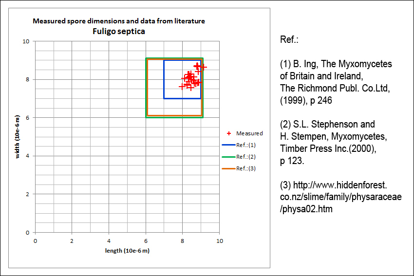
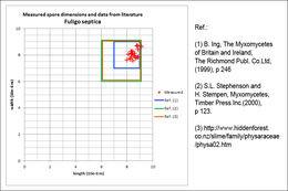
Slo.: reslov cvet - syn. Mucorsepticus L., Reticularia septica (L.) With., Aethalium septicum (L.) Fr., Fuligo varians Sommerf. - Spore statistics and comparison with data from literature. (Fig. 2M). - Habitat: old partly tree overgrown pasture, near mixed wood edge; moderately southeast inclined foot of an old overgrown scree slope; open, dry, sunny place; shallow, skeletal, calcareous ground, exposed to direct rain, average precipitations ~ 3.000 mm/year, average temperature 7-9 deg C, elevation 630 m (2.070 feet), alpine phytogeographical region. - Substratum: a stump of Picea abies cut down three years ago. - Comment: Myxomycetes are poorly known yet very interesting creatures. For decades they have been shuffled back and forth between the animal and plants kingdoms until recognized as separate creatures. They are not animals because they proliferate by spores. They are also not plants since they crumble around (an animal like ability) and fix themselves firmly to substrate only at the end of their life cycle. They don't produce their own food like plants but feed by 'hunting' (actually engulfing) bacteria and tiny bits of other organic matter, which is another animal like feature. The first stage in their development cycle, which is observable in the field, is called plasmodium. Earlier stages (from myxoflagellates, myxoamoebae, to zygote) are microscopic and can be observed only in labs. Plasmodium is a single giant living cell, a clump of protoplasm filled with thousands of cell nuclei, crawling around, eating bacteria and growing. Some of such plasmodia are the largest cells of living creatures known. In some species they can measure several meters across or weight up to 20 kg. That it much larger than ostrich's eggs, which are popularly considered as 'largest living cells'! Plasmodia could be found in the field, some are even brightly colored and easy to spot; however, it is almost impossible to determine to which species they belong. Plasmodium of Fuligo septica is commonly described like disgusting mucus, spilled scrambled eggs, dogs vomiting and other 'benevolent' portrayals. - When the time is right or delicate environmental conditions required for growth worsen plasmodia eventually evolves (usually almost completely) into sporocarps of different forms. These are bodies producing spores and then vanishing. In genus Fuligo sporocarp is a cushion like aethalium sitting on a thin whitish, 'fibrous' layer called hypothallus (Fig.14.). These'cushions' are what one usually finds in the field. But, other Myxomycetes develop also many other forms of sporocarps full of beauty, delicacy and imagination. Aethalia of Fuligo septica are usually covered with a kind of crust called cortex, which is brittle and soon crumbles away. In humid conditions it may not fully develop (Ref.1). Inside a mature aethalium there is a mesh of thin tubes or fibers called capillitium and zillions of dark brown spores. Fuligo septica has characteristic nodes on capillitial tubes, which are clearly seen on Fig. 4M. In due course the aethalium decomposes almost entirely into spore mass (Fig.17., 18.), which are sooner or later blown or washed away (Fig.20. taken about three weeks after the first photo). Size and shape of spores and structure of their surface are important traits for species determination. - Cushion-shaped aethalium measured approximately 14 x 5 cm and was about 3 cm thick. Spores are minutely warty and globose to subglobose. Dimensions: 8 [8,4 ; 8,7] 9,1 x 7,4 [8 ; 8,2] 8,7 microns; Q = [1 ; 1,07] 1,1; N = 25; C = 95%; Me = 8,5 x 8,1 microns; Qe = 1,05. Olympus CH20, NEA 100x/1.25, magnification 1.000 x, oil (spores), NEA 40x/0.65, magnification 400x (capillitium), NEA 10x/0.25, magnification 100x (hypothallus); in water, living material. AmScope MA500 digital camera. Spore sample taken on July 23. 2014. - Herbarium: Mycotheca and lichen herbarium (LJU-Li) of Slovenian Forestry Institute, Vena pot 2, Ljubljana, Index Herbariorum LJF - Ref.: (1) B. Ing, The Myxomycetes of Britain and Ireland, The Richmond Publ. Co.Ltd, (1999), p 246 (2) S.L.Stephenson and H.Stempen, Myxomycetes, Timber Press Inc.(2000), p123. (3) http://www.hiddenforest.co.nz/slime/family/physaraceae/physa02.htm (4) M. Poulain, M. Meyer, J. Bozzonet, Les Myxomycetes, FMBDS (2011), Vol.1., p128; Vol.2., p168.