Comprehensive Description
provided by Smithsonian Contributions to Zoology
Alloposus mollis Verrill, 1880
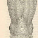
DESCRIPTION.—The mantle is short, gelatinous, and broad (length is nearly equal to width). The mantle opening is wide and reaches roughly to the level of the dorsal edge of the eyes. On the inner surface of the mantle, just medial to the gills and lateral to the median pallial adductor muscle is a distinct transverse fold of the mantle which forms a deep groove. This is the mantle component of the funnel-mantle locking mechanism.
The funnel is very large. It extends far past the eyes and past bases of the ventral arms. Except for a short free tip, it is completely embedded in the gelatinous tissue of the head. The funnel organ is large and W-shape. The posterior lateral edges of the funnel, near the point of attachment of the funnel retractor muscles, fold sharply forward to form the funnel component of the funnel-mantle locking mechanism.
The head is broad and bears large eyes. There is no lateral or dorsal constriction between the mantle and head. Posterior to each eye lies a low, flattened knob which is the “olfactory” organ.
The arms are very large, but gelatinous. They are in the order of I>II>III>IV. The suckers begin in a single but slightly irregular series near the base of each arm and gradually become biserially arranged proximal to the edge of the web.
The gills are huge and bear 18 lamellae, each of which is robust and highly folded. The body wall which covers the ventral surface of the viscera is surprisingly tough and opaque. Very small nephridial papillae project from near the base of the gills. Posterior and lateral to the large median pallial adductor muscle, a pair of oviducts opens through prominent papillae. Each papilla is extremely large and muscular. The largest eggs in the ovary are about 1.5 mm long by 0.5 mm wide. The viscera are essentially as illustrated by Thore (1949). The kidneys are very large and extend to the posterior end of the visceral mass. The crop is situated dorsal to the liver, and the stomach and caecum are dorsal and partially posterior to the liver. The entire dorsal surface of the visceral mass is free from the overlying mantle. The space between the visceral mass and the mantle communicates with the ventral mantle cavity laterally around the visceral mass anterior to the funnel-retractor muscles.
The rachidian tooth of the radula is tricuspid; the first lateral is small and bicuspid, and the second and third laterals are unicuspid. The marginals are simple and broad.
The data for the single specimen captured are: Sex, ; M.L., 115 mm; M.W., 126 mm; H.W., 120 mm; Arm L., I–IV, respectively, 360 mm, 260 mm, 240 mm, 200 mm; Eye D., 38 mm.
TYPE LOCALITY.—South of Newport, Rhode Island, western north Atlantic.
LOCATION OF TYPE.—United States National Museum.
- bibliographic citation
- Young, Richard E. 1972. "The systematics and areal distribution of pelagic cephalopods from the seas off Southern California." Smithsonian Contributions to Zoology. 1-159. https://doi.org/10.5479/si.00810282.97