-
Santos, Norenburg, and Bueno 2006
Nemertea
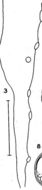
Pointed posterior end of male, with seminal vesicle
-
Santos, Norenburg, and Bueno 2006
Nemertea
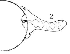
Adult female (arrow) on Libinia spinosa eggs
-
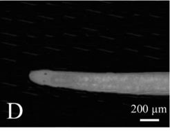
Santos, Norenburg, and Bueno 2006
Nemertea
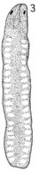
Male with rounded anterior end
-
Santos, Norenburg, and Bueno 2006
Nemertea
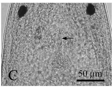
Stylet and basis (arrow) in a female worm
-
Santos, Norenburg, and Bueno 2006
Nemertea
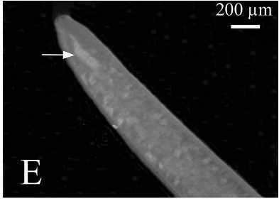
Proboscis aligned in a straight line
-
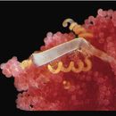
Santos, Norenburg, and Bueno 2006
Nemertea
Anterior end of female
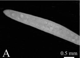
-
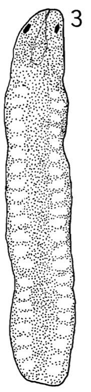
Dorsal view of live C. errans. Ovaries becoming visible as rows of transparent spots on both sides. Scale = 1.0 mm.
-
C. errans feeding on egg of C. magister. Esophagus used in sucking particles of egg yolk into intestine.
-
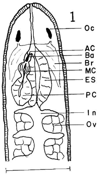
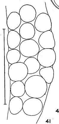
Anterior portion of body of living worm from dorsal surface. Oc, ocellus; Ac, anterior proboscis chamber; Ba, basis and stylet..Anterior portion of body of living worm from dorsal surface. Oc, ocellus; Ac, anterior proboscis chamber; Ba, basis and stylet; Br, brain; Me, middle proboscis chamber; Es, esophagus; Pc, posterior proboscis chamber; In, intestine; Ov, ovary. Scale .5 mm.
-
Chromosomes in mitotic anaphase during division from two to four cell stage in haploid embryo of experimental Carcinonemertes...Chromosomes in mitotic anaphase duringdivision from two to four cell stage in haploid embryo of experimental Carcinonemertes errans. Chromosome number is one-half that of Figure 1b. Magnification: l000x.
-
Chromosomes in mitotic anaphase during division from one to two cell stage in embryo of control Carcinonemertes errans...Chromosomes in mitotic anaphase during division from one to two cell stage in embryo ofcontrol Carcinonemertes errans.The small darkspots to the left of the chromosomes are remains of chromosomes in the two polar bodies. Magnification: l000X
-
Part of a mucous sheath of C. mitsukurii from Charybdis erythrodactyla from the Society Islands. Scale 0.5 mm.
-
Outline of basis of C. mitsukurii as seen in section. Scale 0.02 mm.
-
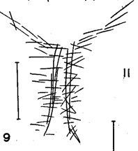
Sketch of cephalic muscle fibers in region of rhynchodaetim in C. mitsukurii. The anterior end is directed toward the bottom
-
Fragment of mucous sheath of C . mitsukurii from egg mass of Charybdis erythrodactyla from Society Islands. Scale 1 mm .
-
Mucous sheath of C. mitsukurii from egg mass of Charybdis cruciata at Hong Kong. Three crab eggs attached. Scale 1 mm.
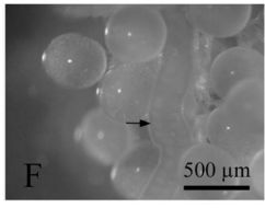
-
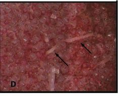
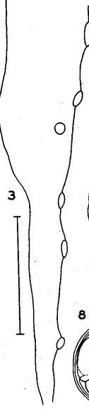
In situ photograph of egg strands (arrows) within egg brood of crab host. Diameter of strands approximately 300 μm
-
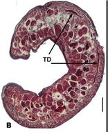
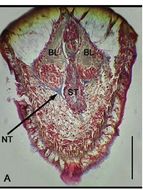
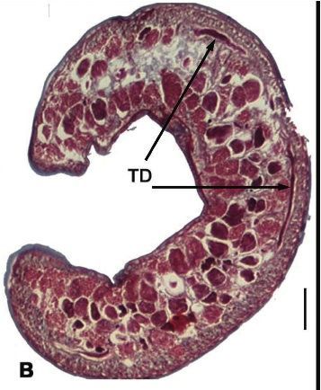
Sperm packed Takakura’s duct system; detail of duct
-
Sperm packed Takakura’s duct system in context of whole worm
-
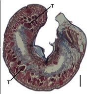
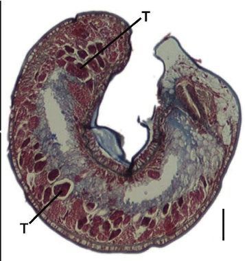
Cross-section showing testes
-
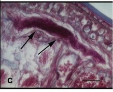
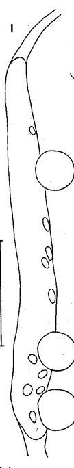
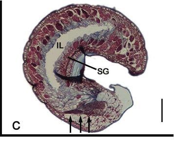
Longitudinal sagittal section showing proboscis alignment (three arrows)
-
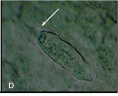
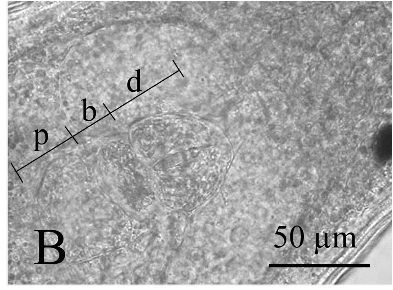
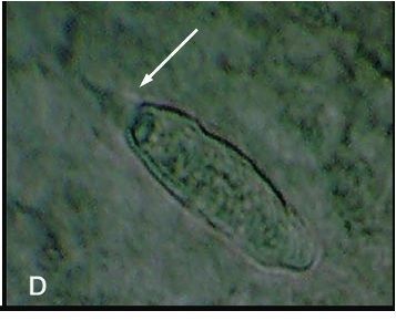
Live squash preparation of stylet (arrow) and basis. Length of stylet approximately 5 μm
-
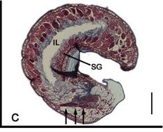
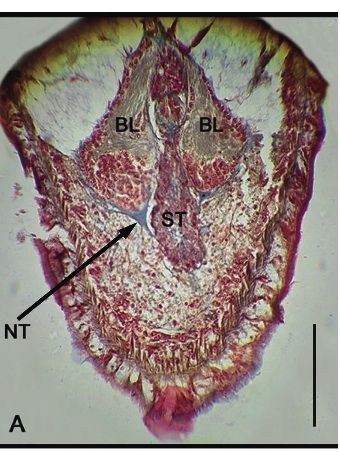
Longitudinal frontal section showing dorsal brain lobe and neuroganglionic tissues
-
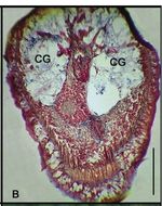
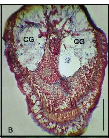
Longitudinal frontal section through cephalic glands