-
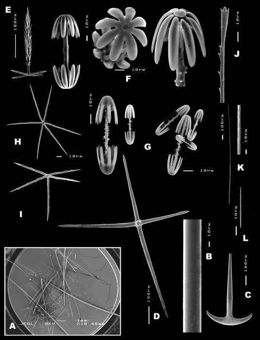
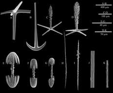
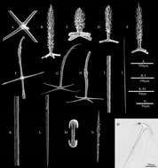
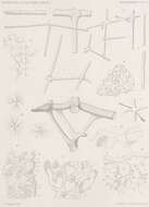
A, the centre part of choanosomal pentactin; B–C, atrial pinular pentactin; D, dermal pinular pentactin; E–F, choanosomal pinular pentactin; G, pinular hexactin; H, stauractin; I, spiny microdiactin; J, smooth middle shaft of marginalia; K, details of the middle shaft of mesouncinate; L, details of the middle shaft of macrouncinate; M, micramphidisc.
-
-
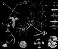
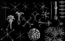
A) choanosomal stauractin; (B, C) atrial pentactins; (D) dermal hexactin; (E) discostauractin; (F) hemi-discohexaster; (G) microhexactin; (H) floricome I; (I) onychohexaster; (J) floricome II; (K) discaster; (L) detail of primary rays of floricome I; (M) detail of primary rays of onychohexaster; (N) regular hexaster; (O) detail of second ray of floricome II; (P) detail of primary rosette of graphiocome; (Q) anchorate discohexaster.
-
-
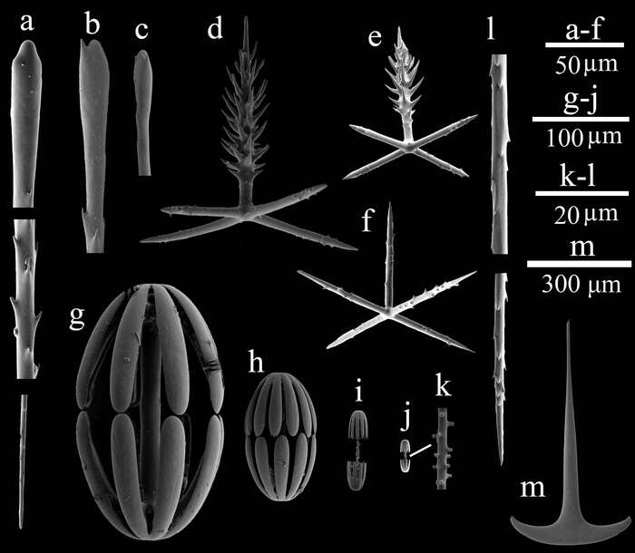
a–c. Clavate monaxon, d. dermal pinular pentactin, e. atrial pinular pentactin, f. micropentactin, g and h. macramphidisc, i. mesamphidisc, j. micramphidiscs, k. details of the shaft of micramphidisc, l. microuncinate, and m. details of the basalia.
-
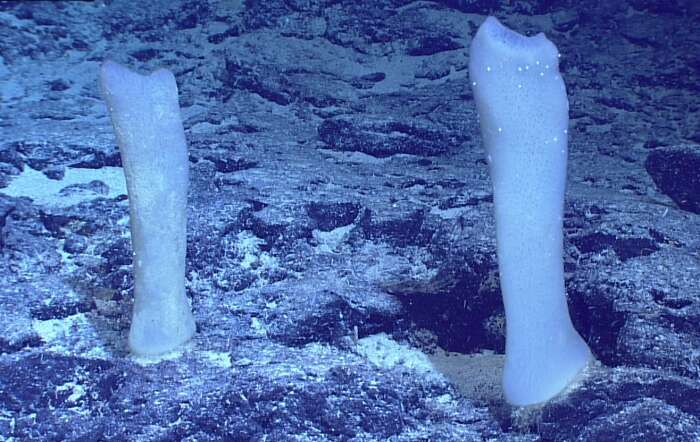
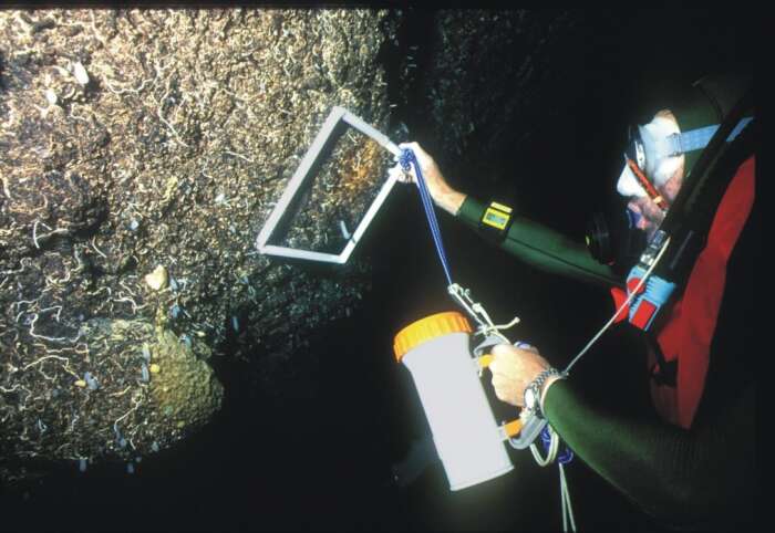
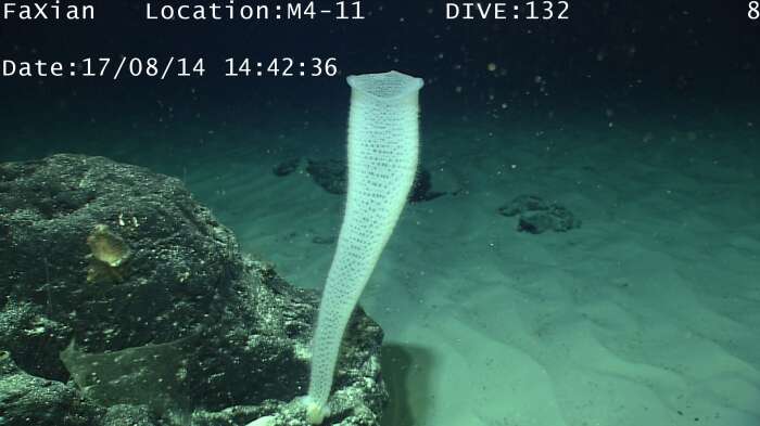
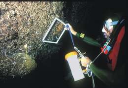
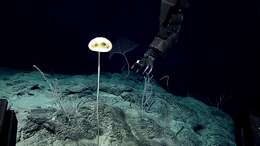
Courtesy of Ocean Networks Canada http://www.oceannetworks.ca/
-
-
-
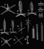
A, choanosomal pentactin; B, the anchor of basalia; C, atrial pinular pentactin; D, dermal pinular pentactin; E–G, micramphidiscs; H, microdiactin I; I, microdiactin II; J, shaft of macrouncinate; K, shaft of mesouncinate;
-
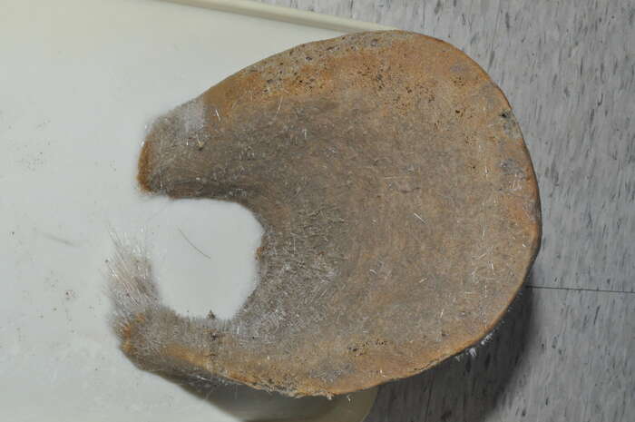
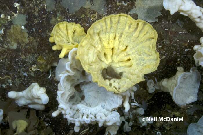
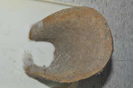
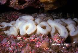
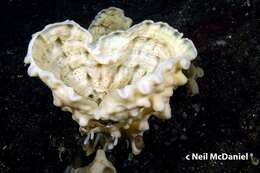
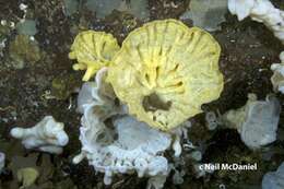
External morphology of dermal areas (scale bar = 5 cm)
-
A, choanosomal pentactin; B–C, dermal pinular pentactins; D–H, atrialia: D–E, pinular pentactins; F, crooked pentactin; G–H, special pentactins; I, shaft of macrouncinate; J, microuncinate; K, shaft of microuncinate; L, terminal of microuncinate; M, micramphidisc; N, microdiactin; O–Q, the anchor of basalia; R–S; marginalia.
-
-
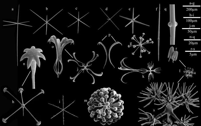
(A–B) dermal hexactins; (C) atrial hexactin; (D) choanosomal pentactin; (E) hexactin of sieve-plate; (F) diactin of sieve-plate; (G) hemidiscohexaster; (H) discohexactin; (I) spiny microhexactin; (J) floricome; (K) drepanocome; (L) codonhexaster; (M) discaster; (N) detail of the disc of a discohexactin; (O) detail of common secondary rays of floricome; (P) detail of claws of secondary rays of floricomes; (Q) detail of tubercles in the middle of sieve-plate diactin; (R) detail of the umbels of a codonhexaster; (S) detail of the middle part of a codonhexaster; (T) detail of the center part of a hemidiscohexaster.
-
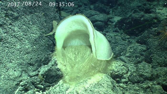
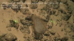
Image courtesy of the NOAA Office of Ocean Exploration and Research, 2016 Deepwater Exploration of the Marianas. Image is part of Figure 8 from the following publication:(https://marinespecies.org/aphia.php?p=sourcedetails&id=382781)
-
-
-
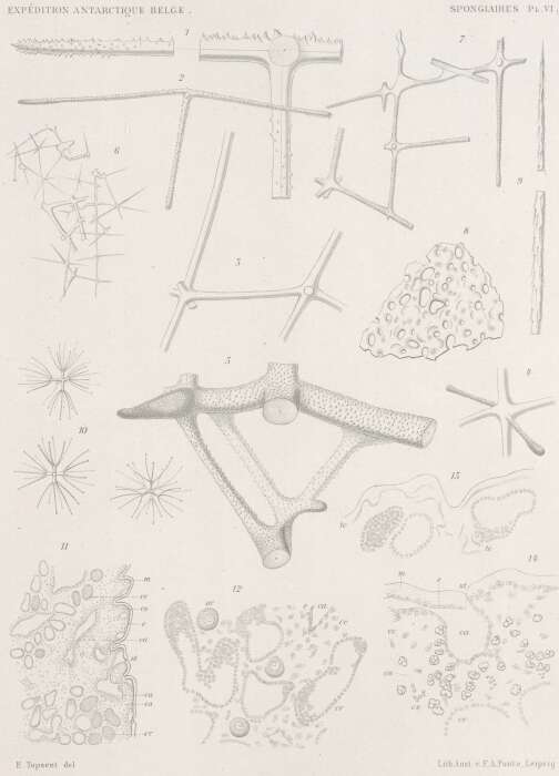
Fig. 1-10. Uncinatera plicata (http://www.marinespecies.org/aphia.php?p=taxdetails&id=171627) sp. (p.41) Centraal deel van het dermaal pentact met een punt van actine. Vergroting: x240 Dermale pentact. Vergroting: x75 Fragment van het dictyonale skelet naar bovenaan de doorsnede. Vergroting: x75 Ander fragment dat het centrum van een hexact met twee vrije actines toont.Vergroting: x75 Fragment van het dictyonale skelet naar onderaan de doorsnede. Vergroting: x75 Opeenhoping van zwak samengegroeide hexacten, aan de basis van de doorsnede. Vergroting: x75 Manier van verbinding van deze hexacten. Vergroting: x240 Deel van de basilaire plaat van de spons. Vergroting: x75 Fragmenten van een uncinete. Vergroting: x160 Discohexasters van 4, 6 en 8 terminale stralen. Vergroting: x400 Fig. 11-14. Halisarca dujardini var. magellanica (http://www.marinespecies.org/aphia.php?p=taxdetails&id=195510) sp., synoniem van Halisarca magellanica (http://www.marinespecies.org/aphia.php?p=taxdetails&id=166042) (p.44) Doorsnede met betrekking tot het ectosoom en het choanosoom. Vergroting: x75 m membraan zonder vaste structuur; e epithelium; st stomion; ca watervoerende kanalen; co collenchym; cv trillende korven Doorsnede in het choanosoom dat de positie van de eicellen toont. Vergroting: x240 ov eicellen; cv trillende korven; ce bindende cellen; ca watervoerende kanalen; e epithelium van deze kanalen Doorsnede aan de rand van een groot watervoerend kanaal, om de bindende fibrillaire kanalen tc te tonen. Vergroting: x240 Doorsnede van de periferie van het lichaam. Vergroting: x240 m membraan zonder vaste structuur; e epithelium; st stomion; ca watervoerende kanalen; co collenchym; cs granulaire cellen; cc bindende cellen; cv trillende korven
-
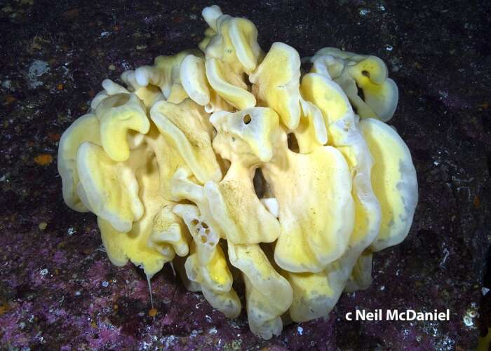
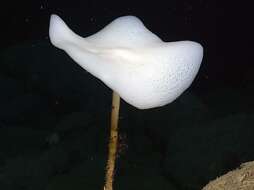
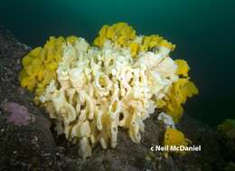
Jervis Inlet, British Columbia
-
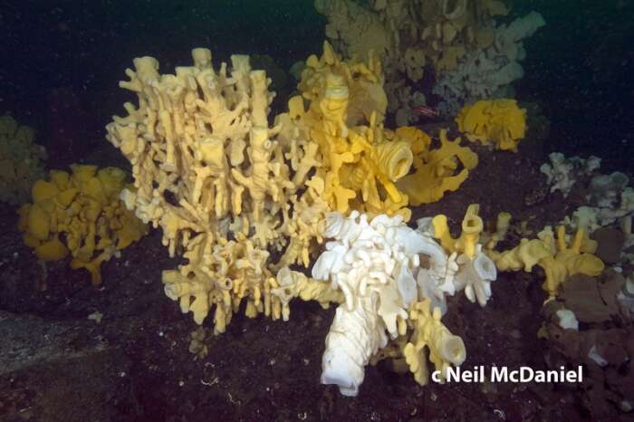
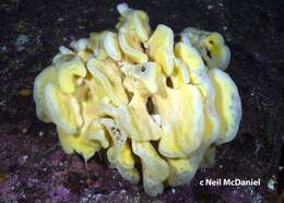
Jervis Inlet, British Columbia
-
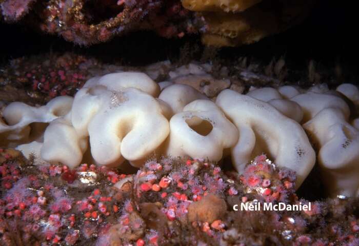
Jervis Inlet, British Columbia
-
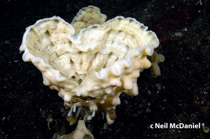
Discovery Passage, British Columbia
-
Jervis Inlet, British Columbia
-
Jervis Inlet, British Columbia
-
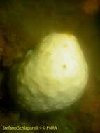
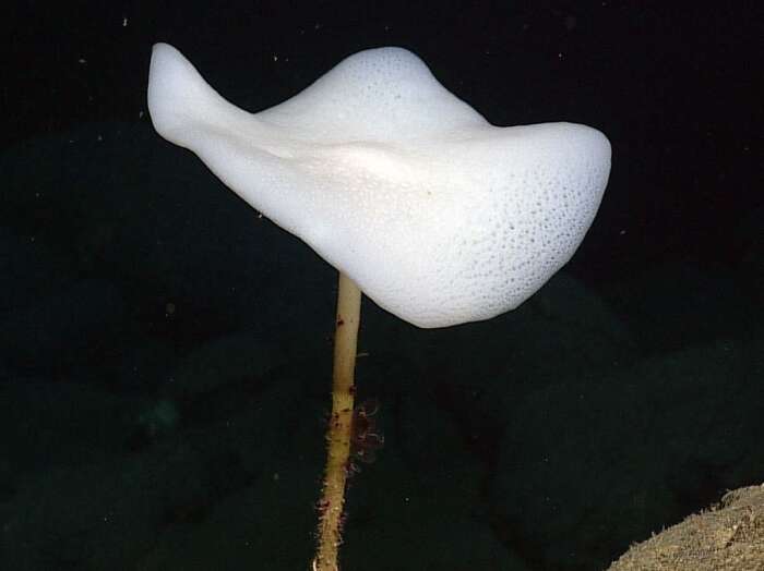
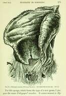
White and amphora- shaped. To me it looks rather like Scolymastra (Anoxycalyx joubini).