-
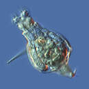
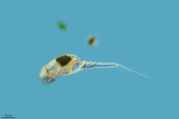
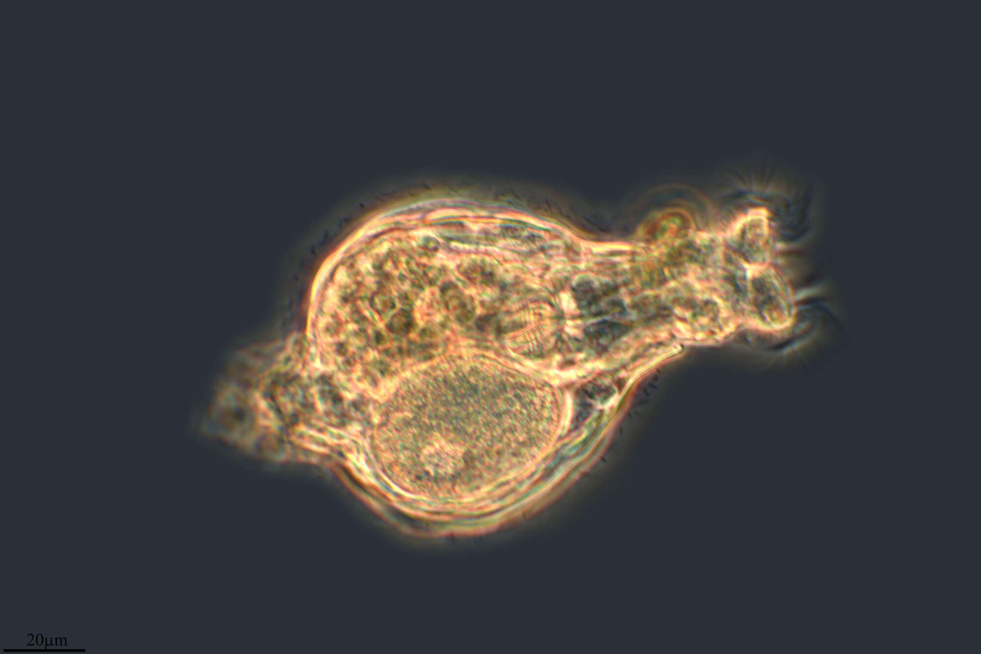
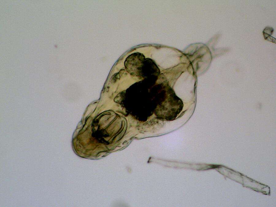
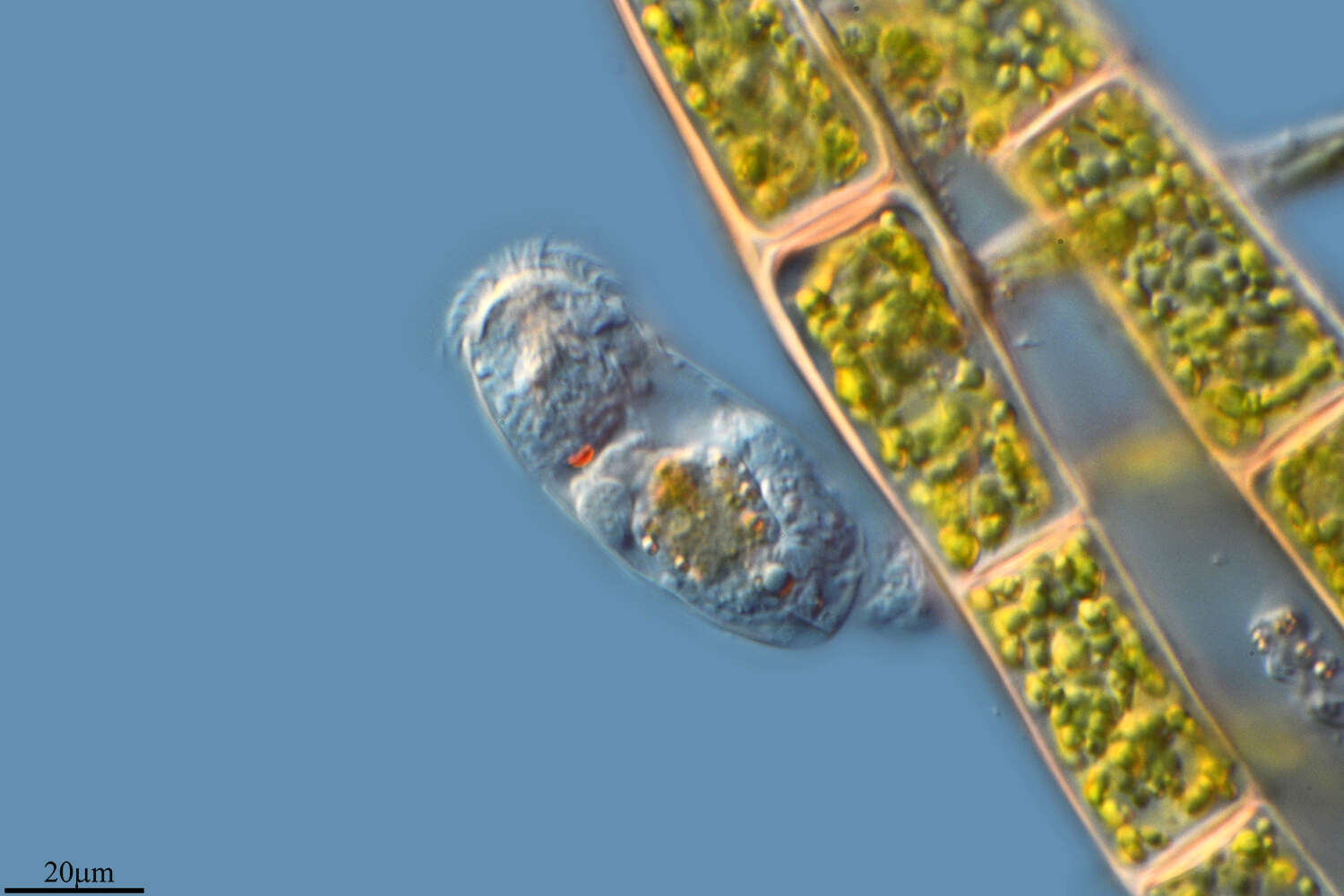
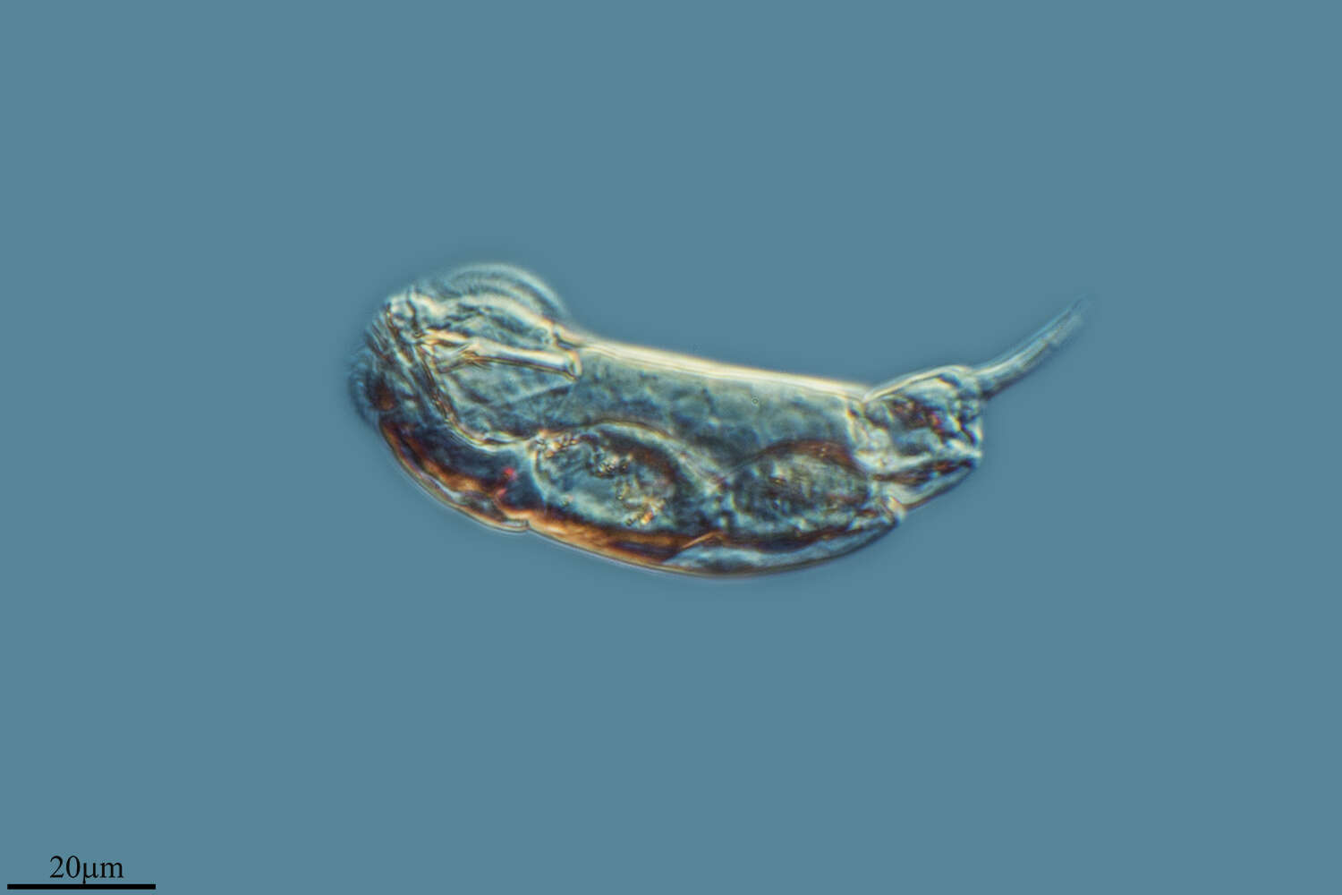
Keratella, a rotifer (metazoa). Rotifers typically have a stiffened body wall (lorica) in segments which can telescope, with a corona of feeding cilia at the anterior end and with toes posteriorly. This genus has no foot nor toe, as an adaptation to a pelagic life. Rotifers are common members of the microbial communities of many aquatic ecosystems. Although they are multicellular animals, they may be only be 100 microns long, and so overlap in size with ciliates. They can be confused with ciliates because they use cilia to capture their food. However, they can be distinguished because they have a lorica, may have podites, and a strong muscular pharynx.
-
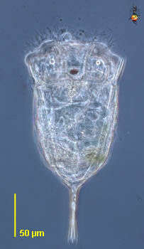
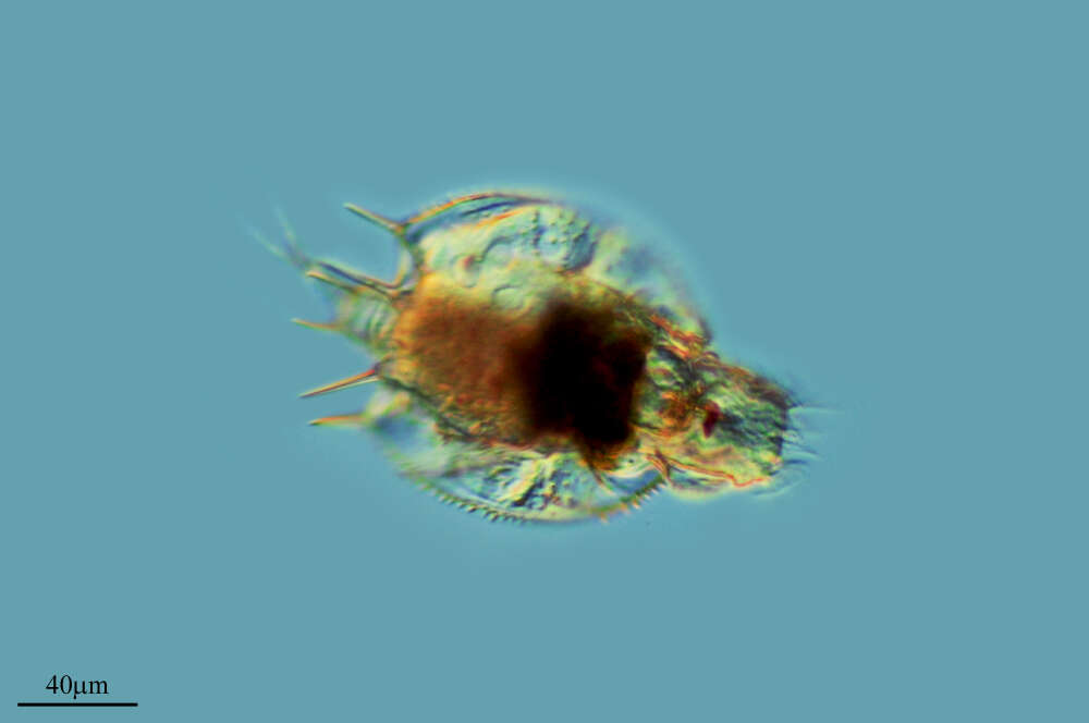
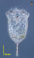
Lepadella. Rotifer observed in sandy and muddy marine sediments in the vicinity of Broome, Western Australia in September 2003. This image was taken using phase contrast optics. This work was supported by the Australian Biological Resources Study.
-
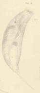
Diurella percellus Gosse, Left side of extended specimen.
-
Diurella rousseleti Voigt. Side view of retracted specimen.
-
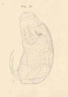
Diurella tenuior Gosse. Larger specimen, dorsal or dorso-dextral view.
-
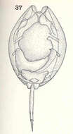
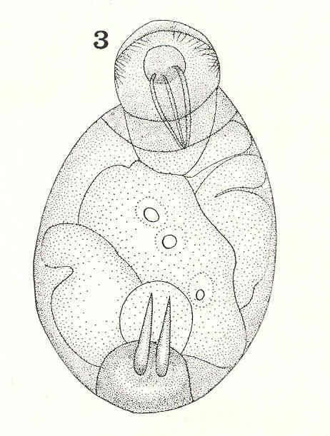
Monostyla cornuta Ehr. Ventral view.
-
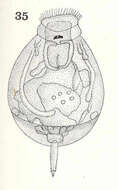
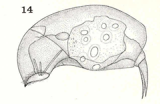
Monostyla bulla Gosse. Dorsal view.
-
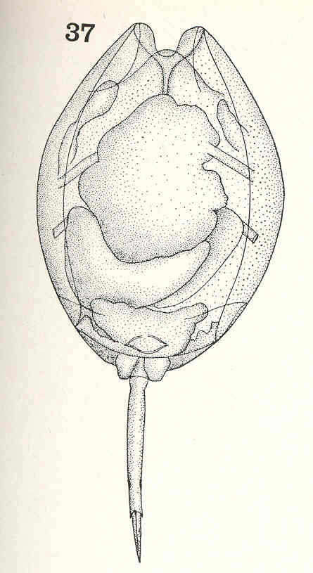
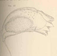
Taphrocampa annulosa Gosse, as seen from above; body curved so that the food is not visible.
-
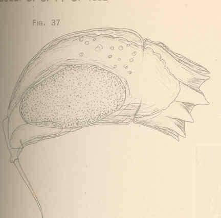
Taphrocampa annulosa Gosse. Side view.
-
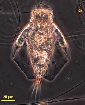
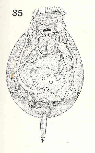
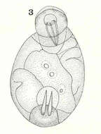
Pleurotrocha parasitica n. sp. Ventral view.
-
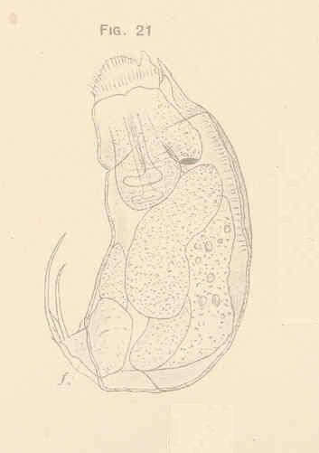
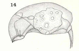
Pleurotrocha parasitica n. sp. Side view.
-
Phuripong Meksuwan, Pornsilp Pholpunthin, Hendrik Segers
Zookeys
Figure 2.A, B Collotheca ferox (A dorsal view B ventral view) C–F Collotheca orchidacea sp. n. (C, E frontal D, F dorsal). Scale bars: A–F = 100 µm (A, B by Rapeepan Jaturapruek).
-
Phuripong Meksuwan, Pornsilp Pholpunthin, Hendrik Segers
Zookeys
Figure 4.A Collotheca heptabrachiata, lateral B Collotheca ornata, ventral C Stephanoceros fimbriatus, lateral D Collotheca stephanochaeta, lateral E Collotheca ambigua, ventral F Collotheca algicola, ventral G, H Collotheca ornata f. cornuta (G dorsal H lateral) I Collotheca campanulata f. longicaudata, attachment stalk J Collotheca ferox, ventral corona margin. Scale bars: A, B, D–H, J = 50 µm, C, I = 100 µm.
-
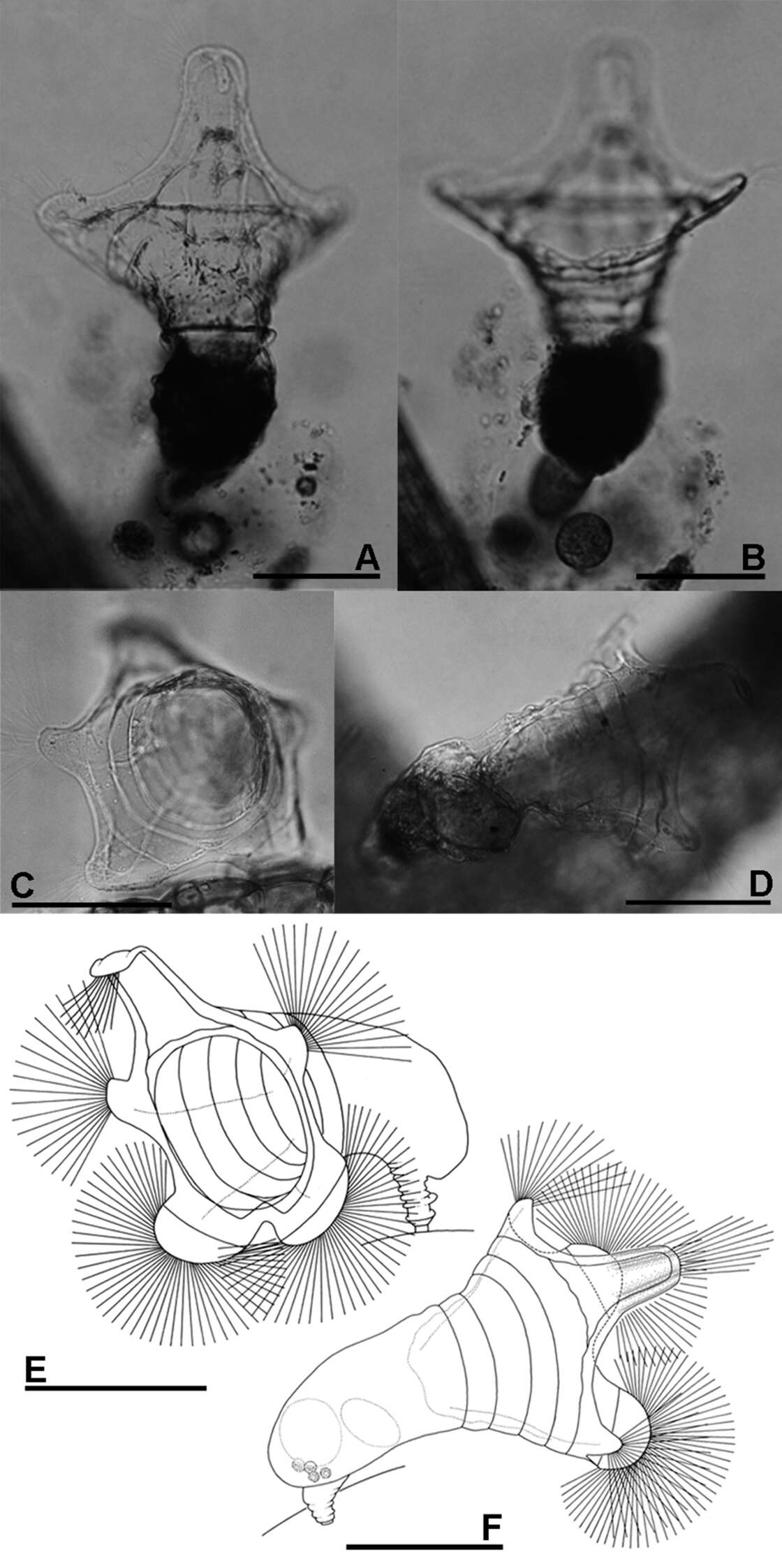
Yongting Luo, Hendrik Segers
Zookeys
Figure 1.Pulchritia dorsicornuta gen. n., comb. n., compound photomicrograph.
-
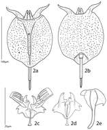
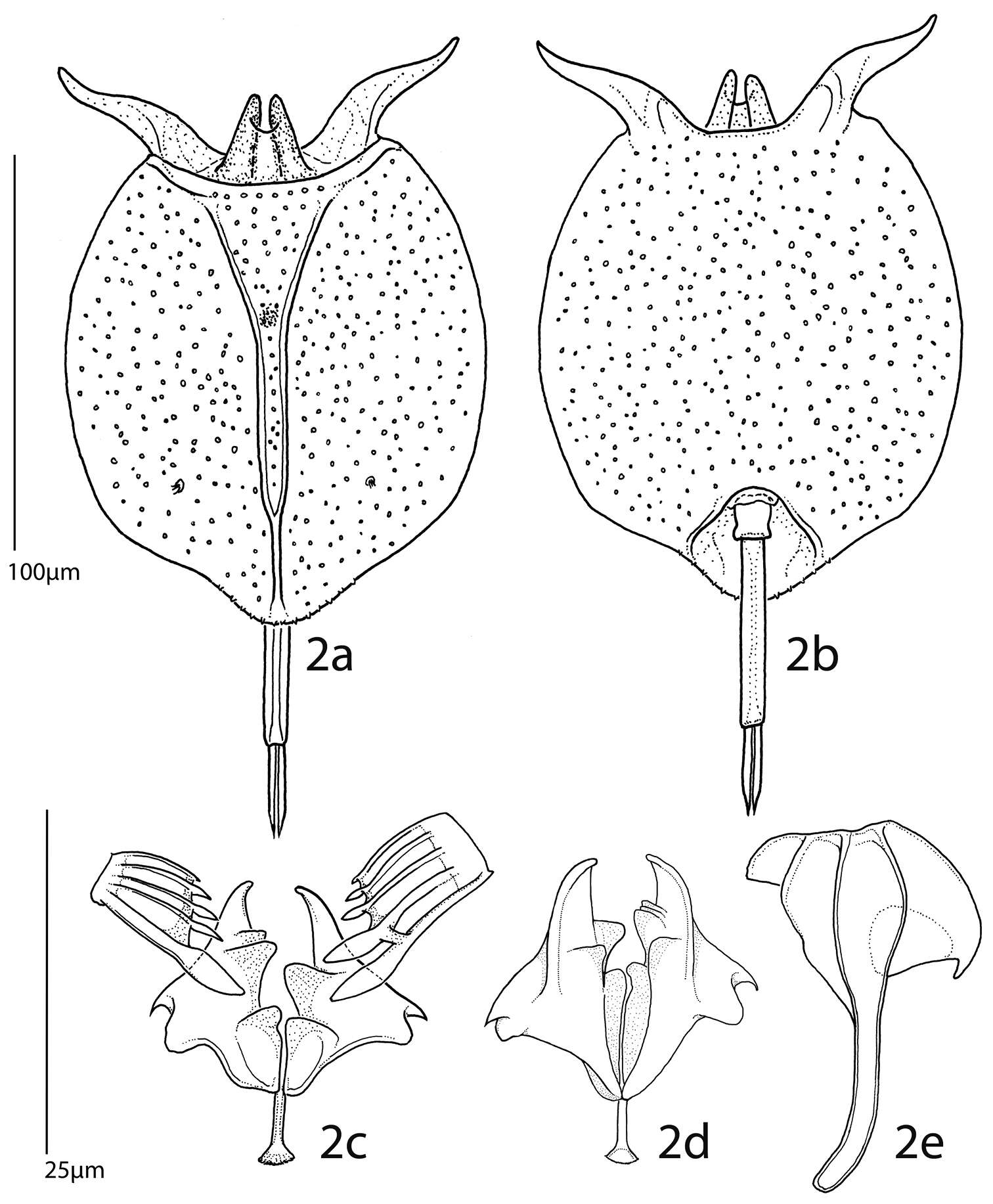
Yongting Luo, Hendrik Segers
Zookeys
Figures 2.Pulchritia dorsicornuta gen. n., comb. n., a habitus, dorsal b habitus, ventral c–e trophi c unci and incus, frontal d incus, caudal e left manubrium, external. Scale bars: a–b= 100µm, c–e= 25µm.
-
Villoslada de Cameros, La Rioja, Spain
-
Melgar de Tera, Castille and Leon, Spain
-
Ribadelago de Franco, Castille and Leon, Spain
-
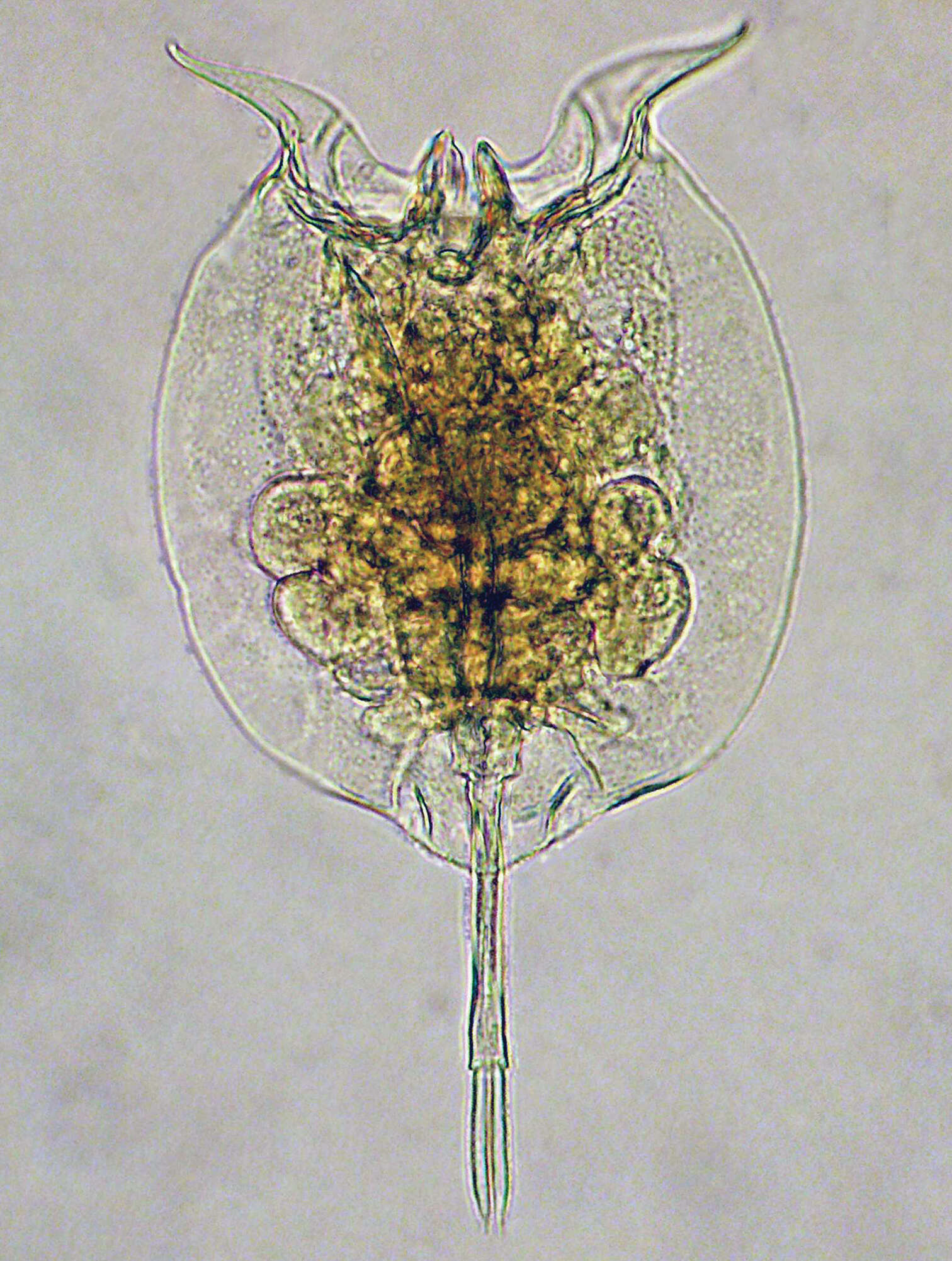
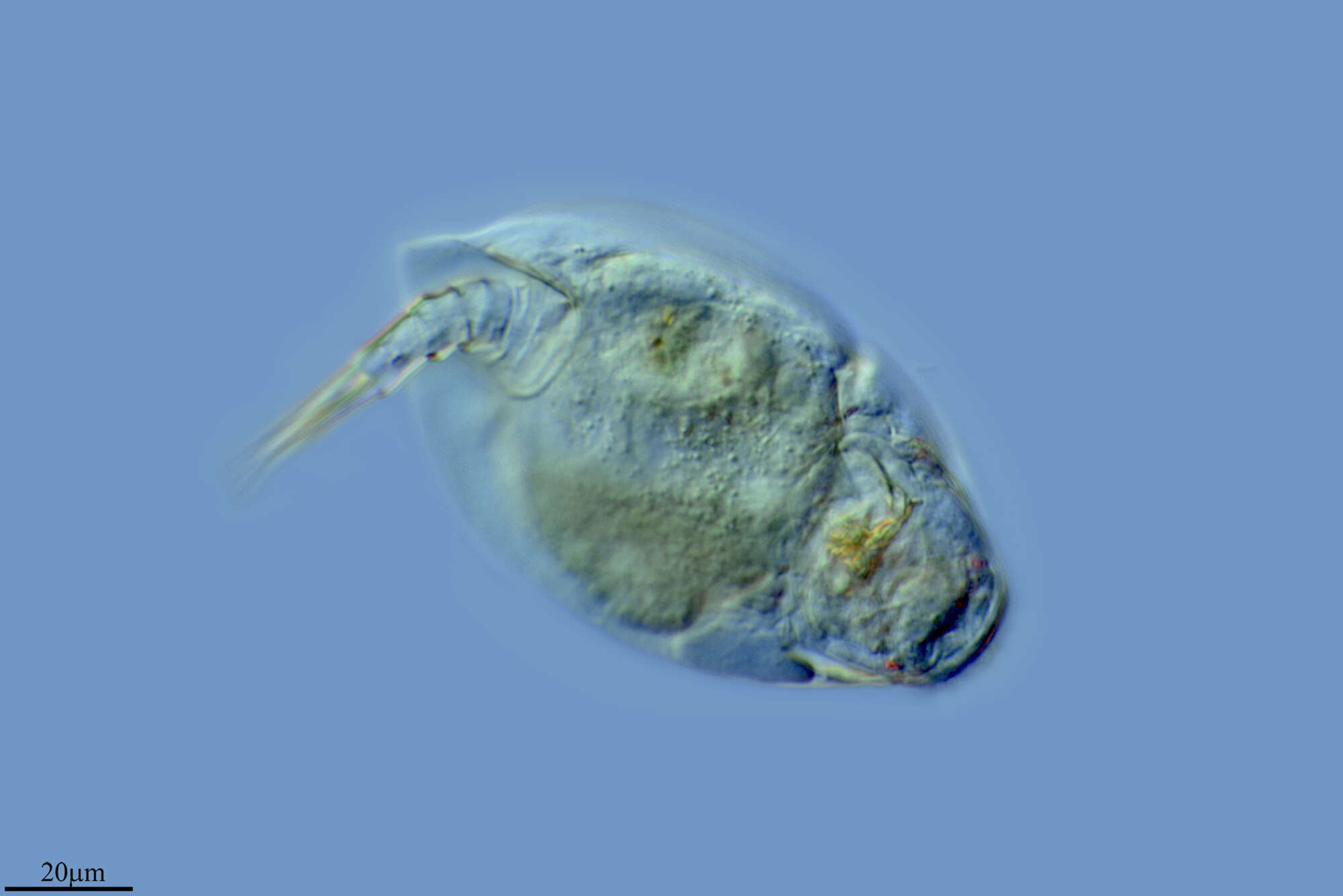
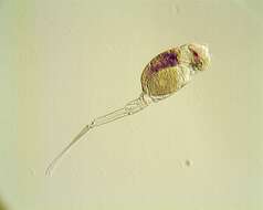
Lorica vase-shaped and thin. Foot long, toes shorter than rest of body.
-
Soba, Cantabria, Spain
-
Soba, Cantabria, Spain
-
It is caracterised by a round tail and short toes.
-
Madrid, Madrid, Spain
-
Ribadelago, Castille and Leon, Spain