-
Jeremy A. Miller, Charles E. Griswold, Nikolaj Scharff, Milan Řezáč, Tamás Szűts, Mohammad Marhabaie
Zookeys
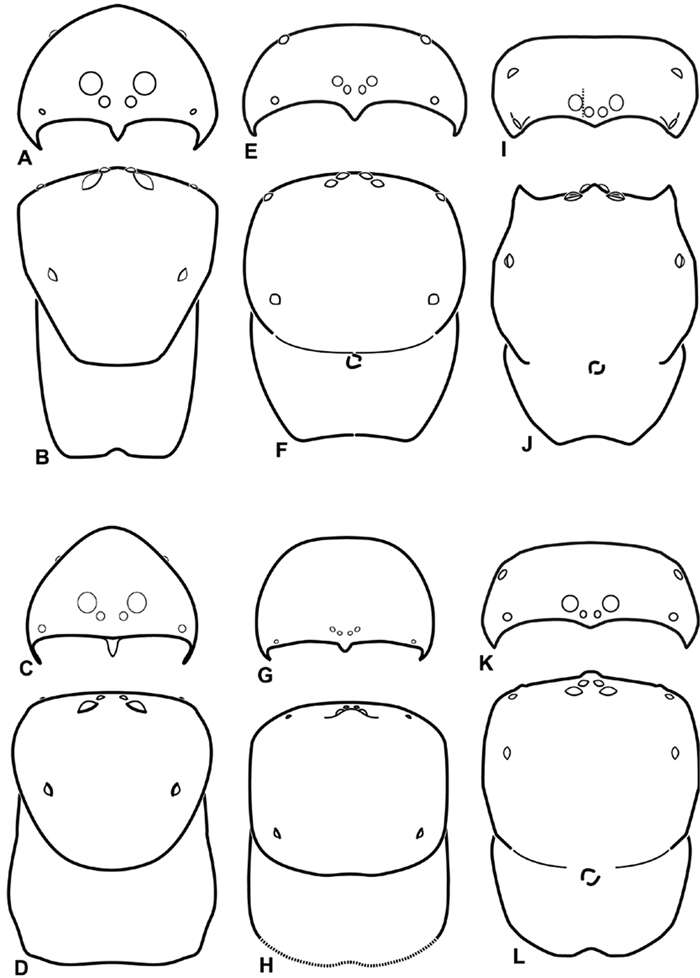
Figure 8.A–L Schematic illustrations of the carapace of assorted eresids A–D Adonea fimbriata E–H Dorceus fastuosus I–L Dresserus sp. A–B, E–F, I–J male C–D, G–H, K–L female A, C, E, G, I, K anterior view B, D, F, H, J, L dorsal view. Dashed line in I drawn tangential to the mesal margin of the PME does not intersect with the AME indicating median eyes separated on vertical axis. Dashed lines at posterior of carapace indicate uncertainty. Not to scale.
-
Jeremy A. Miller, Charles E. Griswold, Nikolaj Scharff, Milan Řezáč, Tamás Szűts, Mohammad Marhabaie
Zookeys
Figure 32.A–F Dorceus fastuosus, male from Mashabin Sand Dunes, Israel (MR006, HUJ), scanning electron micrographs of spinnerets. A overview B left ALS C left PMS D left PLS E vestigial cribellum F modified spigot and flanking aciniform spigot on left PLS. AC aciniform gland spigot ALS anterior lateral spinneret MAP major ampullate gland spigot mAP minor ampullate gland spigot MS modified spigot PI piriform gland spigot PLS posterior lateral spinneret PMS posterior median spinneret.
-
Jeremy A. Miller, Charles E. Griswold, Nikolaj Scharff, Milan Řezáč, Tamás Szűts, Mohammad Marhabaie
Zookeys
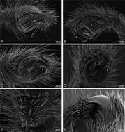
Figure 12.A–L Left male palpi of eresid species, photomicrographs. A–C Adonea fimbriata from Algeria-Morocco (MR012, MR) D–F Dorceus fastuosus from Mashabin Sand Dunes, Israel (MR006, HUJ) G–I Dresserus sp. from Manga Forest Reserve, Tanzania J–L Eresus walckenaeri from Leptokaryas, Greece (MR020, MR) A, D, G, J prolateral view B, E, K retrolateral view H ventral view C, F, I, L expanded palp. BH basal haematodocha MH median haematodocha.
-
Jeremy A. Miller, Charles E. Griswold, Nikolaj Scharff, Milan Řezáč, Tamás Szűts, Mohammad Marhabaie
Zookeys
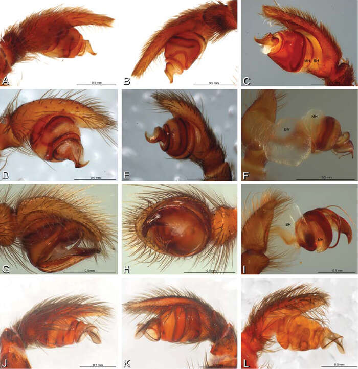
Figure 16.A–L Epigyna of eresid species, photomicrographs. A, D Adonea fimbriata; A from Mehav Am village, Israel (MR003, MR) D from Wadi Mashash, Israel (MR013, HUJ) B, E Dorceus fastuosus from Mashabim sand dunes, Israel (MR002, MR) C, F Dresserus sp. from Klein Kariba, South Africa (CASENT 9025745, CAS) G, J Eresus walckenaeri from 5 km south of Monemvasia, Lakonia, Greece (ZMUC 00012903, ZMUC) H, K Eresus kollari from res. Radotinske udoli, Czechia (MR016, MR) I, L Eresus sandaliatus from SE of Silkeborg, Denmark (CASENT 9039243, CAS) A–C, G–I ventral viewD–F, J–L dorsal view, cleared. CD copulatory duct ML median lobe S spermatheca SH spermathecal head.
-
Jeremy A. Miller, Charles E. Griswold, Nikolaj Scharff, Milan Řezáč, Tamás Szűts, Mohammad Marhabaie
Zookeys
Figure 26.A–J Dorceus fastuosus. A–D, I–J male from Mashabin Sand Dunes, Israel (MR006, HUJ) E–H female from Mashabim sand dunes, Israel (MR002, MR) A–D habitus of male, photomicrographs E–H habitus of female, photomicrographs J, K illustrations of left male palp. A, E dorsal view B, F ventral view C, G anterior view D, H lateral view. I prolateral view. J retrolateral view. C conductor E embolus ST subtegulum T tegulum.
-
Jeremy A. Miller, Charles E. Griswold, Nikolaj Scharff, Milan Řezáč, Tamás Szűts, Mohammad Marhabaie
Zookeys
Figure 27.A–F Dorceus fastuosus from Mashabin Sand Dunes, Israel (MR006, HUJ), scanning electron micrographs of left male palp. A prolateral view B retrolateral view C detail of embolic division, prolateral view D ventral view E detail of embolic division, ventral view F palpal tibia, dorsal view. C conductor E embolus ST subtegulum T tegulum.
-
Jeremy A. Miller, Charles E. Griswold, Nikolaj Scharff, Milan Řezáč, Tamás Szűts, Mohammad Marhabaie
Zookeys
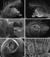
Figure 28.A–D Dorceus fastuosus, male from Mashabin sand dunes, Israel (MR006, HUJ), scanning electron micrographs. A prosoma, anterior view B left chelicerae, lateral view C chelicerae, anterior distal view showing fangs and teeth D epiandrous region.
-
Jeremy A. Miller, Charles E. Griswold, Nikolaj Scharff, Milan Řezáč, Tamás Szűts, Mohammad Marhabaie
Zookeys
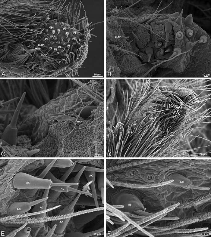
Figure 29.A–F Dorceus fastuosus, female from Mashabim sand dunes, Israel (MR002, MR), scanning electron micrographs. A median eye group B prosoma, dorsal C epigynum, ventral view D cleared vulva, dorsal view E detail, left spermathecal head F detail, right spermatheca. ML median lobe S spermatheca SH spermathecal head.
-
Jeremy A. Miller, Charles E. Griswold, Nikolaj Scharff, Milan Řezáč, Tamás Szűts, Mohammad Marhabaie
Zookeys
Figure 30.A–F Dorceus fastuosus, female from Mashabim sand dunes, Israel (MR002, MR), scanning electron micrographs of spinnerets. A left ALS B left PMS C detail of left PMS D left PLS E aciniform field on left PLS F modified spigot and flanking aciniform gland spigot on left PLS. Unlabeled spigots in B and C thought to be a mixture of aciniform gland spigots and cylindrical gland spigots. AC aciniform gland spigot MAP major ampullate gland spigot mAP minor ampullate gland spigot MS modified spigot PI piriform gland spigot.
-
Jeremy A. Miller, Charles E. Griswold, Nikolaj Scharff, Milan Řezáč, Tamás Szűts, Mohammad Marhabaie
Zookeys
Figure 31.A–F Dorceus fastuosus, female from Mashabim sand dunes, Israel (MR002, MR), scanning electron micrographs. A detail of spigots on left ALS B cribellum C detail cribellar spigots D trichobothrium, left tibia IV E calamistrum, left metatarsus IV F detail, calamistrum seta, left metatarsus IV. MAP major ampullate gland spigot PI piriform gland spigot.
-
Jeremy A. Miller, Charles E. Griswold, Nikolaj Scharff, Milan Řezáč, Tamás Szűts, Mohammad Marhabaie
Zookeys
Figure 8.A–L Schematic illustrations of the carapace of assorted eresids A–D Adonea fimbriata E–H Dorceus fastuosus I–L Dresserus sp. A–B, E–F, I–J male C–D, G–H, K–L female A, C, E, G, I, K anterior view B, D, F, H, J, L dorsal view. Dashed line in I drawn tangential to the mesal margin of the PME does not intersect with the AME indicating median eyes separated on vertical axis. Dashed lines at posterior of carapace indicate uncertainty. Not to scale.
-
Jeremy A. Miller, Charles E. Griswold, Nikolaj Scharff, Milan Řezáč, Tamás Szűts, Mohammad Marhabaie
Zookeys
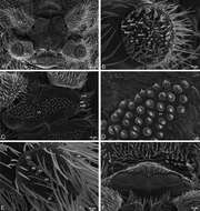
Figure 37.A–F Dresserus sp., scanning electron micrographs. A, C female from Mazumbai, Tanzania (CASENT 9025747, CAS) D, F female from Klein Kariba, South Africa (CASENT 9025745, CAS) A detail of spigots on right ALS B detail of spigots on anterior part of PMS C detail of spigots on anterior part of right PMS D epigynum, ventral view E vulva, dorsal view F detail of pores on right spermathecal head. AC aciniform gland spigot CD copulatory duct CY cylindrical gland spigot FD fertilization duct MAP major ampullate gland spigot mAP minor ampullate gland spigot PI piriform gland spigot S spermatheca SH spermathecal head t tartipore.
-
Jeremy A. Miller, Charles E. Griswold, Nikolaj Scharff, Milan Řezáč, Tamás Szűts, Mohammad Marhabaie
Zookeys
Figure 29.A–F Dorceus fastuosus, female from Mashabim sand dunes, Israel (MR002, MR), scanning electron micrographs. A median eye group B prosoma, dorsal C epigynum, ventral view D cleared vulva, dorsal view E detail, left spermathecal head F detail, right spermatheca. ML median lobe S spermatheca SH spermathecal head.
-
Jeremy A. Miller, Charles E. Griswold, Nikolaj Scharff, Milan Řezáč, Tamás Szűts, Mohammad Marhabaie
Zookeys
Figure 10.A–L Schematic illustrations of the carapace of assorted eresids. A–B Paradonea striatipes C–D Paradonea splendens E–H Paradonea variegata I–L Seothyra henscheli A–D, E–F, I–J male G–H, K–L female. A, C, E, G, I, K anterior view B, D, F, H, J, L dorsal view G illustrates example of median eyes overlapping on horizontal axis. Dashed lines at posterior of carapace indicate uncertainty. Not to scale.
-
Jeremy A. Miller, Charles E. Griswold, Nikolaj Scharff, Milan Řezáč, Tamás Szűts, Mohammad Marhabaie
Zookeys
Figure 11.A–L Schematic illustrations of the carapace of assorted Stegodyphus species. A–D Stegodyphus lineatus E–H Stegodyphus mimosarum I–L Stegodyphus sarasinorum. A–B, E–F, I–J male C–D, G–H, K–L female A, C, E, G, I, K anterior view B, D, F, H, J, L dorsal view A illustrates example of median eyes separated on horizontal axis; E illustrates example of median eyes overlapping on vertical axis. Dashed lines at posterior of carapace indicate uncertainty. Not to scale.
-
Jeremy A. Miller, Charles E. Griswold, Nikolaj Scharff, Milan Řezáč, Tamás Szűts, Mohammad Marhabaie
Zookeys
Figure 36.A–F Dresserus sp., female from Mazumbai, Tanzania (CASENT 9025747, CAS), scanning electron micrographs of spinnerets. A overview B left ALS C right PMS D detail, cylindrical gland spigots on right PMS E left PLS F cribellum. AC aciniform gland spigot ALS anterior lateral spinneret CR cribellum CY cylindrical gland spigot MAP major ampullate gland spigot mAP minor ampullate gland spigot MS modified spigot PI piriform gland spigot PLS posterior lateral spinneret PMS posterior median spinneret.
-
Jeremy A. Miller, Charles E. Griswold, Nikolaj Scharff, Milan Řezáč, Tamás Szűts, Mohammad Marhabaie
Zookeys
Figure 33.A–K Dresserus sp. A–D male from Manga Forest Reserve, Tanzania (ZMUC), image D reversed E–H female from Mazumbai, Tanzania (CASENT 9025747, CAS) I–K male from Mazumbai, Tanzania (CASENT 9025746, CAS) A–D habitus of male, photomicrographs E–H habitus of female, photomicrographs I–K illustrations of left male palp A, E dorsal view B, F ventral view C, G anterior view D, H lateral view I prolateral view J ventral view K retrolateral view. C conductor E embolus ST subtegulum T tegulum.
-
Jeremy A. Miller, Charles E. Griswold, Nikolaj Scharff, Milan Řezáč, Tamás Szűts, Mohammad Marhabaie
Zookeys
Figure 12.A–L Left male palpi of eresid species, photomicrographs. A–C Adonea fimbriata from Algeria-Morocco (MR012, MR) D–F Dorceus fastuosus from Mashabin Sand Dunes, Israel (MR006, HUJ) G–I Dresserus sp. from Manga Forest Reserve, Tanzania J–L Eresus walckenaeri from Leptokaryas, Greece (MR020, MR) A, D, G, J prolateral view B, E, K retrolateral view H ventral view C, F, I, L expanded palp. BH basal haematodocha MH median haematodocha.
-
Jeremy A. Miller, Charles E. Griswold, Nikolaj Scharff, Milan Řezáč, Tamás Szűts, Mohammad Marhabaie
Zookeys
Figure 34.A–F Dresserus sp. A–E male from Mazumbai, Tanzania (CASENT 9025746, CAS), scanning electron micrographs of right palp, images reversed to appear as left palp F female from Klein Kariba, South Africa (CASENT 9025745, CAS), scanning electron micrographs of left chelicera A prolateral view B retrolateral view C ventral view D apical view E palpal tibia, dorsal view F distal part of chelicerae showing fang and teeth. C conductor E embolus T tegulum.
-
Jeremy A. Miller, Charles E. Griswold, Nikolaj Scharff, Milan Řezáč, Tamás Szűts, Mohammad Marhabaie
Zookeys
Figure 55.A–F Gandanameno sp. from Harare, Zimbabwe (AcAT 2005/123, NCA), scanning electron micrographs, right male palp, images reversed to appear as left palp. A prolateral view B retrolateral view C ventral view D apical view E detail of distal tip of conductor F palpal tibia, dorsal view. C conductor E embolus ST subtegulum T tegulum.
-
Jeremy A. Miller, Charles E. Griswold, Nikolaj Scharff, Milan Řezáč, Tamás Szűts, Mohammad Marhabaie
Zookeys
Figure 16.A–L Epigyna of eresid species, photomicrographs. A, D Adonea fimbriata; A from Mehav Am village, Israel (MR003, MR) D from Wadi Mashash, Israel (MR013, HUJ) B, E Dorceus fastuosus from Mashabim sand dunes, Israel (MR002, MR) C, F Dresserus sp. from Klein Kariba, South Africa (CASENT 9025745, CAS) G, J Eresus walckenaeri from 5 km south of Monemvasia, Lakonia, Greece (ZMUC 00012903, ZMUC) H, K Eresus kollari from res. Radotinske udoli, Czechia (MR016, MR) I, L Eresus sandaliatus from SE of Silkeborg, Denmark (CASENT 9039243, CAS) A–C, G–I ventral viewD–F, J–L dorsal view, cleared. CD copulatory duct ML median lobe S spermatheca SH spermathecal head.
-
Jeremy A. Miller, Charles E. Griswold, Nikolaj Scharff, Milan Řezáč, Tamás Szűts, Mohammad Marhabaie
Zookeys
Figure 1.A–H Habitus of living Eresidae, photographs. A, B Adonea fimbriata A juvenile female (photo by Martin Forman) B adult male from Israel (photo by Martin Forman) C Dresserus kannemeyeri, adult female from Ndumo Game Reserve, South Africa (photo Stanislav Macík) D Dresserus sp., adult malefrom Namibia, between the towns Aus and Helmeringhausen (26°13.049'S, 16°36.063'E; photo by Martin Forman) E, F Gandanameno sp. E subadult female from Cape Town, South Africa (Stanislav Macík) F adult male from Anysberg Nature Reserve, Western Cape Province, South Africa (photo Martin Forman) G, H adult male Loureedia annulipes; G from Tel Krayot, Israel (photo by Martin Forman) H from Arad, Israel (photo by Martin Forman).
-
Jeremy A. Miller, Charles E. Griswold, Nikolaj Scharff, Milan Řezáč, Tamás Szűts, Mohammad Marhabaie
Zookeys
Figure 37.A–F Dresserus sp., scanning electron micrographs. A, C female from Mazumbai, Tanzania (CASENT 9025747, CAS) D, F female from Klein Kariba, South Africa (CASENT 9025745, CAS) A detail of spigots on right ALS B detail of spigots on anterior part of PMS C detail of spigots on anterior part of right PMS D epigynum, ventral view E vulva, dorsal view F detail of pores on right spermathecal head. AC aciniform gland spigot CD copulatory duct CY cylindrical gland spigot FD fertilization duct MAP major ampullate gland spigot mAP minor ampullate gland spigot PI piriform gland spigot S spermatheca SH spermathecal head t tartipore.
-
Jeremy A. Miller, Charles E. Griswold, Nikolaj Scharff, Milan Řezáč, Tamás Szűts, Mohammad Marhabaie
Zookeys
Figure 38.A–F Dresserus sp., female from Klein Kariba, South Africa (CASENT 9025745, CAS), scanning electron micrographs of legs A tarsal organ, left leg I B trichobothrium, left leg I C tarsal claw, left leg I setae removed D left metatarsus IV, retrolateral view, showing calamistrum E detail of calimistrum F detail of teeth on calimistrum setae.