-
Jeremy A. Miller, Charles E. Griswold, Nikolaj Scharff, Milan Řezáč, Tamás Szűts, Mohammad Marhabaie
Zookeys
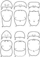
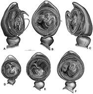
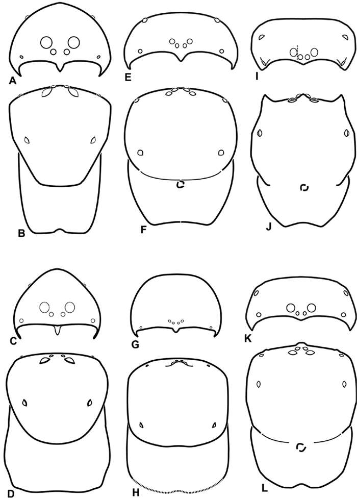
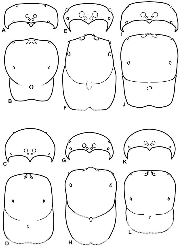
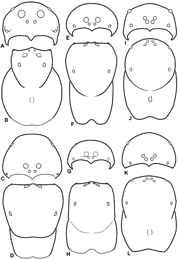
Figure 8.A–L Schematic illustrations of the carapace of assorted eresids A–D Adonea fimbriata E–H Dorceus fastuosus I–L Dresserus sp. A–B, E–F, I–J male C–D, G–H, K–L female A, C, E, G, I, K anterior view B, D, F, H, J, L dorsal view. Dashed line in I drawn tangential to the mesal margin of the PME does not intersect with the AME indicating median eyes separated on vertical axis. Dashed lines at posterior of carapace indicate uncertainty. Not to scale.
-
Jeremy A. Miller, Charles E. Griswold, Nikolaj Scharff, Milan Řezáč, Tamás Szűts, Mohammad Marhabaie
Zookeys
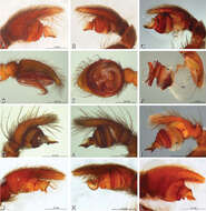
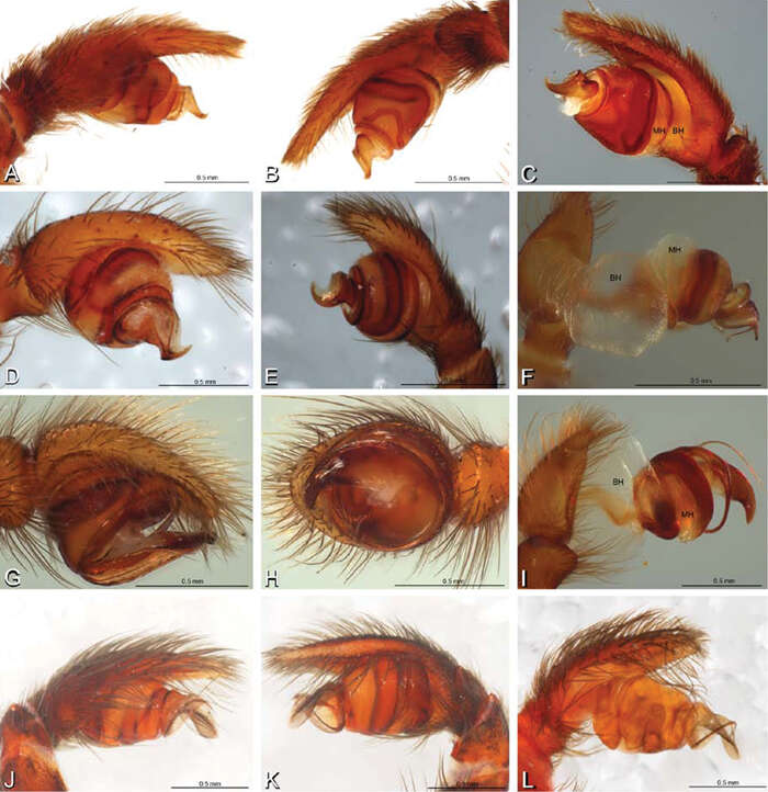
Figure 12.A–L Left male palpi of eresid species, photomicrographs. A–C Adonea fimbriata from Algeria-Morocco (MR012, MR) D–F Dorceus fastuosus from Mashabin Sand Dunes, Israel (MR006, HUJ) G–I Dresserus sp. from Manga Forest Reserve, Tanzania J–L Eresus walckenaeri from Leptokaryas, Greece (MR020, MR) A, D, G, J prolateral view B, E, K retrolateral view H ventral view C, F, I, L expanded palp. BH basal haematodocha MH median haematodocha.
-
Jeremy A. Miller, Charles E. Griswold, Nikolaj Scharff, Milan Řezáč, Tamás Szűts, Mohammad Marhabaie
Zookeys
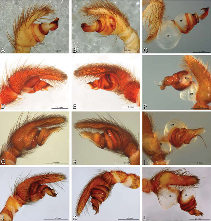
Figure 13.A–L Left male palpi of eresid species, photomicrographs. A–C Eresus kollari from res. Radotinske udoli, Czechia (MR007, MR) D–F Gandanameno sp. from Van Riebeeck Park, Western Cape, South Africa (CASENT 9023763, CAS) G–I Loureedia annulipes from Haluqim Ridge, Israel (PET03, MR) J, K Paradonea striatipes from Otjivasandu (NMN), Namibia L Paradonea splendens from Sunnyside, South Africa (C1076, SAM) A, D, G, J, L prolateral view B, H, K retrolateral view E ventral view C, F, I expanded palp. BH basal haematodocha MH median haematodocha.
-
Jeremy A. Miller, Charles E. Griswold, Nikolaj Scharff, Milan Řezáč, Tamás Szűts, Mohammad Marhabaie
Zookeys
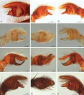
Figure 15.A–L Left male palpi of eresid species, photomicrographs. A–C Seothyra henscheli from Gobabeb Station, Namibia (SMN 40828, NMN) D, F Stegodyphus lineatus D–E from Negev, Israel (MR) F from Nengrahar, Afghanistan (MR010, MR) G–I Stegodyphus mimosarum from Forêt d'Analalava, Fianarantsoa, Madagascar (CASENT 9015950, CAS) J–L Stegodyphus sarasinorum from 7.5 km E PwintPhyu, Magway Division, Myanmar (CASENT 9019370, CAS) A, D, G, J prolateral view B, E, H, K retrolateral view C, F, I, L expanded palp. BH basal haematodocha MH median haematodocha.
-
Jeremy A. Miller, Charles E. Griswold, Nikolaj Scharff, Milan Řezáč, Tamás Szűts, Mohammad Marhabaie
Zookeys
Figure 14.A–L Left male palpi of Paradonea species, photomicrographs. A Paradonea splendens from Sunnyside, South Africa (C1076, SAM) B, C Paradonea variegata from Breekkierie Dunes, Northern Cape, South Africa (C1062, SAM) D–I Paradonea parva D–F holotype from junction of Marico and Crocodile Rivers, South Africa (B3701, SAM) G–I from 4 km N of Hopetown, Northern Cape, South Africa (AcAT 97/988, NCA) J–L Paradonea presleyi sp. n. holotype from Falcon College, Zimbabwe (CASENT 9039236, CAS) A, C, F, I, L retrolateral view B, D, G, J prolateral view E, H, K ventral view.
-
Jeremy A. Miller, Charles E. Griswold, Nikolaj Scharff, Milan Řezáč, Tamás Szűts, Mohammad Marhabaie
Zookeys
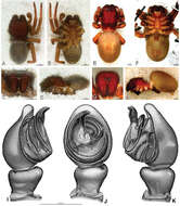
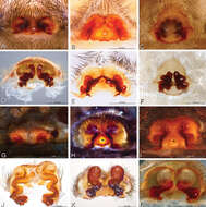
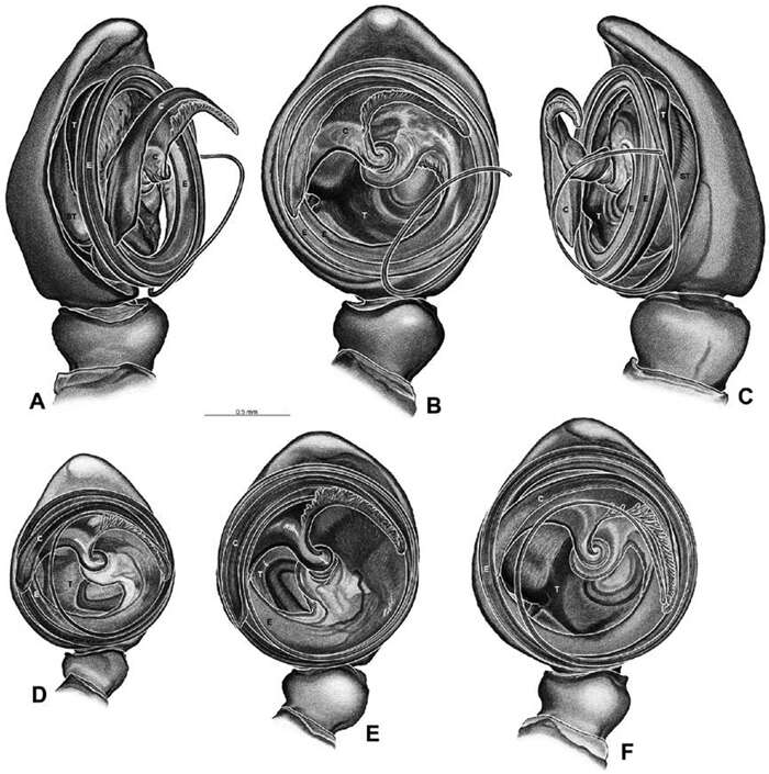
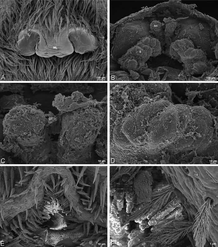
Figure 33.A–K Dresserus sp. A–D male from Manga Forest Reserve, Tanzania (ZMUC), image D reversed E–H female from Mazumbai, Tanzania (CASENT 9025747, CAS) I–K male from Mazumbai, Tanzania (CASENT 9025746, CAS) A–D habitus of male, photomicrographs E–H habitus of female, photomicrographs I–K illustrations of left male palp A, E dorsal view B, F ventral view C, G anterior view D, H lateral view I prolateral view J ventral view K retrolateral view. C conductor E embolus ST subtegulum T tegulum.
-
Jeremy A. Miller, Charles E. Griswold, Nikolaj Scharff, Milan Řezáč, Tamás Szűts, Mohammad Marhabaie
Zookeys
Figure 48.A–F Gandanameno sp., illustrations of left male palp. A–C from Naauwpoort, North West Province, South Africa (SAM 1600, SAM) D from Van Riebeeck Park, Western Cape, South Africa (CASENT 9023763, CAS) E from Graaff-Reinet, Eastern Cape, South Africa (SAM 12571, SAM) F from Hanover, South Africa (SAM 9465, SAM) A obliquely retrolateral view B, D–F ventral view C obliquely prolateral view. All images at the same scale. C conductor E embolus ST subtegulum T tegulum.
-
Jeremy A. Miller, Charles E. Griswold, Nikolaj Scharff, Milan Řezáč, Tamás Szűts, Mohammad Marhabaie
Zookeys
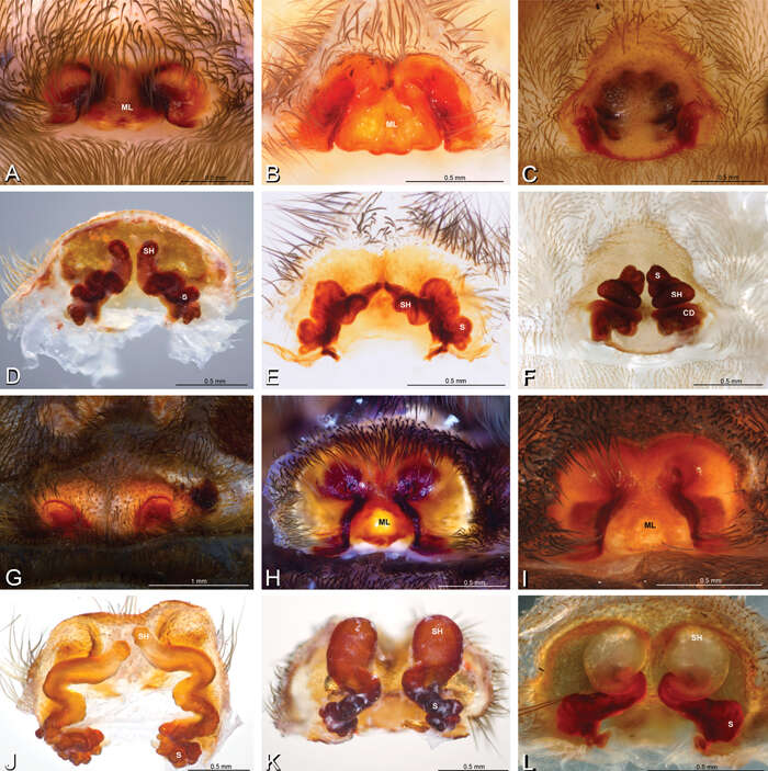
Figure 16.A–L Epigyna of eresid species, photomicrographs. A, D Adonea fimbriata; A from Mehav Am village, Israel (MR003, MR) D from Wadi Mashash, Israel (MR013, HUJ) B, E Dorceus fastuosus from Mashabim sand dunes, Israel (MR002, MR) C, F Dresserus sp. from Klein Kariba, South Africa (CASENT 9025745, CAS) G, J Eresus walckenaeri from 5 km south of Monemvasia, Lakonia, Greece (ZMUC 00012903, ZMUC) H, K Eresus kollari from res. Radotinske udoli, Czechia (MR016, MR) I, L Eresus sandaliatus from SE of Silkeborg, Denmark (CASENT 9039243, CAS) A–C, G–I ventral viewD–F, J–L dorsal view, cleared. CD copulatory duct ML median lobe S spermatheca SH spermathecal head.
-
Jeremy A. Miller, Charles E. Griswold, Nikolaj Scharff, Milan Řezáč, Tamás Szűts, Mohammad Marhabaie
Zookeys
Figure 17.A–F Epigyna of Gandanameno sp., photomicrographs. A, D from Iringa, Tanzania (ZMUC 19970517, ZMUC) B, E from Kommetjie, Western Cape, South Africa (CASENT 9039241, CAS), note broken embolus left in female reproductive system C, F from Port Elizabeth, South Africa (port-3325, ZMHB) A–C ventral view D–F dorsal view, cleared. CD copulatory duct S spermatheca SH spermathecal head.
-
Jeremy A. Miller, Charles E. Griswold, Nikolaj Scharff, Milan Řezáč, Tamás Szűts, Mohammad Marhabaie
Zookeys
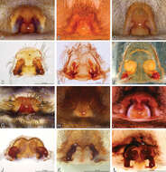
Figure 18.A–L Epigyna of eresid species, photomicrographs. A, D Loureedia annulipes from Wadi Mashash, Negev, Israel (MR019, MR) B, E Paradonea variegata from Steinkopf, Northern Cape, South Africa (ZMB 26964, ZMHB) C, F Seothyra henscheli; C from Kuiseb River, Gobabeb, Namibia (SMN 46627, NMN) F from Sout Rivier, Namibia (CASENT 9039242, CAS) G, J Stegodyphus lineatus from Belkis, near Birecor, Turkey (MR015, MR) H, K Stegodyphus mimosarum H from Forêt d'Analalava, Fianarantsoa, Madagascar (CASENT 9015950, CAS) K from Réserve Spéciale de Cap Sainte Marie, Toliara, Madagascar (CASENT 9012844, CAS) I, L Stegodyphus sarasinorum from 7.5 km E PwintPhyu, Magway Division, Myanmar (CASENT 9019370, CAS) A–C, G–I ventral view D–F, J–L dorsal view, cleared. AL anterior lobe ML median lobe S spermatheca SH spermathecal head.
-
Jeremy A. Miller, Charles E. Griswold, Nikolaj Scharff, Milan Řezáč, Tamás Szűts, Mohammad Marhabaie
Zookeys
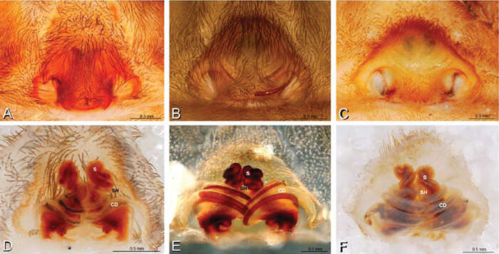
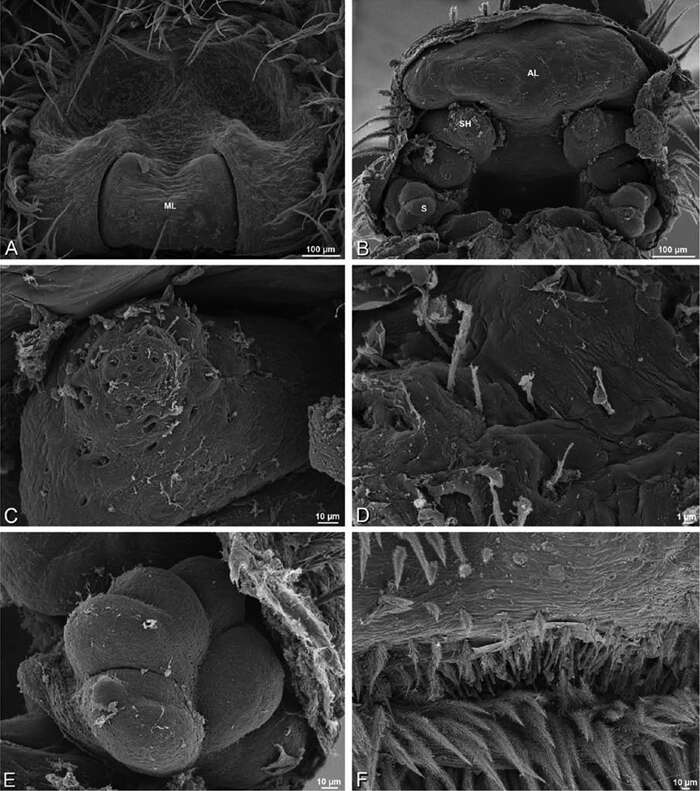
Figure 22.A–F Adonea fimbriata, scanning electron micrographs. A female from Mehav Am village, Israel (MR003, MR) B–D female from Wadi Mashash, Israel (MR013, HUJ) E, F male from Algeria-Morocco (MR012, MR) A–D vulva E, F epiandrous region A epigynum, ventral view B cleared vulva, dorsal view C detail, spermathecal heads D detail, right spermatheca E epiandrous region F detail of epiandrous gland spigots. ML median lobe S spermatheca SH spermathecal head.
-
Jeremy A. Miller, Charles E. Griswold, Nikolaj Scharff, Milan Řezáč, Tamás Szűts, Mohammad Marhabaie
Zookeys
Figure 9.A–L Schematic illustrations of the carapace of assorted eresids. A–D Eresus kollari E–H Gandanameno sp. I–L Loureedia annulipes A–B, E–F, I–J male C–D, G–H, K–L female A, C, E, G, I, K anterior view B, D, F, H, J, L dorsal view. Dashed lines at posterior of carapace indicate uncertainty. Not to scale.
-
Jeremy A. Miller, Charles E. Griswold, Nikolaj Scharff, Milan Řezáč, Tamás Szűts, Mohammad Marhabaie
Zookeys
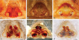
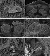
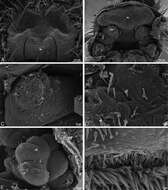
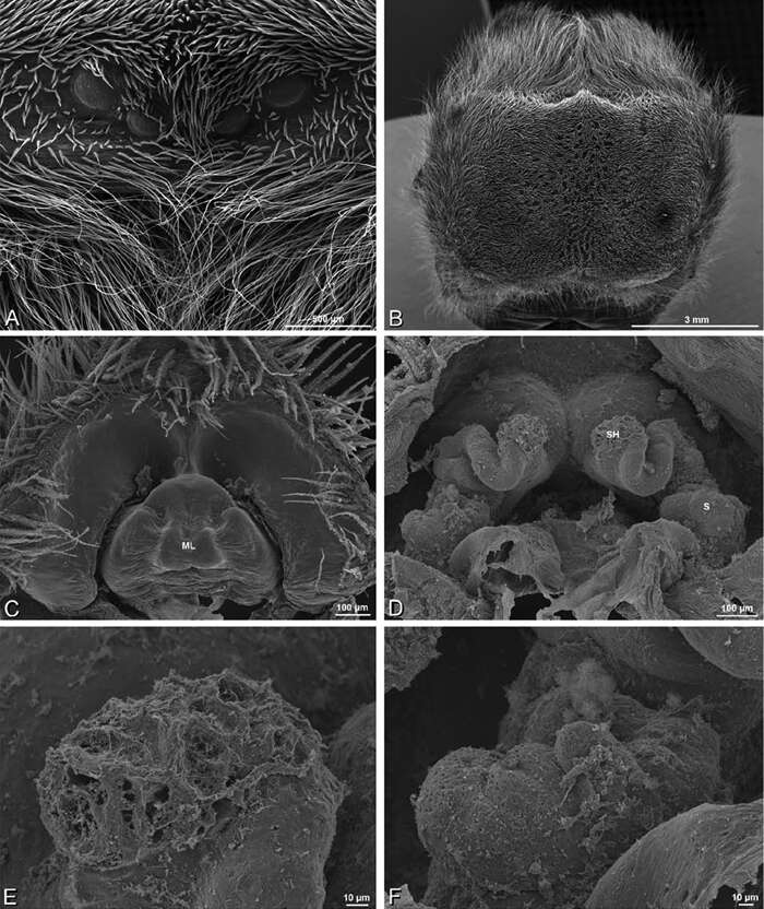
Figure 29.A–F Dorceus fastuosus, female from Mashabim sand dunes, Israel (MR002, MR), scanning electron micrographs. A median eye group B prosoma, dorsal C epigynum, ventral view D cleared vulva, dorsal view E detail, left spermathecal head F detail, right spermatheca. ML median lobe S spermatheca SH spermathecal head.
-
Jeremy A. Miller, Charles E. Griswold, Nikolaj Scharff, Milan Řezáč, Tamás Szűts, Mohammad Marhabaie
Zookeys
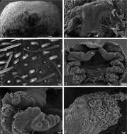
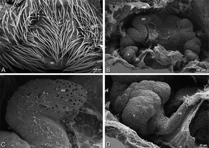
Figure 37.A–F Dresserus sp., scanning electron micrographs. A, C female from Mazumbai, Tanzania (CASENT 9025747, CAS) D, F female from Klein Kariba, South Africa (CASENT 9025745, CAS) A detail of spigots on right ALS B detail of spigots on anterior part of PMS C detail of spigots on anterior part of right PMS D epigynum, ventral view E vulva, dorsal view F detail of pores on right spermathecal head. AC aciniform gland spigot CD copulatory duct CY cylindrical gland spigot FD fertilization duct MAP major ampullate gland spigot mAP minor ampullate gland spigot PI piriform gland spigot S spermatheca SH spermathecal head t tartipore.
-
Jeremy A. Miller, Charles E. Griswold, Nikolaj Scharff, Milan Řezáč, Tamás Szűts, Mohammad Marhabaie
Zookeys
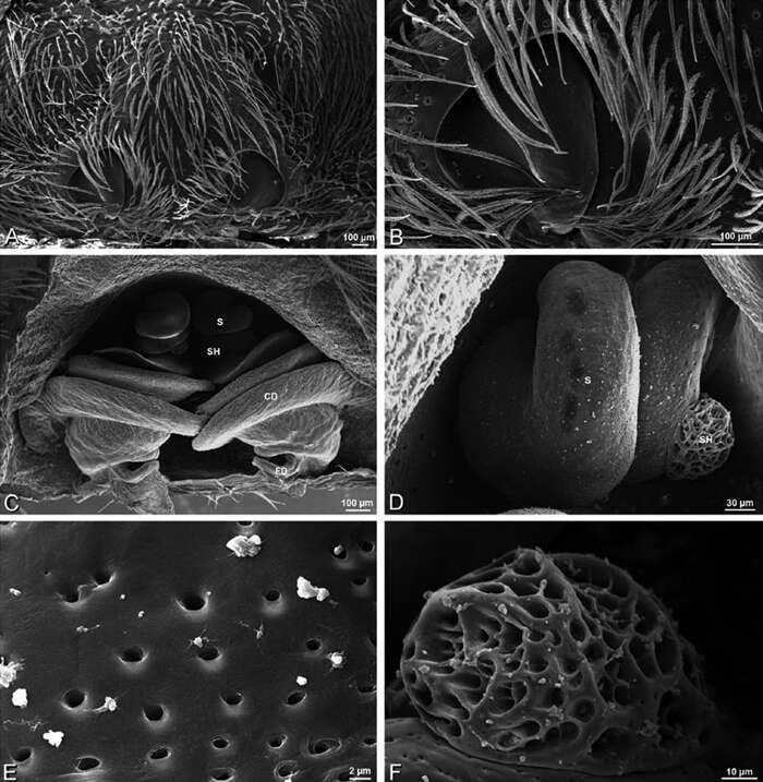
Figure 42.A–F Eresus walckenaeri female from 5 km south of Monemvasia, Lakonia, Greece (ZMUC 00012903), scanning electron micrographs. A prosoma, anterior view B epigynum C tarsal organ, left leg I. ventral view D vulva, dorsal view E left spermatheca F left spermathecal head. S spermatheca SH spermathecal head.
-
Jeremy A. Miller, Charles E. Griswold, Nikolaj Scharff, Milan Řezáč, Tamás Szűts, Mohammad Marhabaie
Zookeys
Figure 59.A–F Scanning electron micrographs of epigynum and vulva of Gandanameno sp. A, B from Iringa, Tanzania (ZMUC 19970530, ZMUC) C–F from Kommetjie, Cape Town, South Africa (CASENT 9039241, CAS) A epigynum, ventral view B detail of right copulatory opening, ventral view C cleared vulva, dorsal view D detail of right spermatheca and spermathecal head E detail, right spermatheca F detail, right spermathecal head. CD copulatory duct FD fertilization duct S spermatheca SH spermathecal head.
-
Jeremy A. Miller, Charles E. Griswold, Nikolaj Scharff, Milan Řezáč, Tamás Szűts, Mohammad Marhabaie
Zookeys
Figure 65.A–F Loureedia annulipes, scanning electron micrographs. A–E vulva of female from Wadi Mashash, Negev, Israel (MR019, MR) F male from Haluqim Ridge, Israel (MR008, HUJ) A epigynum, ventral view B cleared vulva, dorsal view C, D detail, left spermathecal head E detail, right spermatheca. F epiandrous region. AL anterior lobe on atrium ML median lobe S spermatheca SH spermathecal head.
-
Jeremy A. Miller, Charles E. Griswold, Nikolaj Scharff, Milan Řezáč, Tamás Szűts, Mohammad Marhabaie
Zookeys
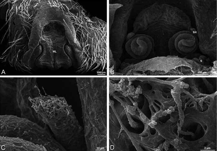
Figure 76.A–F Seothyra henscheli from Sout Rivier, Namibia (CASENT 9039242, CAS), scanning electron micrographs of epigynum A epigynum, ventral view B vulva, dorsal view C, D spermathecal head. ML median lobe S spermatheca SH spermathecal head.
-
Jeremy A. Miller, Charles E. Griswold, Nikolaj Scharff, Milan Řezáč, Tamás Szűts, Mohammad Marhabaie
Zookeys
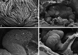
Figure 86.A–D Stegodyphus mimosarum female from Forêt d'Analalava, Fianarantsoa, Madagascar (CASENT 9015950, CAS), scanning electron micrographs of genitalia. A epigynum, ventral view B vulva, dorsal view C spermathecal head D spermatheca and fertilization duct. FD fertilization duct ML median lobe S spermatheca SH spermathecal head.
-
Jeremy A. Miller, Charles E. Griswold, Nikolaj Scharff, Milan Řezáč, Tamás Szűts, Mohammad Marhabaie
Zookeys
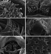
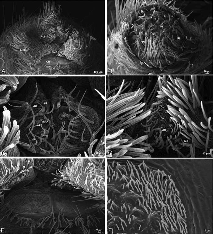
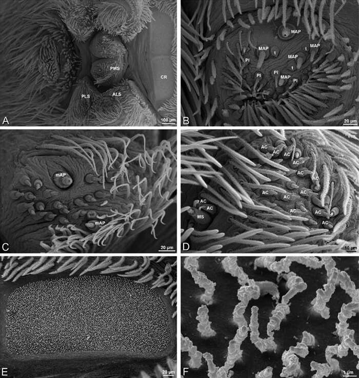
Figure 77. A–F Seothyra henscheli from Kuiseb River, Gobabeb, Namibia (SMN 46627, NMN), scanning electron micrographs of female spinnerets. A overview B left ALS C left and right PMS D right PLS E cribellum F cribellar spigots. AC aciniform gland spigot ALS anterior lateral spinneret CR cribellum CY cylindrical gland spigot mAP minor ampullate gland spigot MS modified spigot PI piriform gland spigot PLS posterior lateral spinneret PMS posterior median spinneret.
-
Jeremy A. Miller, Charles E. Griswold, Nikolaj Scharff, Milan Řezáč, Tamás Szűts, Mohammad Marhabaie
Zookeys
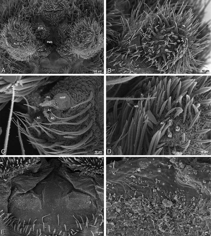
Figure 78.A–F Seothyra henscheli from Gobabeb Station, Namibia (SMN 40828, NMN), scanning electron micrographs of male spinnerets. A overview B right ALS C left PMS D left PLS E vestigial cribellum F detail of vestigial cribellum. AC aciniform gland spigot ALS anterior lateral spinneret mAP minor ampullate gland spigot MS modified spigot PI piriform gland spigot PLS posterior lateral spinneret PMS posterior median spinneret.
-
Jeremy A. Miller, Charles E. Griswold, Nikolaj Scharff, Milan Řezáč, Tamás Szűts, Mohammad Marhabaie
Zookeys
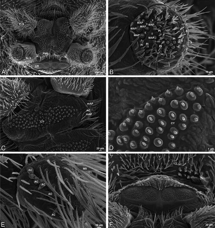
Figure 36.A–F Dresserus sp., female from Mazumbai, Tanzania (CASENT 9025747, CAS), scanning electron micrographs of spinnerets. A overview B left ALS C right PMS D detail, cylindrical gland spigots on right PMS E left PLS F cribellum. AC aciniform gland spigot ALS anterior lateral spinneret CR cribellum CY cylindrical gland spigot MAP major ampullate gland spigot mAP minor ampullate gland spigot MS modified spigot PI piriform gland spigot PLS posterior lateral spinneret PMS posterior median spinneret.
-
Jeremy A. Miller, Charles E. Griswold, Nikolaj Scharff, Milan Řezáč, Tamás Szűts, Mohammad Marhabaie
Zookeys
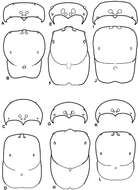
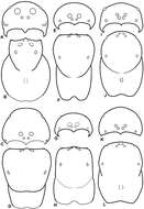
Figure 10.A–L Schematic illustrations of the carapace of assorted eresids. A–B Paradonea striatipes C–D Paradonea splendens E–H Paradonea variegata I–L Seothyra henscheli A–D, E–F, I–J male G–H, K–L female. A, C, E, G, I, K anterior view B, D, F, H, J, L dorsal view G illustrates example of median eyes overlapping on horizontal axis. Dashed lines at posterior of carapace indicate uncertainty. Not to scale.
-
Jeremy A. Miller, Charles E. Griswold, Nikolaj Scharff, Milan Řezáč, Tamás Szűts, Mohammad Marhabaie
Zookeys
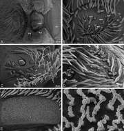
Figure 94.A–F Stegodyphus sarasinorum, scanning electron micrographs of spinnerets of female from 7.5 km E PwintPhyu, Magway Division, Myanmar (CASENT 9019370, CAS). A overview B right ALS C left PMS D right PLS E cribellum F cribellar spigots. Unlabeled spigots in C thought to be a mixture of aciniform gland spigots and cylindrical gland spigots. AC aciniform gland spigot ALS anterior lateral spinneret CR cribellum MAP major ampullate gland spigot mAP minor ampullate gland spigot MS modified spigot n nubbin PI piriform gland spigot PLS posterior lateral spinneret PMS posterior median spinneret t tartipore.