-
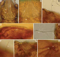
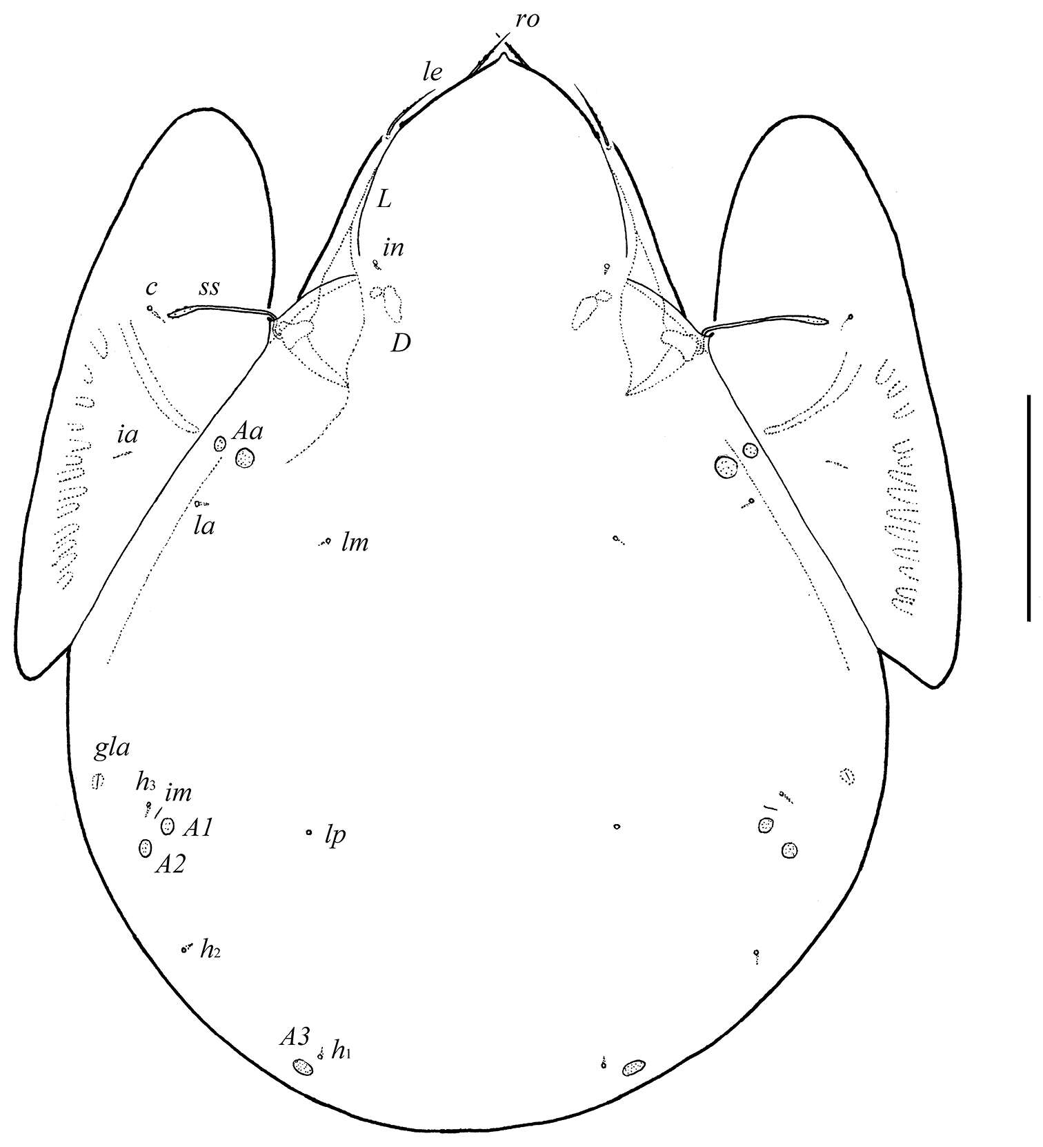
Figure 1.Conchogneta glabrisensillata sp. n. A Dorsal view of idiosoma B Ventral view of idiosoma C Prodorsum D Sensillus and bothridium, lateral view E Slight variation of sensillus, lateral view.
-
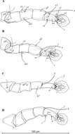
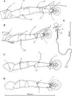
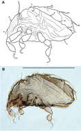
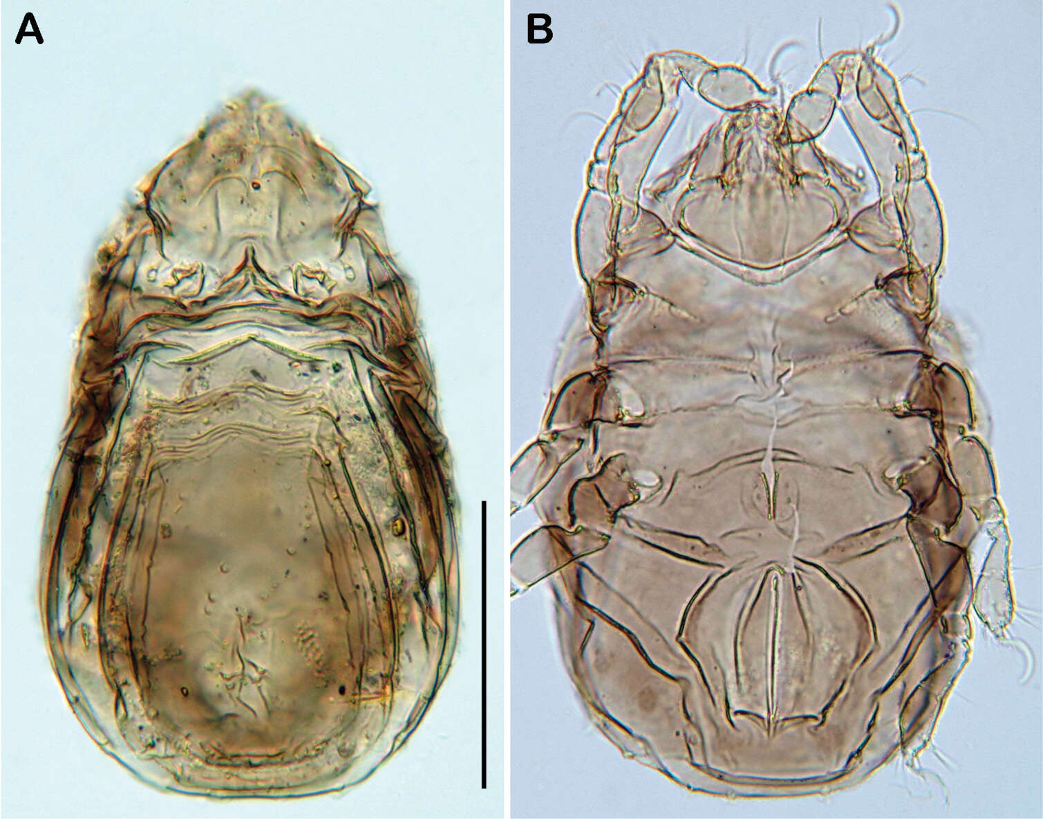
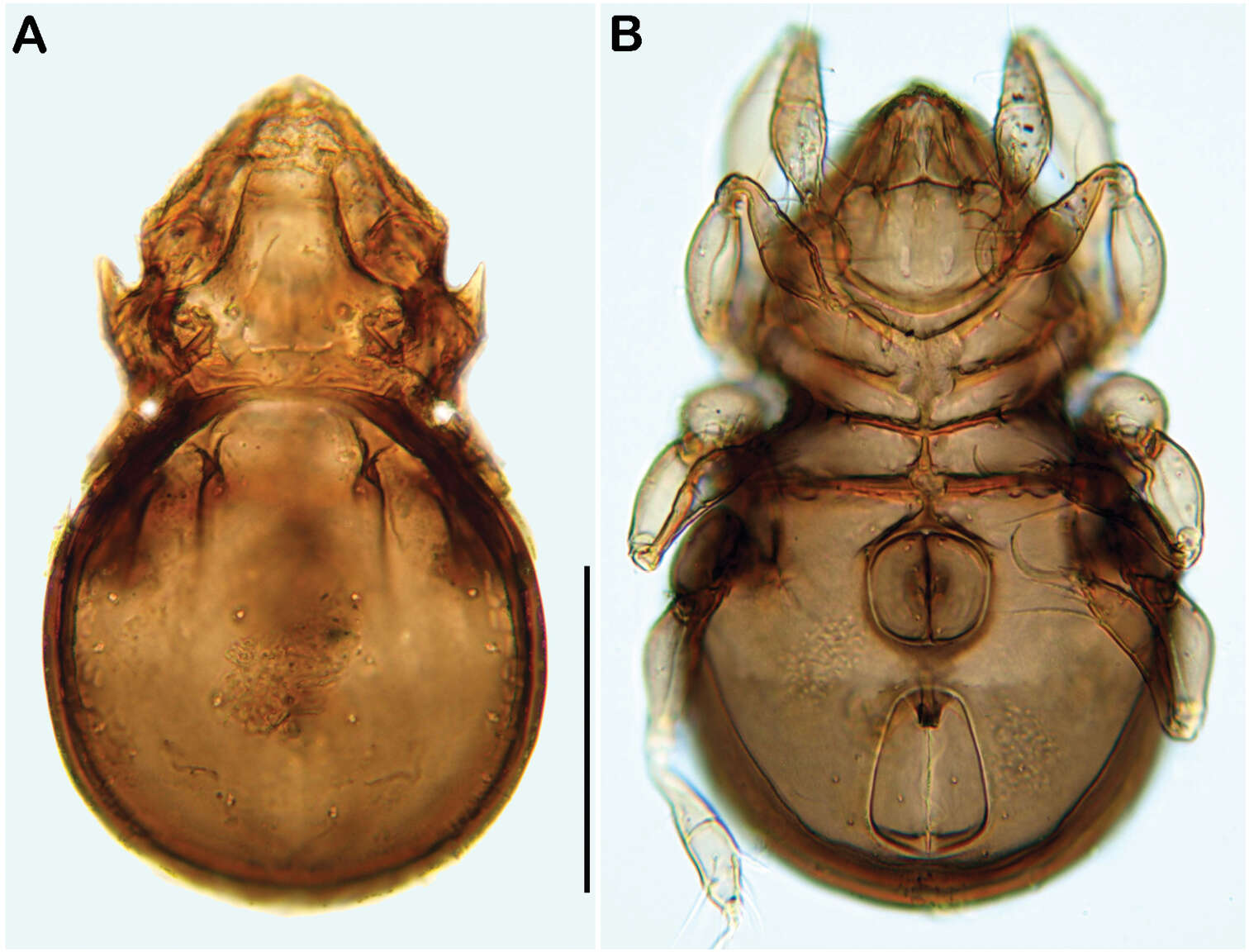
Figure 3.Selenoribates quasimodo sp. n. adult micrographs layered from 5–10 sequentially focused images. A dorsal view B ventral view C lateral view. Scale bars = 100 µm.
-
Ioana Cristina Constantinescu, Gabriel Chişamera, D. Khlur B. Mukhim, Costică Adam
Zookeys
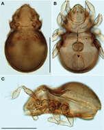
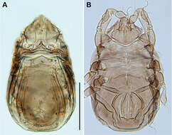
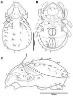
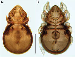
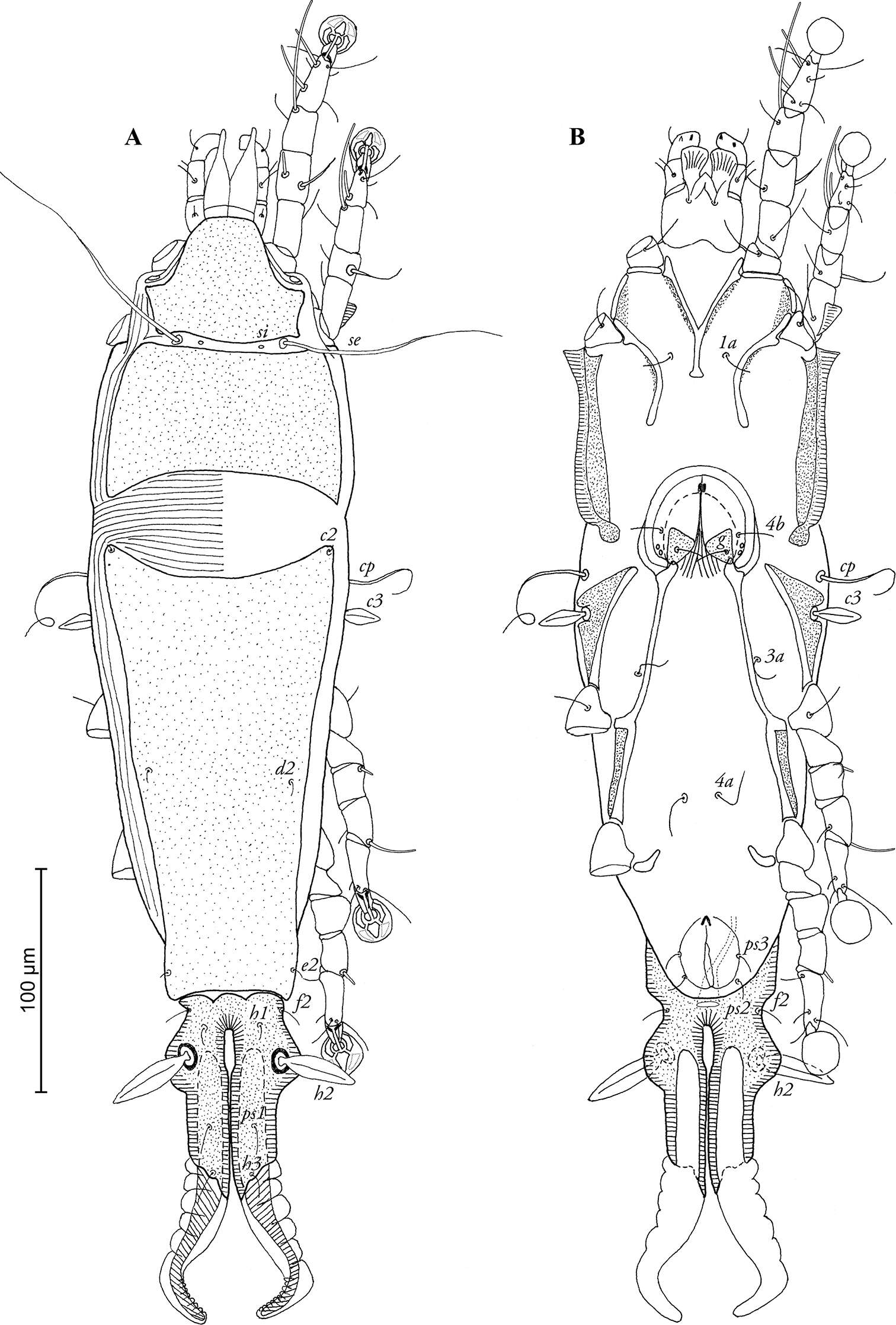
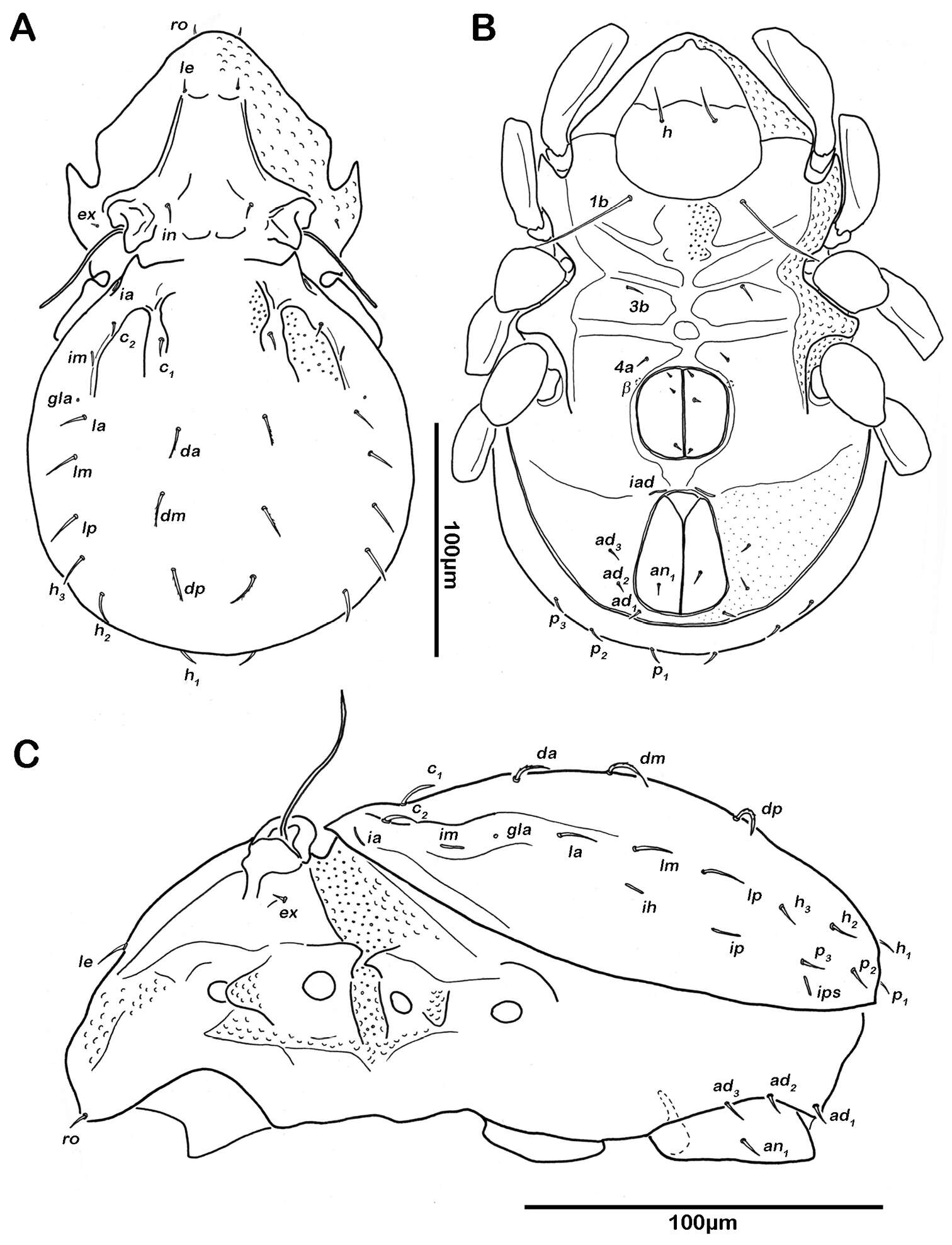
Figure 1.Pedanodectes angustilobus sp. n., male holotype: A dorsal view of idiosoma B ventral view of idiosoma.
-
Sergey G. Ermilov, Olman Alvarado-Rodríguez, Axel P. Retana-Salazar
Zookeys
Figure 1.Pergalumna elongatiporosa sp. n.: dorsal view. Scale bar 100 μm.
-
Figure 2.Conchogneta glabrisensillata sp. n. A Lateral view of prodorsum and anterior part of notogaster B Humeral region, showing tubercles Ha and Hp C Palp, right, antiaxial view D Leg I, right, antiaxial view E Genu and tibia of leg II, right, antiaxial view F Genu and tibia of leg III, right, antiaxial view G Leg IV, right, antiaxial view.
-
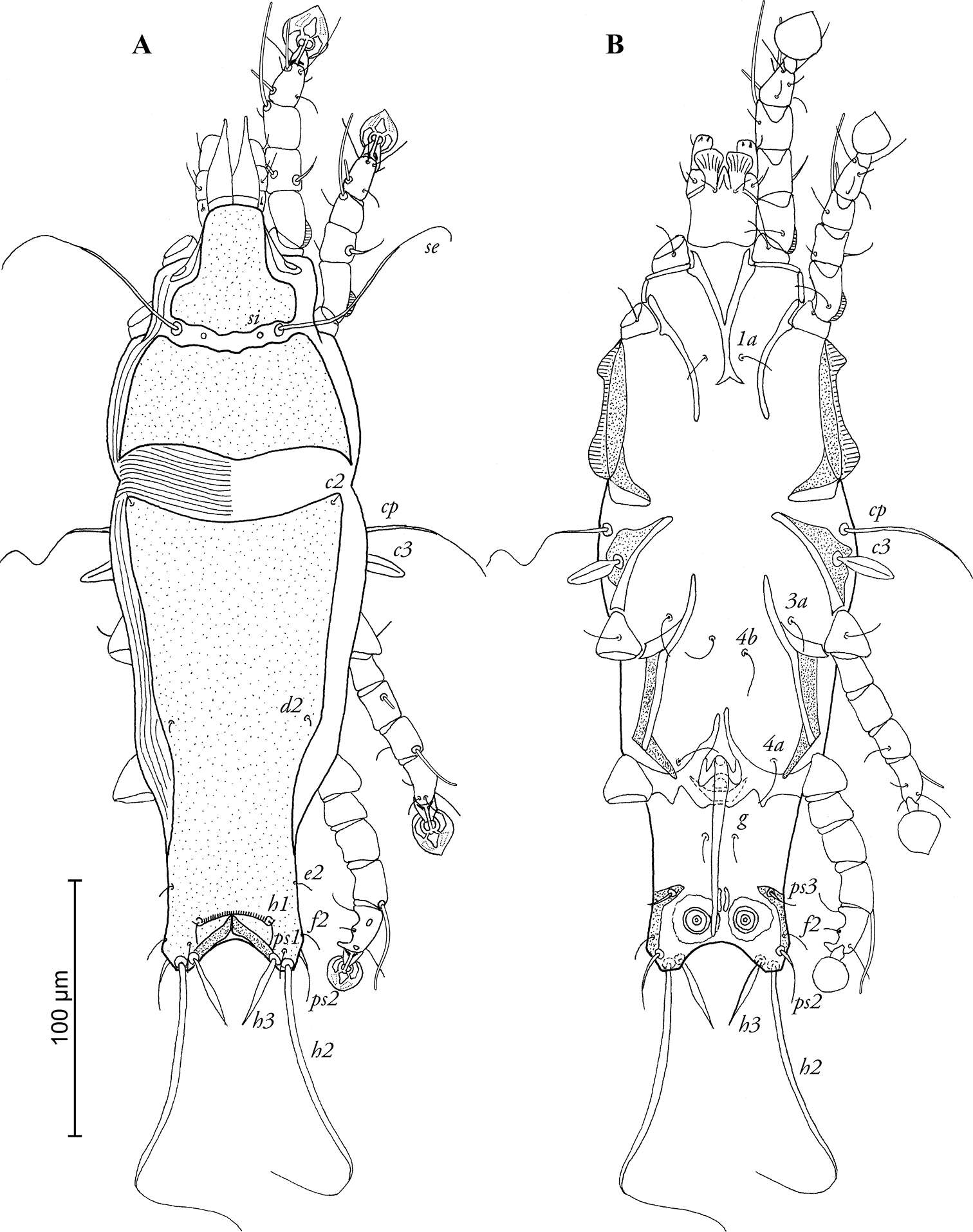
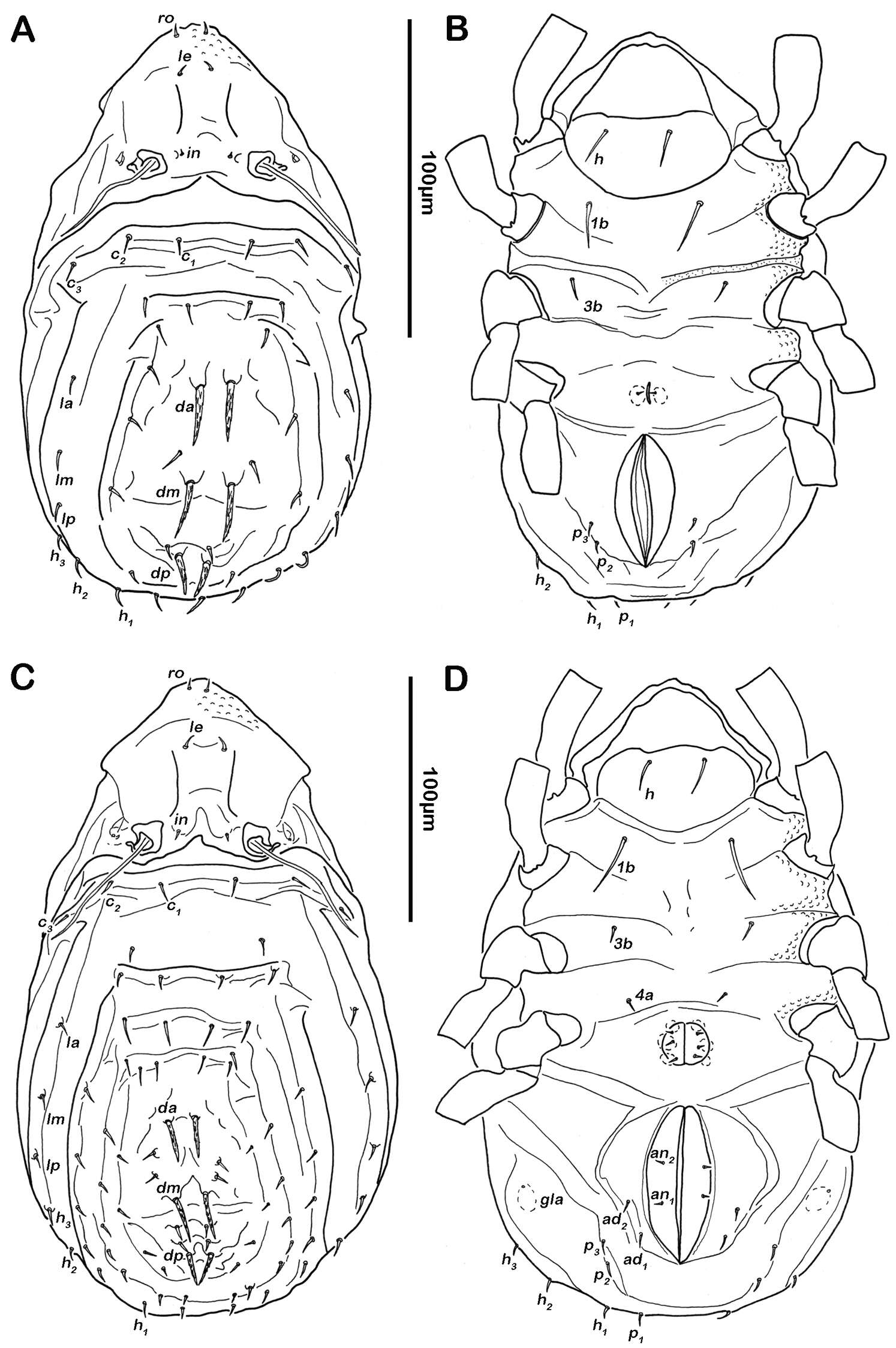
Figure 4.Selenoribates quasimodo sp. n. nymphs. A protonymph dorsal view B protonymph ventral view C tritonymph dorsal view D tritonymph ventral view.
-
Ioana Cristina Constantinescu, Gabriel Chişamera, D. Khlur B. Mukhim, Costică Adam
Zookeys
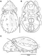
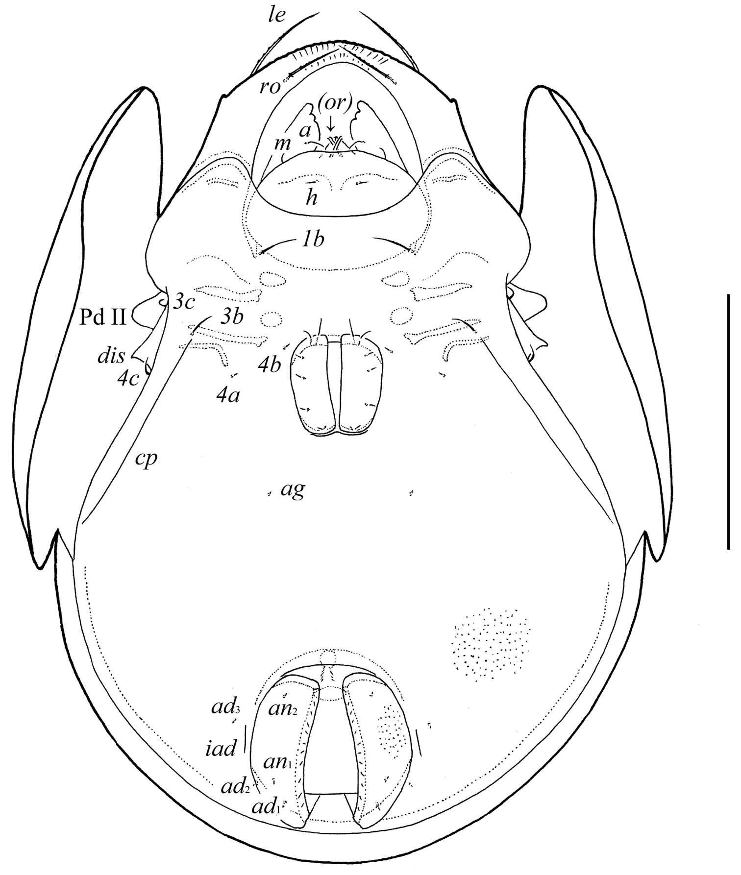
Figure 2.Pedanodectes angustilobus sp. n., female paratype: A dorsal view of idiosoma B ventral view of idiosoma.
-
Sergey G. Ermilov, Olman Alvarado-Rodríguez, Axel P. Retana-Salazar
Zookeys
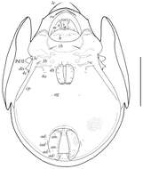
Figure 2.Pergalumna elongatiporosa sp. n.: ventral view (legs not illustrated). Scale bar 100 μm.
-
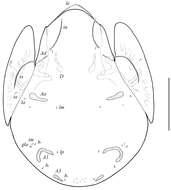
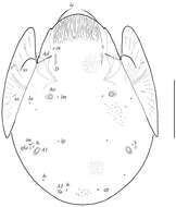
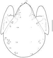
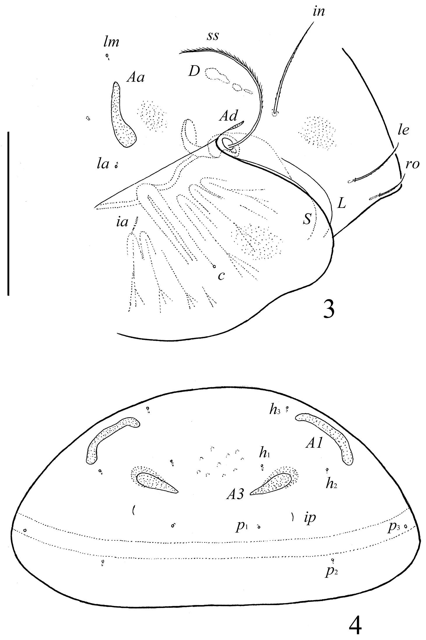
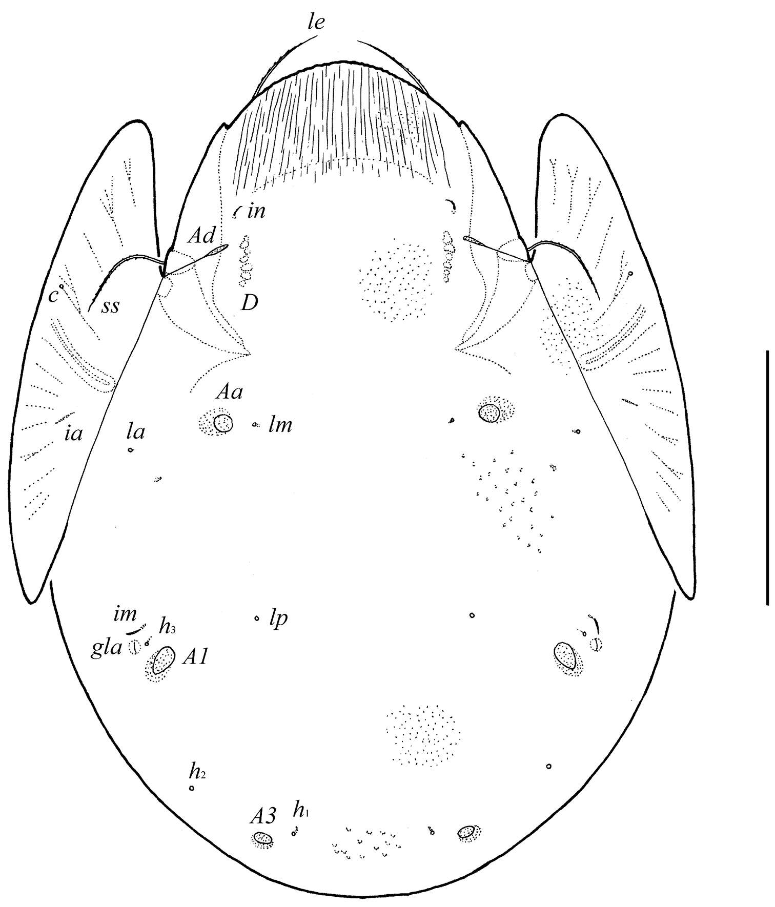
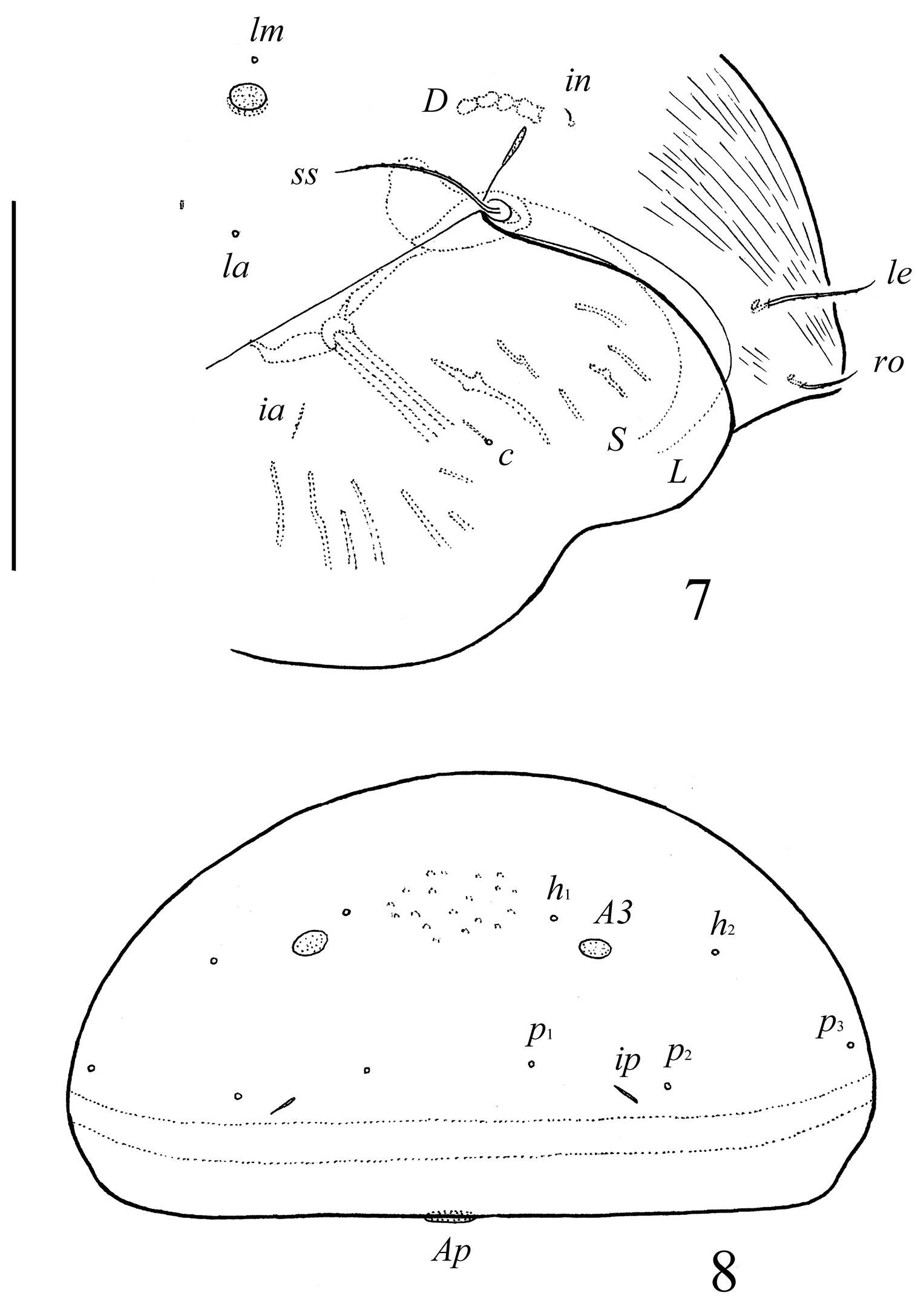
Figure 3.Conchogneta glabrisensillata sp. n. A Prodorsum, showing enantiophysis E, costula and bothridium B Central part of prodorsum, showing alveolus of interlamellar seta and interbothridial tubercle (indicated by arrow) C Part of laterial view of prodorsum, showing sensillus and granular tubercles on humeral region D Lateral view of prodorsal costula E Sensillus, lateral view F Slight variation of sensillus, lateral view G Granular tubercles on lateral part of prodorsum H Humeral region, showing tubercles Ha and Hp.
-
Figure 5.Selenoribates quasimodo sp. n. tritonymph, micrographs layered from 5–10 sequentially focused images. A dorsal view B ventral view. Scale bar = 100 µm.
-
Ioana Cristina Constantinescu, Gabriel Chişamera, D. Khlur B. Mukhim, Costică Adam
Zookeys
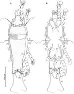
Figure 3.Pedanodectes angustilobus sp. n., details: A–D legs I–IV of male, respectively, dorsal view.
-
Sergey G. Ermilov, Olman Alvarado-Rodríguez, Axel P. Retana-Salazar
Zookeys
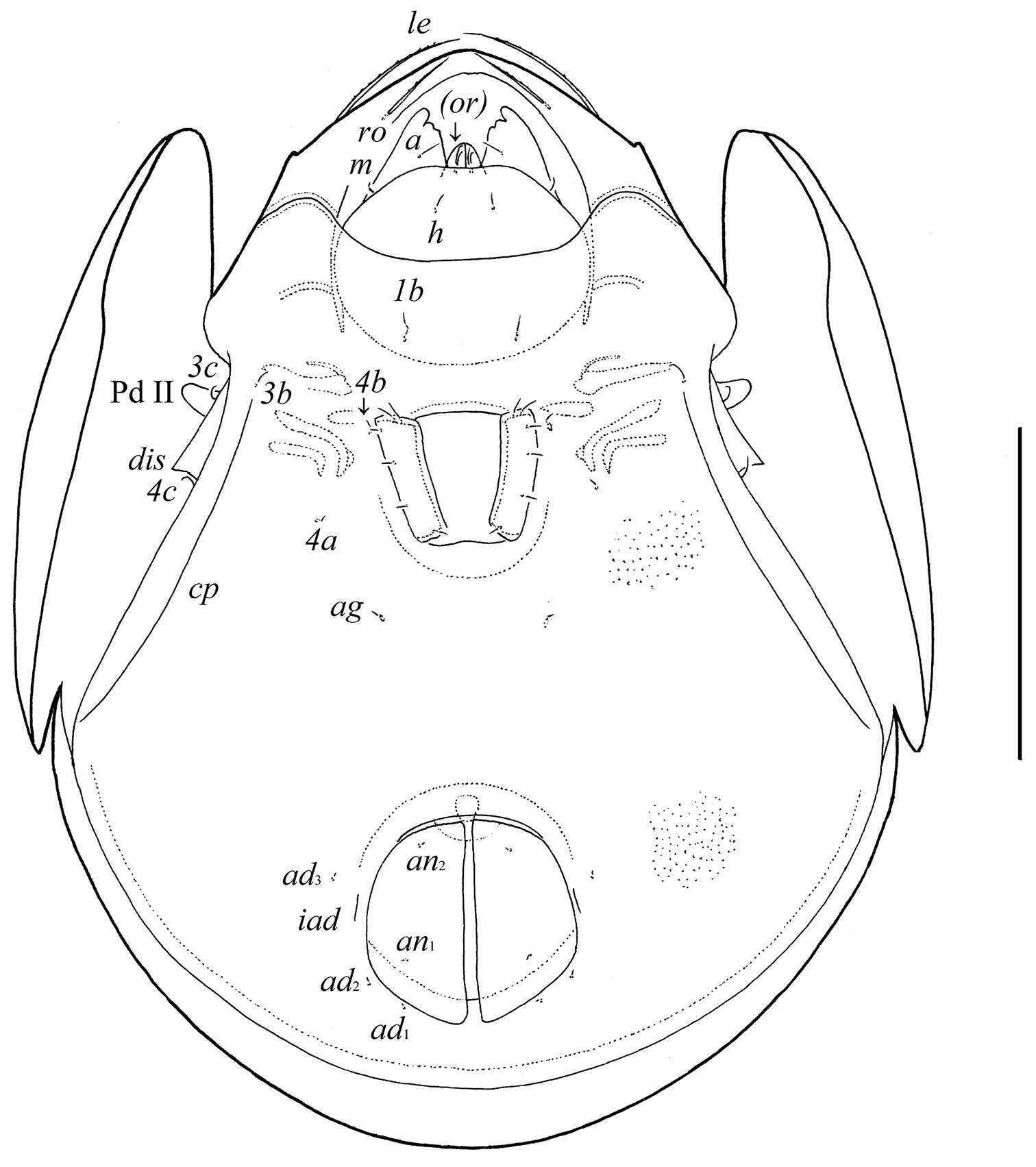
Figures 3–4.Pergalumna elongatiporosa sp. n.: 3 dorso-lateral view of prodorsum and anterior part of notogaster and pteromorph (gnathosoma and legs not illustrated) 4 posterior view of notogaster. Scale bars 100 μm.
-
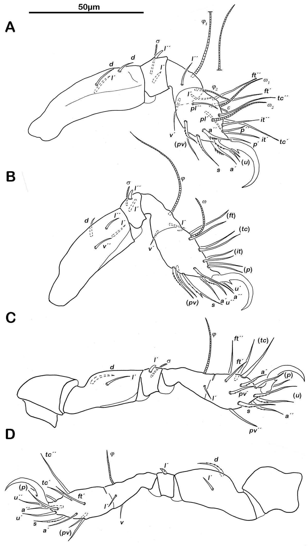
Figure 6.Selenoribates quasimodo sp. n. tritonymph legs. A right leg I antiaxial view B right leg II antiaxial view C left leg III antiaxial view D right leg IV antiaxial view.
-
Ioana Cristina Constantinescu, Gabriel Chişamera, D. Khlur B. Mukhim, Costică Adam
Zookeys
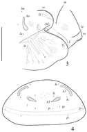
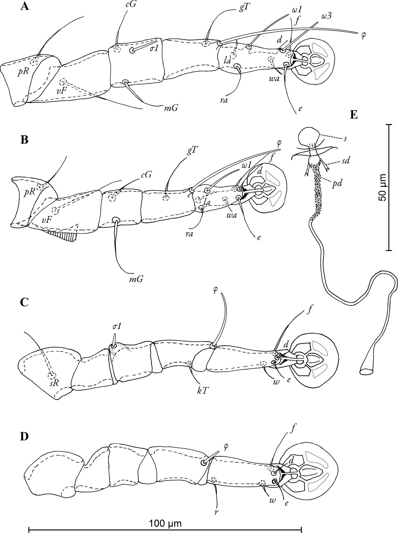
Figure 4.Pedanodectes angustilobus sp. n., details: A–D legs I–IV of female, respectively E spermatheca and spermaducts, dorsal view. Abbreviations: pd - primary spermaduct; s - spermatheca; sd - secondary spermaduct.
-
Sergey G. Ermilov, Olman Alvarado-Rodríguez, Axel P. Retana-Salazar
Zookeys
Figure 5.Pergalumna striatiprodorsum sp. n.: dorsal view. Scale bar 200 μm.
-
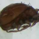
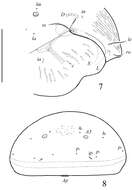
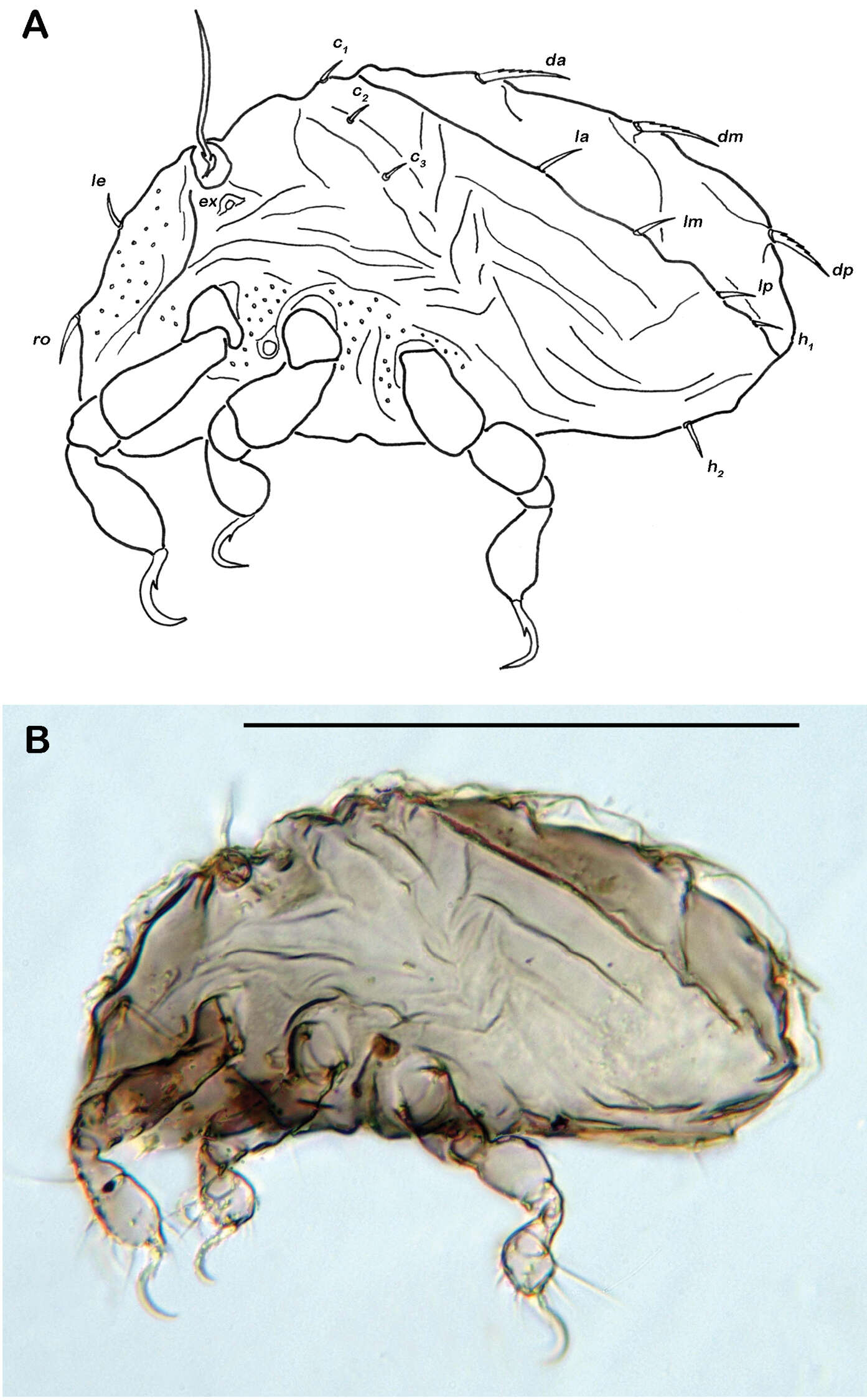
Figure 7.Selenoribates satanicus sp. n. adult. A dorsal view B ventral view C lateral view.
-
Sergey G. Ermilov, Olman Alvarado-Rodríguez, Axel P. Retana-Salazar
Zookeys
Figure 6.Pergalumna striatiprodorsum sp. n.: ventral view (legs not illustrated). Scale bar 200 μm.
-
Figure 8.Selenoribates satanicus sp. n. adult micrographs layered from 5–10 sequentially focused images. A dorsal view B ventral view. Scale bar = 100 µm.
-
Sergey G. Ermilov, Olman Alvarado-Rodríguez, Axel P. Retana-Salazar
Zookeys
Figures 7–8.Pergalumna striatiprodorsum sp. n.: 7 dorso-lateral view of prodorsum and anterior part of notogaster and pteromorph (gnathosoma and legs not illustrated) 8 posterior view of notogaster. Scale bars 200 μm.
-
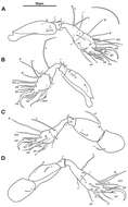
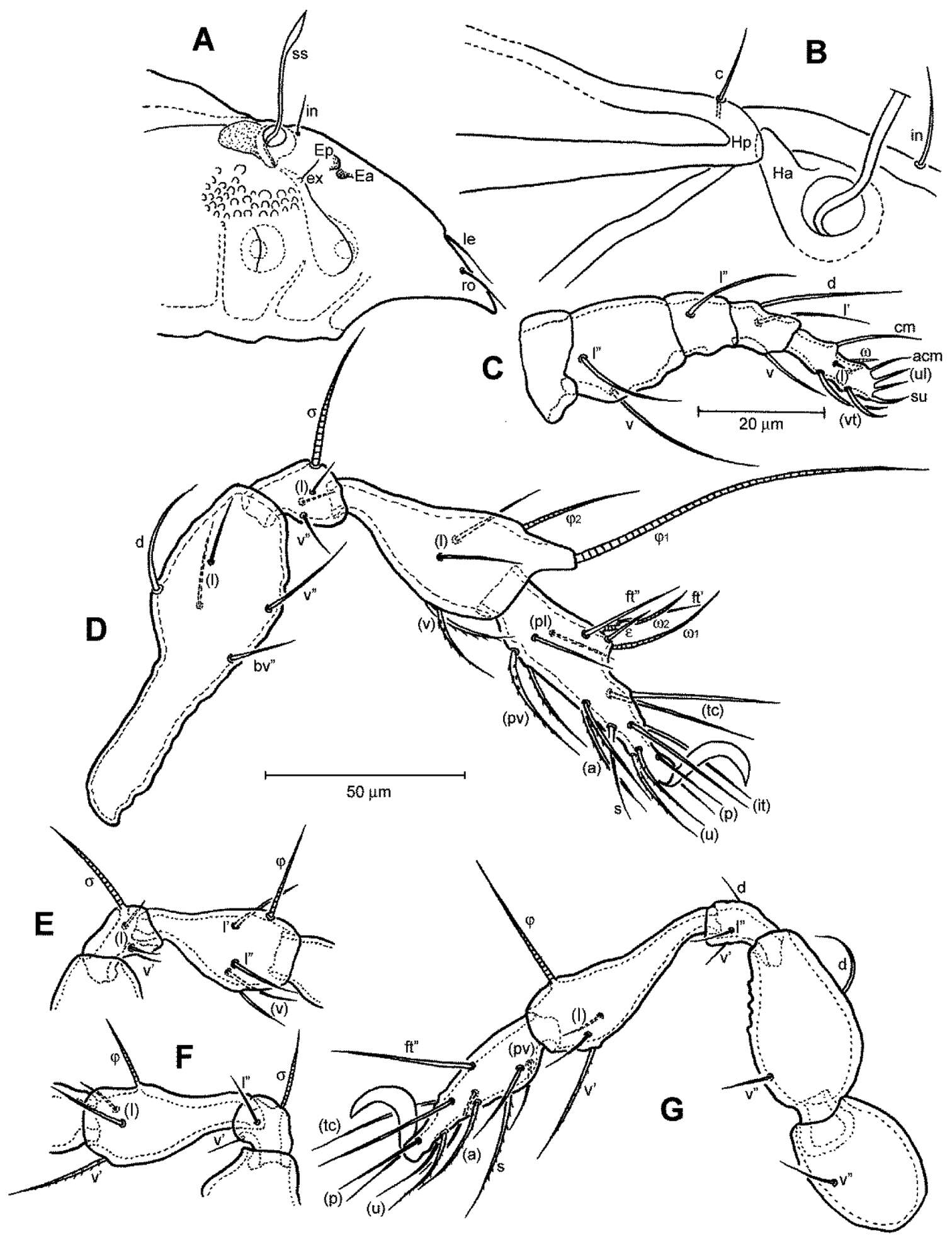
Figure 9.Selenoribates satanicus sp. n. legs. A right leg I antiaxial view B right leg II axial view C right leg III antiaxial view D left leg IV antiaxial view.
-
Sergey G. Ermilov, Jochen Martens, Andrei V. Tolstikov
Zookeys
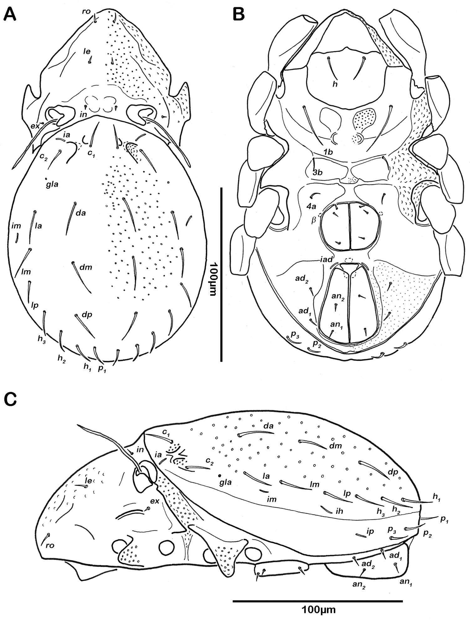
Figure 1.Galumna tetraporosa sp. n., adult: dorsal view. Scale bar 200 μm.
-
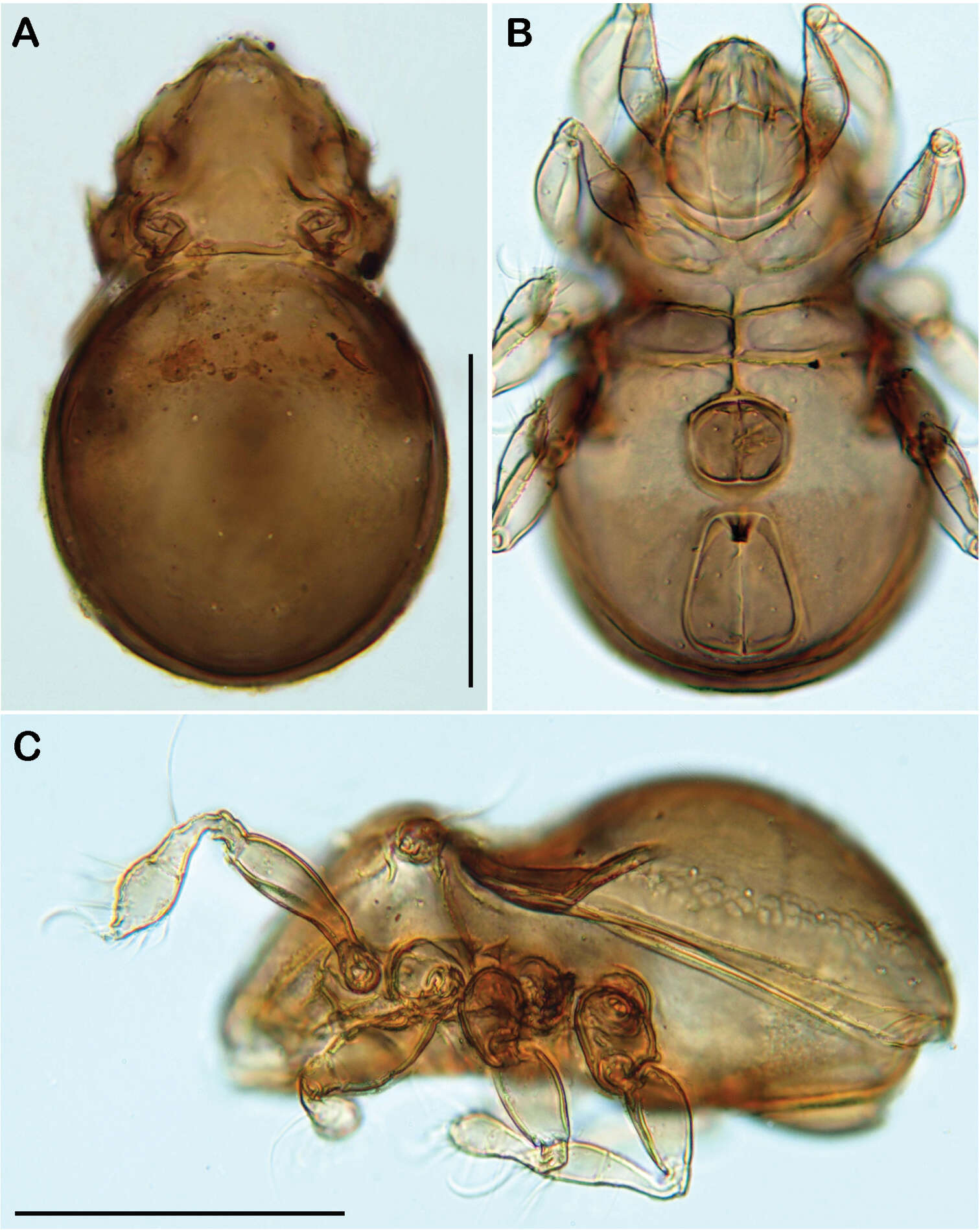
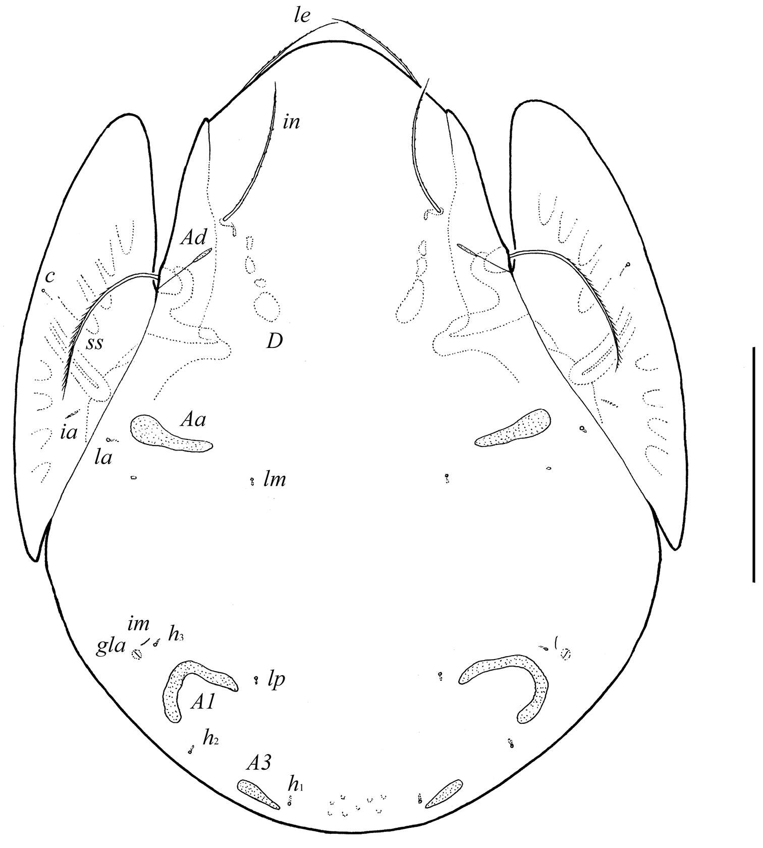
Figure 10.Selenoribates satanicus sp. n. larva. A lateral view B lateral view, micrograph layered from 5–10 sequentially focused images. Scale bar = 100 µm.
-
Sergey G. Ermilov, Jochen Martens, Andrei V. Tolstikov
Zookeys
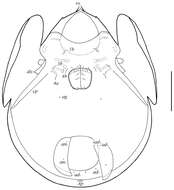
Figure 2.Galumna tetraporosa sp. n., adult: ventral view (gnathosoma and legs not illustrated). Scale bar 200 μm.
-
Figure 11.Selenoribates elegans sp. n. adult. A dorsal view B ventral view C lateral view.