portrait
Description:
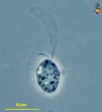
Paraphysomonas (para-fie-sew-moan-ass) a heterotrophic stramenopile (related to Ochromonas and organisms traditionally referred to as chrysophytes). It is distinguished because the body surface is coated with a fine layer of scales, although in most species (this one is an exception) the scales cannot be seen with the light microscope. There are two flagella, a long one with hairs (the hair are not visible with the light microscope) but which beats with an undulating motion and draws fluid and suspended food particles to the surface of the cell. This photograph was taken with a lengthened exposure, and the envelope of the flagellar beat is visible. Phase contrast.
Included On The Following Pages:
- Life (creatures)
- Cellular (cellular organisms)
- Eukaryota (eukaryotes)
- SAR (Stramenopiles, Alveolates, Rhizaria)
- Stramenopiles (heterokont)
- Ochrophyta (Ochrophyte)
- Chrysophyceae (golden algae)
- Chromulinales
- Paraphysomonadaceae
- Paraphysomonas
- Oomycota (oomycetes)
- Chrysista
This image is not featured in any collections.
Source Information
- license
- cc-by-nc
- author
- D. J. Patterson.
- provider
- micro*scope
- original
- original media file
- visit source
- partner site
- micro*scope
- ID