Drawing
Description:
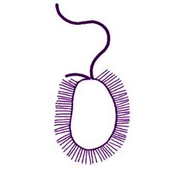
Paraphysomonas vestita (Stokes, 1885) De Saedeleer, 1929. Ovoid to elongate cells, from 6 to 15 microns long. Two flagella insert in an apical depression. One flagellum is very short and held laterally while the other, two to three times the length of the cell, is held anteriorly and beats with short wavelength and high amplitude wave pattern. Cells often contain granules. Scales are visible in light microscopy as delicate spines. In electron microscopy, the scales have a plain circular base (1-2 microns) with a well-marked rim and a tapering spine 3 to 5 microns long.
Included On The Following Pages:
- Life (creatures)
- Cellular (cellular organisms)
- Eukaryota (eukaryotes)
- SAR (Stramenopiles, Alveolates, Rhizaria)
- Stramenopiles (heterokont)
- Ochrophyta (Ochrophyte)
- Chrysophyceae (golden algae)
- Chromulinales
- Paraphysomonadaceae
- Paraphysomonas
- Paraphysomonas vestita
- Oomycota (oomycetes)
- Chrysista
This image is not featured in any collections.
Source Information
- license
- cc-by-nc
- author
- Won Je Lee
- provider
- micro*scope
- original
- original media file
- visit source
- partner site
- micro*scope
- ID