Drawing
Description:
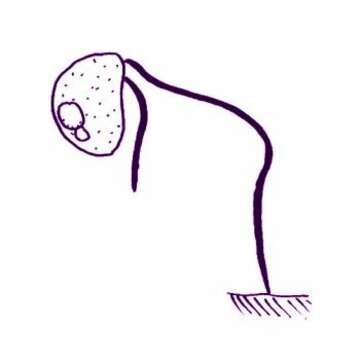
Cafeteria minuta (Ruinen, 1938) Larsen and Patteron, 1990. Cells are almost globular, 3.5-4 microns but with a subapical ventral depression from which the flagella emerge. The hairy or feeding flagellum (the hairs are not visible with the light microscope), which projects in front of the swimming cell, measures 10-12 microns, and may beat with more than one complete sine wave along its length. Second flagellum about 5 microns, curves over the side of the body and may attach by its tip to the substratum.
Included On The Following Pages:
- Life (creatures)
- Cellular (cellular organisms)
- Eukaryota (eukaryotes)
- SAR (Stramenopiles, Alveolates, Rhizaria)
- Stramenopiles (heterokont)
- Bigyra
- Bicosoecida
- Cafeteriaceae
- Cafeteria
- Cafeteria minuta
- Bicosoecia
- Bicoecea
- Bicosoecida
- Cyathobodoniae
This image is not featured in any collections.
Source Information
- license
- cc-by-nc
- author
- Won Je Lee
- provider
- micro*scope
- original
- original media file
- visit source
- partner site
- micro*scope
- ID