Drawing
Description:
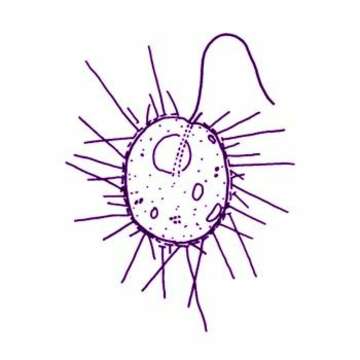
Thaumatomastix salina (Birch-Andersen) Beech and Moestrup, 1986. Cells are ovoid (7-12 microns x 8-15 microns), slightly compressed dorso-ventrally, and have a long flagellum 3/4-5/4 of the cell length. A short flagellum, which is rarely visible, emerges together with the long flagellum from what appears to be a very slight groove or depression located latero-anteriorly. A furrow-like structure is often noted running from the flagellar bases to the cell midline. Cells are solitary, and are most often observed attached to pieces of detritus. Cells occasionally move in a creeping motion, with the long flagellum trailing and gliding over the coverslip. In some cases cells swim freely with the long flagellum making irregular, arhythmical flicking motions. The cell cytoplasm has a granular appearance and is devoid of any kind of chloroplast, a diffuse area of a pale orange colour can often be noticed in the central part of the cell when phase contrast oil immersion optics are used. One cell was noted in an early stage of division where both flagella had replicated. Spine scales, varying in length, radiate from the entire cell surface. Flattened cells slough off their scales and scales of a second type, spineless body scales, can then be seen to be elliptical in outline.
Included On The Following Pages:
- Life (creatures)
- Cellular (cellular organisms)
- Eukaryota (eukaryotes)
- SAR (Stramenopiles, Alveolates, Rhizaria)
- Rhizaria (rhizarians)
- Cercozoa (cercozoans)
- Imbricatea
- Silicofilosea
- Thaumatomonadida
- Thaumatomastigidae
- Thaumatomastix
- Thaumatomastix salina
This image is not featured in any collections.
Source Information
- license
- cc-by-nc
- author
- Won Je Lee
- provider
- micro*scope
- original
- original media file
- visit source
- partner site
- micro*scope
- ID