Surface detail, in vivo
Description:
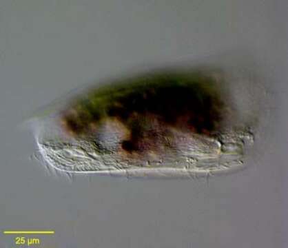
Surface detail of the marine Phyllopharyngeid ciliate, Dysteria brasiliensis Da Cunha, De Faria & Pinto, 1922. This is one of the largest species of this genus (100-130 um).The posterior terminates in a sharp spinous process (slightly out of focus here) not to be confused with the ventral posterior podite by which the cell attaches to the substrate. The podite is angled anteriorly in this image (the the viewer's right).Collected from a commercial saltwater aquarium in Boise, Idaho. March 2004. DIC.
Included On The Following Pages:
- Life (creatures)
- Cellular (cellular organisms)
- Eukaryota (eukaryotes)
- SAR (Stramenopiles, Alveolates, Rhizaria)
- Alveolata (alveolates)
- Ciliophora (ciliates)
- Intramacronucleata
- Phyllopharyngea
- Phyllopharyngia
- Dysteriida
- Dysteriidae
- Dysteria
- Dysteria brasiliensis
This image is not featured in any collections.
Source Information
- license
- cc-by-nc
- author
- Bill Bourland
- provider
- micro*scope
- original
- original media file
- visit source
- partner site
- micro*scope
- ID