Portrait
Description:
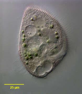
Portrait of the medium-size pigmented heterotrich ciliate, Blepharisma lateritium (Ehrenberg,1831; Stein, 1859). The cell is teardrop-shaped and pale pink in color. The peristome extends about 2/3 the cell length along the left side. The peristome is bordered on the left by serial polykinetids forming an adoral zone of membranelles and on the right by a narrow undulating membrane which is less conspicuous. The longitudinal somatic kineties bend to the left margin on the dorsal surface. The single ellipsoid to spherical macronucleus is located left of midline in the cell center. There are multiple very small micronuclei which are difficult to discern in vivo. These become swollen and densely stained in silver impregnated specimens. The contractile vacuole is at the posterior end. There are rows of pink cortical pigment granules between the somatic kineties. The pigment, blepharismin, is structurally similar to hypericin. When exposed to light, blepharismin causes a change in the cell's membrane potential and thus direction of ciliary beat causing light avoidance or photodispersal. Exposure to bright light for even short periods causes cell lysis. This is often observed in illuminated Blepharisma under the microscope. Blepharismin probably has a defensive function against predators such as the haptorid ciliate, Dileptus. Collected from an artificial freshwater dredge pond near Boise, Idaho October 2004. DIC optics.
Included On The Following Pages:
- Life (creatures)
- Cellular (cellular organisms)
- Eukaryota (eukaryotes)
- SAR (Stramenopiles, Alveolates, Rhizaria)
- Alveolata (alveolates)
- Ciliophora (ciliates)
- Postciliodesmatophora
- Heterotrichea
- Heterotrichida
- Blepharismidae
- Blepharisma
- Blepharisma lateritium
This image is not featured in any collections.
Source Information
- license
- cc-by-nc
- author
- William Bourland
- provider
- micro*scope
- original
- original media file
- visit source
- partner site
- micro*scope
- ID