Infraciliature in division
Description:
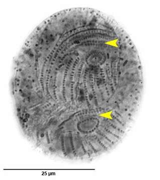
Ventral surface of Chlamydonella alpestris (Foissner, 1979) in mid-division. C. alpestris is a small hypostome ciliate. The body is strongly curved dorsally and flattened ventrally. The right side is convex and the left is straight. The cell shape has been distorted by fixation. Ciliature is limited to the ventral surface except for a small dorsal anterior tuft on the left. Kineties curve anterior to the cytostome on the right. About 10 evenly spaced longitudinal kineties extend from the level of the cytostome to the posterior end. A flattened transverse Y-shaped kinety just anterior to the cytostome is considered distinctive (yellow arrowheads). A second short curved kinety lies just anterior to this. The circular oral aperture is supported by trichites. The single round macronucleus is located in the mid-body (not visible here). There are two contractile vacuoles, one anterior and one posterior. This individual has consumed a diatom and green alga. Collected from freshwater pond near Boise, Idaho October 2003. Silver carbonate stain (see Foissner, W.Europ. J. Protistol.27,313-330;1991). Black and white.Brightfield optics.
Included On The Following Pages:
- Life (creatures)
- Cellular (cellular organisms)
- Eukaryota (eukaryotes)
- SAR (Stramenopiles, Alveolates, Rhizaria)
- Alveolata (alveolates)
- Ciliophora (ciliates)
- Intramacronucleata
- Phyllopharyngea
- Phyllopharyngia
- Chlamydodontida
- Lynchellidae
- Chlamydonella
- Chlamydonella alpestris
This image is not featured in any collections.
Source Information
- license
- cc-by-nc
- author
- William Bourland
- provider
- micro*scope
- original
- original media file
- visit source
- partner site
- micro*scope
- ID